The landscape of subatomic imaging is poised for a profound transformation as an interdisciplinary team of researchers in Austria successfully lays the groundwork for a hybrid technology that merges conventional electron microscopy with trapped-ion quantum computing. Spearheaded by institutions including the Vienna University of Technology (TU Wien), alongside the University of Vienna, Johannes Kepler University (JKU) Linz, and the University of Innsbruck, this ambitious scientific undertaking seeks to extract previously inaccessible quantum data carried by electrons. By capturing and processing this overlooked information, scientists anticipate overcoming longstanding physical thresholds that have historically constrained the imaging of delicate biological structures.
As construction of the world’s first quantum computer-enhanced electron microscope gets underway at TU Wien, the scientific community is taking a closer look at a methodology that promises higher-resolution imaging under lower radiation dosages. This breakthrough development could redefine observational limits in structural biology, materials science, and quantum information science.
The Core Innovation: Unlocking Hidden Quantum Data
Standard transmission electron microscopes operate on a conceptually straightforward yet physically punishing premise: a focused beam of electrons is directed at a sample, and the particles that pass through or scatter are counted to construct a high-resolution image. Over decades of refinement, these instruments have achieved extraordinary feats, allowing researchers to visualize individual atoms, crystalline lattices, and complex macromolecular assemblies with astonishing clarity.
However, this high-resolution capability comes with a severe trade-off. To resolve fine atomic details, instruments must project a high density of electrons onto the specimen. While robust inorganic materials can withstand this intense bombardment, delicate biological specimens—such as isolated proteins, viral capsids, and cellular membranes—are rapidly degraded, ionized, or structurally destroyed by the energy transfer. This limitation creates a persistent dilemma in structural biology: samples are frequently damaged before a clear, high-resolution image can be fully realized.
The Austrian research consortium addresses this fundamental bottleneck by shifting attention away from mere electron counting and toward the intrinsic quantum properties of the beam itself. While traditional detectors register only the arrival position or intensity of electrons, each individual particle also carries nuanced quantum mechanical information that is routinely discarded. By routing the electron beam through a system integrated with a trapped-ion quantum computer, the research team aims to intercept, entangle, and process this hidden data, turning weak signals that would typically vanish into background noise into distinct, actionable imagery.
Chronology and Collaborative Development
The genesis of this hybrid architecture represents a multi-year convergence of distinct scientific disciplines, drawing heavily upon Austria’s robust national infrastructure for quantum research. The foundational concepts underpinning the project were initially formulated through theoretical analyses conducted jointly by theoretical physicists at TU Wien and algorithmic specialists at JKU Linz.
The collaborative effort was officially catalyzed under the umbrella of the quantA Cluster of Excellence, a major research initiative funded by the Austrian Science Fund (FWF) alongside backing from the Gordon and Betty Moore Foundation. This funding framework enabled physicists, quantum information theorists, and microscopy experts from four separate Austrian academic institutions to pool their resources and specialized hardware.
In the initial theoretical phase, researchers utilized advanced mathematical modeling to prove that entangling imaging electrons with trapped ions could theoretically boost signal-to-noise ratios without increasing the total number of incident particles. Following the publication of these mathematical frameworks, the project transitioned into an engineering and development phase.
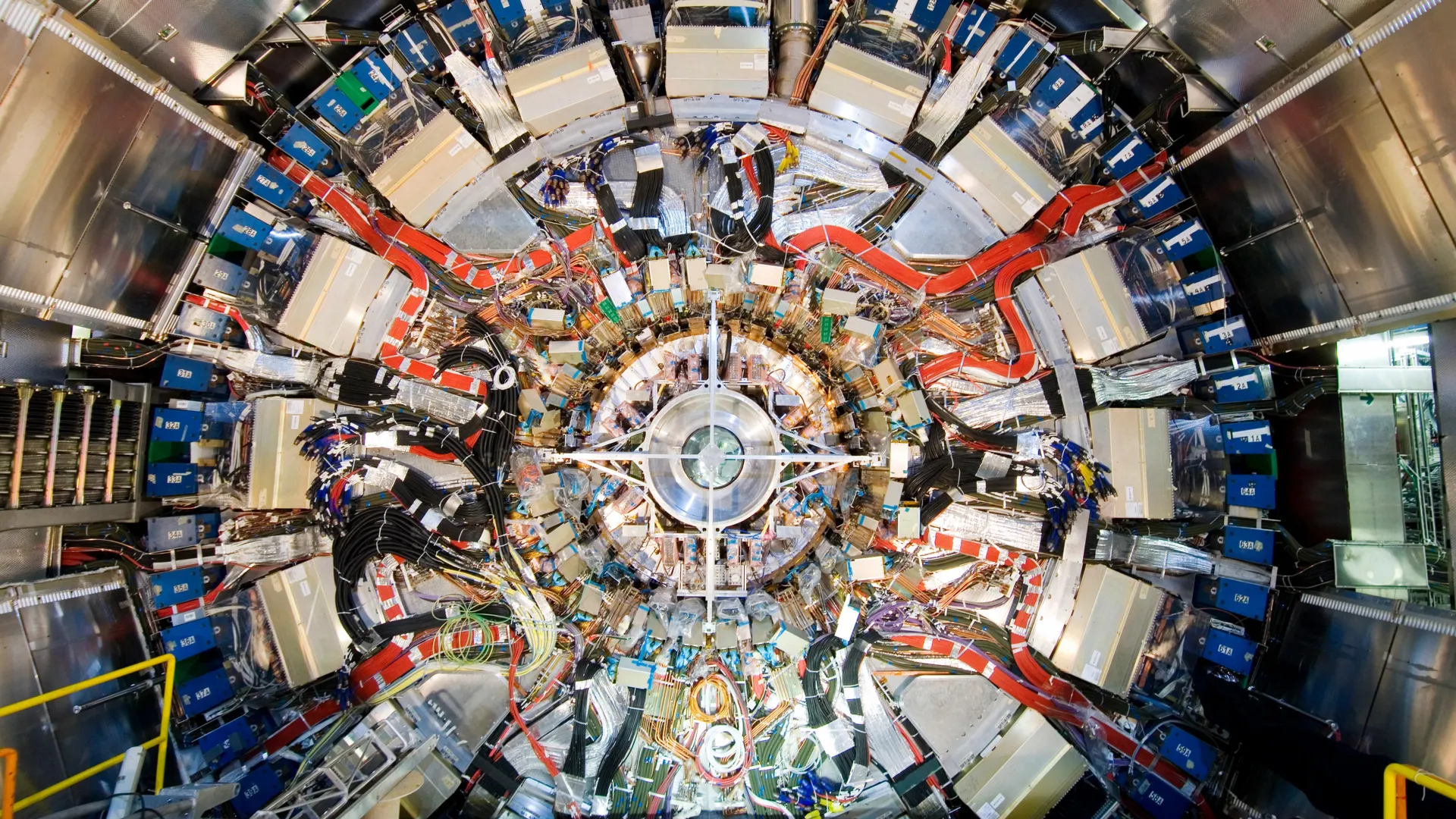
The quantum processing core of the new instrument—specifically an advanced ion-trap quantum computing module developed under the direction of Philipp Schindler’s research group at the University of Innsbruck—is currently being physically integrated into an electron microscope at TU Wien’s University Service Center for Transmission Electron Microscopy (USTEM). This hardware integration phase marks a pivotal transition from theoretical physics to experimental validation, with the consortium aiming to capture its first hybrid quantum-enhanced images in the near future.
Mechanics of the Hybrid System: Entanglement and Signal Enhancement
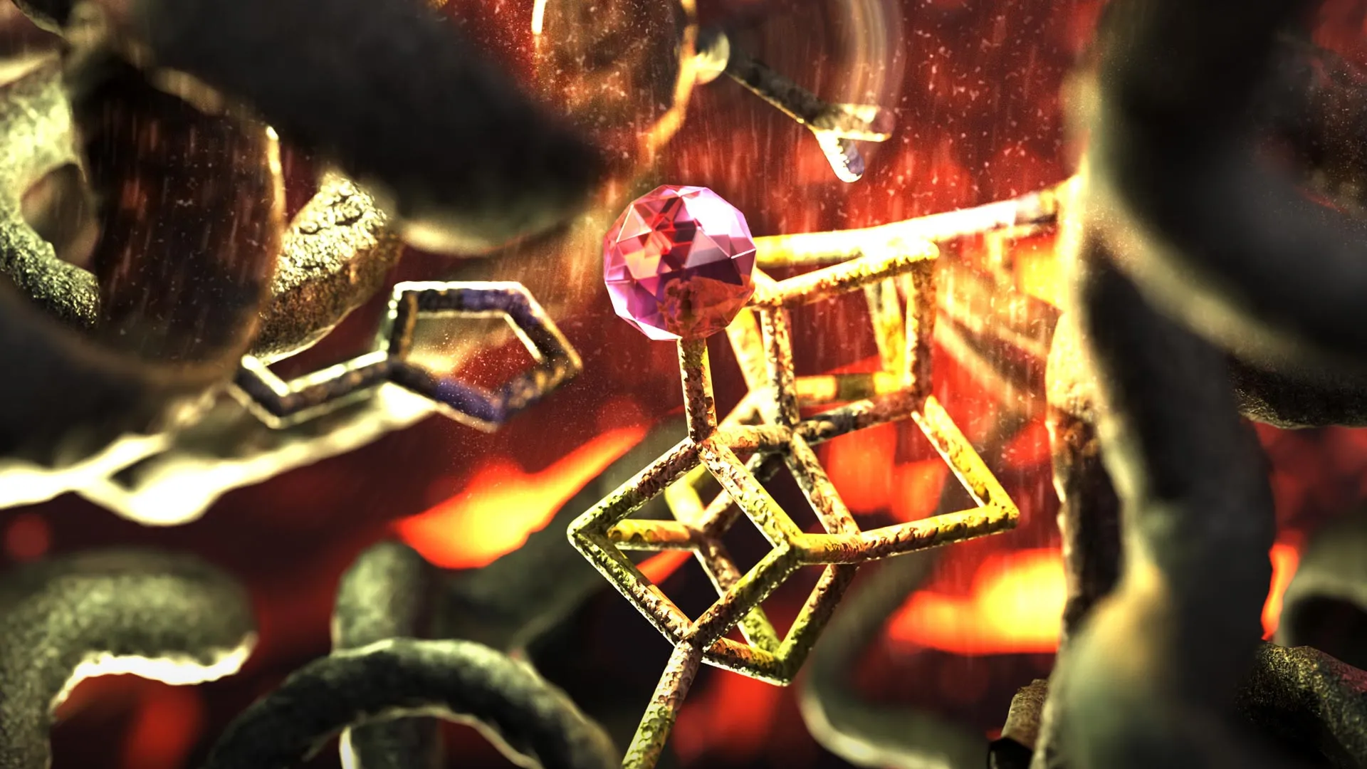
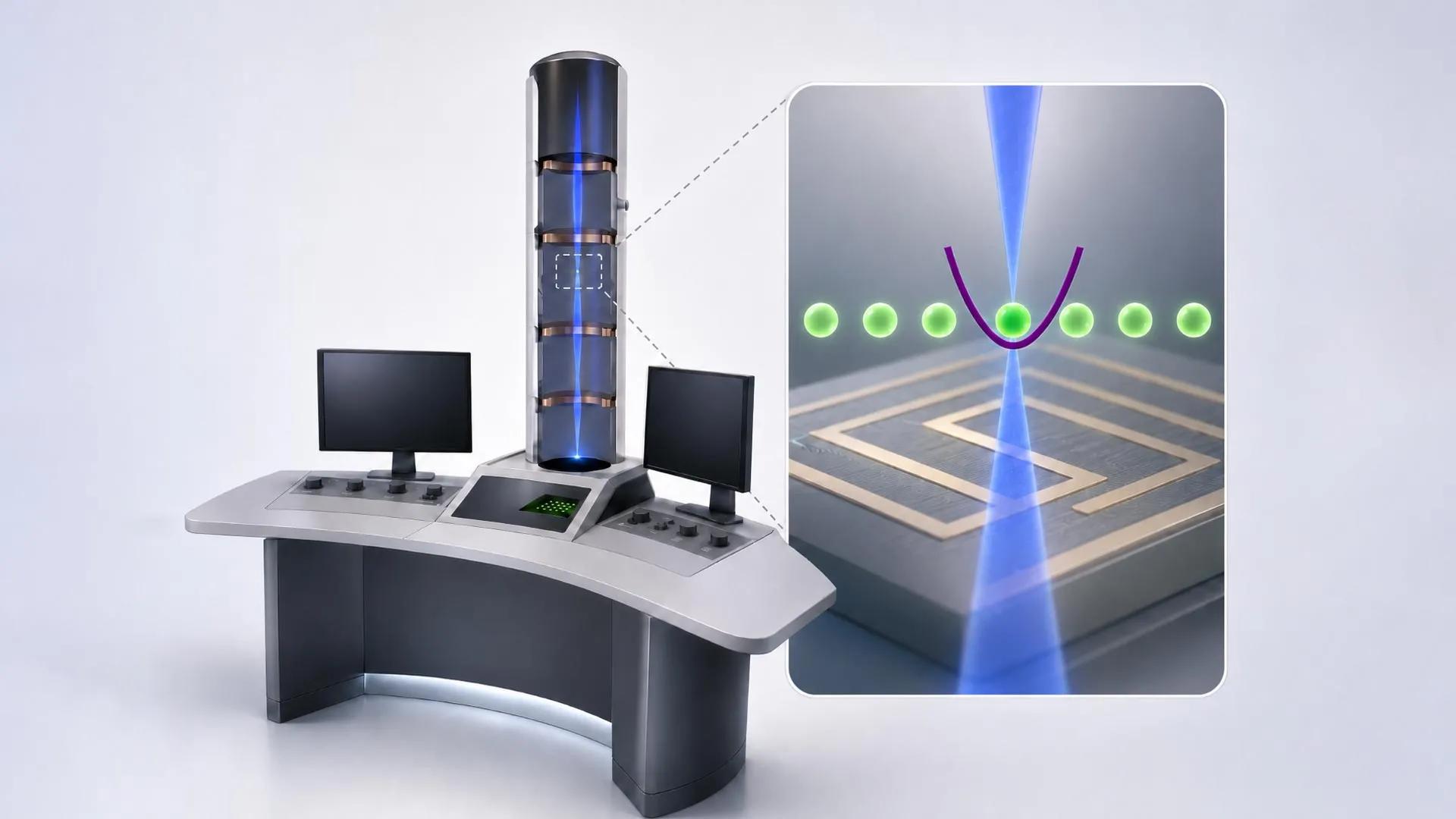
To understand how a quantum computer can enhance an electron microscope, one must examine the physical interaction between the electron beam and the trapped ions. The proposed apparatus aligns individual ions along the trajectory of the microscope’s electron beam. As an electron traverses this path, it interacts electrostatically with the trapped ions, inducing a state of quantum entanglement where the physical states of both the electron and the ion become inextricably linked.
Through this entanglement, quantum information pertaining to the electron’s trajectory, phase, and scattering characteristics is safely transferred to and stored within the ion. Crucially, the protocol does not stop with a single particle. Subsequent electrons can be directed through the system to interact sequentially with the quantum memory architecture of the trapped ions.
By executing a series of carefully tailored quantum algorithms developed in collaboration with Johannes Kofler’s team at JKU Linz, the system can coherently combine the information gathered across multiple electrons. This cumulative processing yields a signal of maximum strength derived from a remarkably sparse population of primary particles.
By utilizing quantum mechanics to bypass standard statistical noise barriers, the instrument effectively transforms faint, ambiguous scattering events into high-fidelity data points. Consequently, scientists can reconstruct sharp, detailed structural images of sensitive samples while exposing those specimens to a fraction of the electron dose traditionally required.
Perspectives from the Research Team
The diverse team behind the project emphasizes that the fundamental utility of the electron beam as an imaging probe remains unchanged, while the analytical backend undergoes a paradigm shift.
"Today, we can image tiny details on the atomic scale," remarks Philipp Haslinger of the Institute of Atomic and Subatomic Physics at TU Wien. "However, this requires a large number of electrons. And not every sample can be exposed to so many electrons without being damaged. This is often a problem, particularly when imaging biological samples such as individual proteins."
Elias Pescoller, a doctoral student at TU Wien and first author of the pivotal study detailing the concept, elaborates on the mechanics of the system: "Our idea is to combine the electrons with a quantum computer. We let them interact with ions that are held in place along the path of the electron beam. This can, for example, create quantum entanglement between the electron and the quantum computer. The electron and the ion then share a joint quantum state."
Pescoller further notes the broader methodological advantage: "Quantum physics allows us to overcome the statistical limits that constrain conventional electron microscopes."
Addressing the signal processing aspect, Dennis Rätzel of TU Wien explains the efficiency of the multi-electron approach: "If we perform very specific quantum-computing operations each time, we can optimally combine the information from several electrons so that we obtain a signal of maximum strength even though we use only a relatively small number of electrons."
Iva Březinová of the Institute for Theoretical Physics at TU Wien highlights the interpretive transformation enabled by the quantum backend: "The electrons themselves are used to image small objects, just as in any other electron microscope. But by processing the quantum information carried by these electrons in a quantum computer, we can extract significantly more information from the process. What would previously have been indistinguishable from random noise can thus become a clear signal."
Looking toward the broader institutional synergy, Thomas Juffmann of the University of Vienna underscores the collaborative significance of the undertaking: "It is really exciting that, within the quantA Cluster of Excellence, we can combine the expertise in quantum information, quantum computing and electron microscopy available at the different universities in Austria. This allows us to launch a unique project."
Broader Implications and Future Applications
The successful deployment of a quantum computer-enhanced electron microscope could yield profound implications across multiple scientific disciplines. In structural biology, the ability to obtain high-resolution tomographic and spatial data from fragile macromolecules without inducing severe radiation damage would open unprecedented avenues for drug discovery, virology, and cellular pathology. Researchers could potentially visualize dynamic biological processes and delicate protein conformations that currently degrade too rapidly under standard imaging protocols to be accurately recorded.
In materials science, the technology could facilitate the non-destructive analysis of beam-sensitive quantum materials, polymers, and delicate nanostructures. As industries push toward smaller manufacturing tolerances and more complex nanoscale devices, the demand for non-invasive analytical tools continues to grow exponentially.
Beyond microscopy, the successful coupling of free-electron beams with trapped-ion quantum computers establishes a novel technological platform for quantum networking and hybrid quantum systems. By demonstrating that quantum information can be reliably transferred between free-flying electrons and stationary matter qubits, the Austrian research consortium has charted a new methodological pathway that extends well beyond the boundaries of traditional imaging.
As the construction of the hybrid microscope advances at USTEM, the international scientific community awaits experimental verification of the team’s theoretical models. Should the hardware perform as projected, physics and biology textbooks may soon reflect a new generation of microscopes—instruments that see deeper into the atomic realm not by casting a harsher light, but by listening more carefully to the subtle quantum whispers of each passing electron.