A landmark study conducted by a collaborative team of researchers from Japan has fundamentally reshaped our understanding of female reproductive biology, specifically identifying an overlooked yet crucial role for microtubules in the intricate communication between oocytes and granulosa cells within ovarian follicles. The research, spearheaded by Masamitsu Sato and Mika Toya from Waseda University and Kyoto University respectively, focused on the microtubule-stabilizing protein Camsap3, revealing its indispensable function in organizing these cellular structures which are vital for successful ovulation and fertility. Utilizing sophisticated knockout mouse models and advanced imaging techniques, the team meticulously examined transzonal projections (TZPs), the cellular bridges facilitating communication, and unveiled that microtubules, previously underestimated, are core components of these structures, with Camsap3 acting as a critical regulator.
The Foundation of Female Fertility: Ovarian Follicle Dynamics
Female fertility is an exquisitely complex biological process, hinging on the successful development and maturation of oocytes (eggs) encapsulated within ovarian follicles. These follicles, microscopic sacs located in the ovaries, undergo a series of precise developmental stages – from primordial to primary, secondary, antral, and finally, the mature Graafian follicle – each step critical for producing a viable egg ready for fertilization. Central to this process is the dynamic interplay between the developing oocyte and its surrounding somatic cells, known as granulosa cells. These support cells are instrumental in providing essential nutrients, signaling molecules, and structural integrity, creating a microenvironment conducive to oocyte growth and maturation. Disruptions at any stage of this delicate cellular ballet can lead to significant fertility challenges, affecting millions of individuals globally. According to the World Health Organization, an estimated 1 in 6 people worldwide experience infertility, underscoring the urgent need for deeper scientific understanding of its underlying mechanisms.
Communication between the oocyte and granulosa cells is primarily facilitated by specialized cellular extensions called transzonal projections (TZPs). These thin, elongated structures traverse the zona pellucida, a thick glycoprotein layer surrounding the oocyte, forming direct physical connections. Historically, TZPs were largely conceptualized as actin-based structures, with the contribution of microtubules within them remaining poorly understood or largely dismissed. Actin filaments are known for their role in cell motility and shape, while microtubules, components of the cytoskeleton, are crucial for maintaining cell structure, intracellular transport, and cell division. The prevailing view had therefore limited the perceived structural complexity and functional potential of TZPs, particularly concerning long-distance intracellular transport.
Unveiling Camsap3’s Pivotal Role
Driven by the ambition to clarify the precise contribution of microtubules to ovarian follicle development, the research group embarked on an investigation into Camsap3, a known microtubule-stabilizing protein. The team comprised prominent researchers including Akihiro Aikawa, Takao Tsurumaki, and Erina Kuranaga from Kyoto University, alongside Junya Ito from Azabu University, collectively bringing diverse expertise to this challenging biological puzzle. Their central hypothesis posited that if microtubules were indeed critical in TZPs, then a protein responsible for their stability, like Camsap3, would play a significant role in follicle communication and overall fertility.
To rigorously test this hypothesis, the researchers employed a comprehensive experimental strategy. The cornerstone of their approach involved the generation of Camsap3-knockout (KO) mice, genetically engineered to lack the functional Camsap3 protein. These KO mice were then meticulously compared against wild-type mice, serving as the control group, to isolate the specific effects of Camsap3 deficiency. The evaluation of fertility and ovulation involved both natural mating experiments and hormone-induced superovulation assays, providing a robust assessment of reproductive capacity.
Further layers of analysis included detailed histological examination of ovarian follicles at various developmental stages, allowing for a microscopic assessment of follicular health and progression. To visualize the intricate cellular architecture of TZPs and their cytoskeletal components, the team utilized cutting-edge super-resolution microscopy alongside immunofluorescence staining techniques. This advanced imaging allowed for the precise localization of actin filaments, microtubules, and Camsap3 itself within TZPs, pushing the boundaries of what could be observed at the subcellular level. Complementary experiments were conducted to assess granulosa cell apoptosis (programmed cell death) and proliferation, factors crucial for follicular integrity and growth. Finally, follicle reconstitution assays were performed, a sophisticated method to dissect the individual contributions of granulosa cells and oocytes to the overall follicular development, providing granular insights into their interdependent roles. This multi-faceted approach ensured a thorough and reliable investigation into Camsap3’s function and the broader implications for reproductive health.
Breakthrough Findings: Infertility and Reimagined TZPs
The rigorous investigation yielded several groundbreaking findings that significantly advance the field of reproductive biology. Perhaps the most striking observation was the complete infertility of female Camsap3-KO mice. Despite exhibiting normal estrous cycles, indicating an intact hormonal regulatory system, these mice consistently failed to ovulate. This crucial detail strongly suggested that the observed infertility was not due to a systemic hormonal imbalance but rather a localized defect at the cellular level within the ovarian follicles, directly implicating Camsap3’s role in the physical and functional integrity of the reproductive machinery.
Further histological analysis illuminated the specific developmental roadblock encountered by the Camsap3-KO mice. Follicle development was severely disrupted during the critical transition from the secondary to the antral stage. This arrested development led to a marked increase in follicular degeneration, a process known as atresia, where follicles prematurely break down. Consequently, there was a drastic reduction in the number of mature Graafian follicles, the final stage before ovulation, effectively preventing the release of viable eggs.
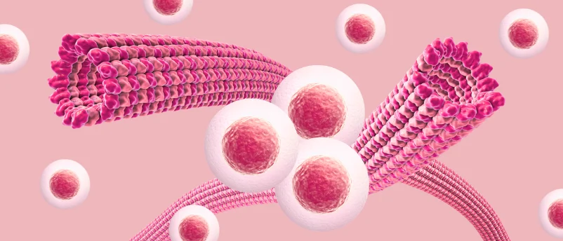
However, it was the application of super-resolution imaging that delivered the most paradigm-shifting discovery. This advanced microscopy technique unequivocally demonstrated that over 80% of TZPs contained both microtubules and actin filaments. This finding directly challenged and effectively overturned the long-standing scientific dogma that TZPs were predominantly actin-based structures. The presence of microtubules in such high prevalence within TZPs indicated a previously underestimated, yet profoundly significant, role for these cytoskeletal elements in mediating granulosa cell-oocyte communication.

Masamitsu Sato elaborated on the profound implications of this revelation, stating, "Super-resolution microscopy revealed that microtubules are present within TZPs that connect the oocyte and surrounding granulosa cells at a higher frequency than previously detected, highlighting an underestimated role for microtubules in oocyte-granulosa cell communication." This statement underscores the critical shift in understanding necessitated by the study’s findings, moving away from a simplified view of TZP structure towards a more complex, dual-cytoskeletal model.
The Molecular Mechanism of Impaired Communication
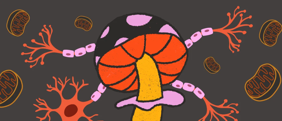
The absence of Camsap3 proved to be the root cause of severe disorganization of microtubules within TZPs. In the Camsap3-KO mice, not only was the structural integrity of microtubules compromised, but there was also a significant reduction in the overall number of TZPs. Furthermore, the study identified the disappearance of specialized TNT-like TZPs, referring to structures resembling tunneling nanotubes. These particular TZP subtypes are thought to facilitate the transport of larger molecules and even organelles, such as mitochondria, between cells. The loss of these specialized transport conduits in Camsap3-deficient mice indicated a severe impairment in the capacity for efficient intercellular exchange.
As a direct consequence of these structural and numerical deficiencies in TZPs, the communication pathways between granulosa cells and the oocyte were significantly compromised. This breakdown in vital communication ultimately prevented the proper maturation of ovarian follicles and, critically, inhibited successful ovulation. Mika Toya further elucidated the molecular underpinnings of these observations, adding, "The study demonstrated that Camsap3 stabilizes microtubules within the TZP, revealing the molecular mechanism by which impaired communication between the oocyte and granulosa cells leads to infertility and follicular atresia." Her statement succinctly captures the essence of the discovery: Camsap3 is a molecular linchpin, whose absence destabilizes TZP microtubules, thereby sabotaging the essential communication network required for fertility.
Broader Impact and Future Horizons
This groundbreaking discovery offers unprecedented insights into the cellular mechanisms governing female fertility. By definitively establishing microtubules as crucial structural and functional components of transzonal projections, the study fundamentally expands the current understanding of ovarian follicle biology. Moreover, the identification of Camsap3 as a critical regulator of microtubule organization within TZPs marks it as a pivotal player in reproductive function, opening new avenues for research and clinical intervention.
The implications of these findings are far-reaching, particularly in the realm of infertility diagnosis and treatment. Currently, many cases of infertility remain unexplained or are attributed to complex multifactorial causes. This research provides a novel cellular target, suggesting that defects in Camsap3 function or microtubule organization within TZPs could be underlying causes of previously undiagnosed female infertility. Future diagnostic approaches might include assessing Camsap3 expression or microtubule integrity in follicular cells.
Furthermore, these insights could pave the way for innovative therapeutic strategies. For instance, pharmaceutical interventions aimed at enhancing Camsap3 activity or stabilizing microtubules within TZPs could potentially restore or improve communication between granulosa cells and oocytes, thereby increasing fertility rates. This opens up exciting possibilities for developing new pharmacological agents or refining existing ones to address specific cellular dysfunctions in the ovary.
Beyond direct clinical treatments, the study’s findings hold significant promise for advancing assisted reproductive technologies (ART), such as in vitro fertilization (IVF). A deeper understanding of the optimal conditions for oocyte maturation is crucial for improving the success rates of IVF cycles. The knowledge that Camsap3-mediated microtubule organization is essential for healthy follicular development can inform the design of more effective in vitro follicular culture systems. By mimicking the natural microenvironment more accurately, including factors that promote robust TZP formation and microtubule stability, it may be possible to produce higher quality oocytes for ART procedures, ultimately leading to better outcomes for individuals seeking to overcome infertility.
Looking ahead, this research also sets the stage for numerous future investigations. Scientists can now explore other microtubule-associated proteins that might interact with Camsap3 or contribute to TZP stability and function. The precise mechanisms of transport facilitated by TNT-like TZPs, particularly for large molecules and organelles, warrant further detailed study. Translating these findings from mouse models to human fertility will be a critical next step, involving comparative studies to confirm the conservation of these mechanisms in human ovarian physiology.
In conclusion, the meticulous work of the Japanese research team has unveiled a previously underestimated, yet profoundly essential, role for microtubules in ovarian follicle development. By demonstrating that Camsap3-mediated microtubule organization is indispensable for maintaining robust granulosa cell-oocyte communication, this study provides a crucial missing piece in the puzzle of successful ovulation and female fertility. This discovery not only enhances fundamental biological understanding but also offers tangible pathways for improving infertility diagnosis, developing novel treatments, and refining assisted reproductive technologies for the benefit of countless individuals worldwide.