In a significant advancement for neurodegenerative research, scientists at Johns Hopkins Medicine have unveiled new evidence suggesting that small clusters of lab-grown brain tissue, known as organoids, can serve as a predictive tool for how individuals with Alzheimer’s disease respond to psychiatric medications. This research, published in the prestigious journal Alzheimer’s & Dementia: The Journal of the Alzheimer’s Association, marks a pivotal shift toward precision medicine in the treatment of dementia, a condition that currently affects more than 7 million Americans and remains one of the most significant challenges in modern healthcare.
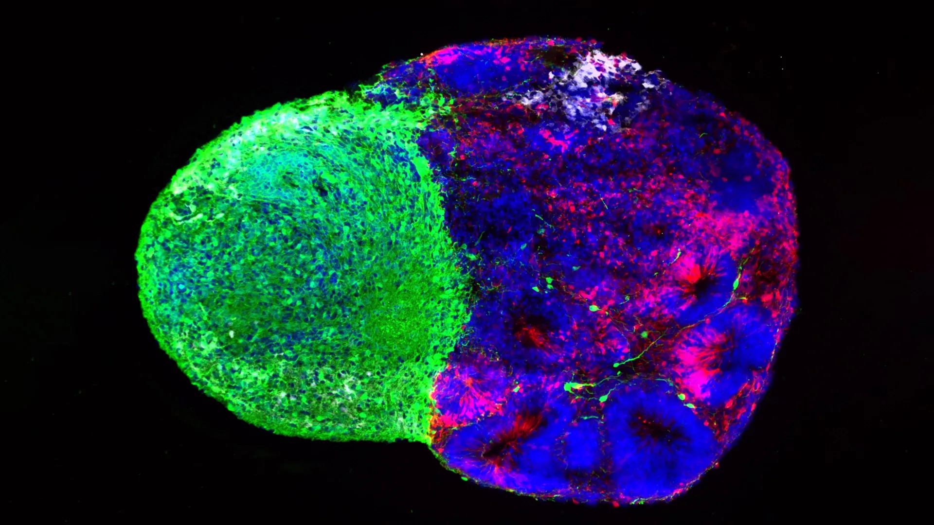
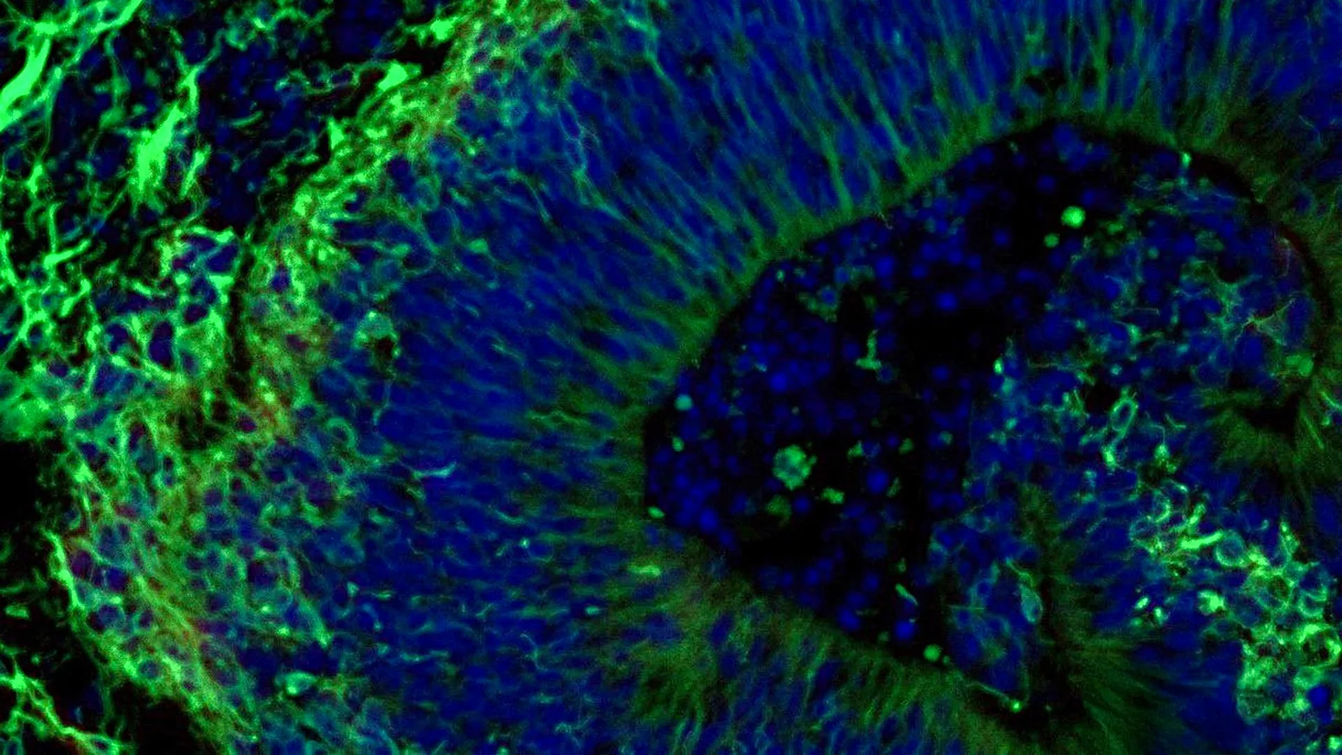
The study centers on the use of patient-derived organoids—miniature, three-dimensional models of brain tissue—to simulate the complex biological environment of the human brain. By utilizing these models, researchers were able to observe how specific medications, such as selective serotonin reuptake inhibitors (SSRIs), interact with diseased tissue on a molecular level. This approach offers a potential solution to the long-standing problem of "trial and error" in prescribing psychiatric medications for Alzheimer’s patients, who often suffer from debilitating symptoms such as anxiety, depression, and agitation.
The Evolution of Brain Organoids and Personalized Diagnostics
The foundation of this research lies in the ability to reprogram adult cells into a state of pluripotency. The Johns Hopkins team began by collecting blood samples from participants at the NIH-funded Johns Hopkins Alzheimer’s Disease Research Center. Through a process involving induced pluripotent stem cells (iPSCs), the researchers reverted these blood cells to a stem-cell-like state, which allows them to be differentiated into any cell type in the body—in this case, specialized neurons found in the hindbrain.
The hindbrain is a critical region located at the back of the skull, responsible for regulating autonomic functions such as heart rate, sleep cycles, and respiration. Crucially for this study, the hindbrain is also a primary site for the production of serotonin, a neurotransmitter that plays a vital role in mood regulation. By guiding these iPSCs to organize themselves into pea-sized clusters, the team created hundreds of organoids that mirrored the genetic and molecular makeup of the individual patients from whom the blood was drawn.
Dr. Vasiliki Machairaki, an associate professor of genetic medicine at the Johns Hopkins University School of Medicine and the study’s lead author, noted that this large-scale approach is one of the most comprehensive organoid studies conducted in the field of Alzheimer’s research. The ability to create "patient-specific" brain models allows scientists to move beyond generalized treatments and toward a framework where a patient’s own biology dictates their clinical care plan.
Bridging the Gap in Symptom Management
While there is currently no cure for Alzheimer’s disease, the management of its neuropsychiatric symptoms is a cornerstone of patient care. Nearly all patients with the condition experience some form of psychological distress, which can lead to a rapid decline in quality of life and increased caregiver burden. SSRIs, specifically escitalopram oxalate (commonly known as Lexapro), are frequently prescribed to manage these symptoms. However, the clinical response to these drugs is notoriously inconsistent.
The Johns Hopkins study sought to determine if organoids could reveal why some patients benefit from SSRIs while others do not. When the researchers treated the Alzheimer’s-derived organoids with escitalopram, they observed a distinct divergence in molecular responses. In a subset of the organoids, the medication successfully increased the production of proteins involved in serotonin signaling and synaptic communication. In others, the drug elicited little to no change.
This variation mirrors the real-world clinical experience of physicians and suggests that the organoid model can accurately reflect a patient’s internal response to a drug before it is even administered. "On a large-scale level, our model may eventually be used to identify subgroups of patients, based on underlying molecular mechanisms, who are more likely to respond to certain drugs," Dr. Machairaki explained. This could significantly reduce the time patients spend on ineffective medications, minimizing side effects and accelerating the path to symptom relief.
Extracellular Vesicles: The Potential for a Liquid Biopsy
One of the most promising discoveries in the study involves extracellular vesicles (EVs)—tiny, membrane-bound particles released by the organoids into their surrounding environment. These vesicles act as cellular "mail," carrying proteins, lipids, and genetic information from the parent cells. The researchers found that these particles provide a window into the health and activity of the brain tissue without requiring invasive procedures.
By analyzing the contents of the EVs, the team identified several key proteins that were significantly altered in Alzheimer’s-derived organoids compared to those from healthy controls. Specifically, levels of proteins such as RAB3A, NSF, and ATCAY—all of which are essential for normal communication between neurons and memory function—were found to be depleted in the Alzheimer’s models.
Furthermore, the researchers observed that when the organoids responded positively to SSRI treatment, the protein profiles within the EVs shifted accordingly. This suggests that EVs could eventually serve as biomarkers in a "liquid biopsy" for the brain. In a clinical setting, a simple blood or cerebrospinal fluid test could analyze these vesicles to determine the stage of a patient’s Alzheimer’s disease, identify their specific disease subtype, and predict which medications would be most effective.
Data and Molecular Insights
The data generated by the study highlights the profound molecular differences between healthy aging and Alzheimer’s pathology. The researchers utilized advanced proteomic analysis to compare the protein landscapes of the various organoid groups. Key findings included:
- Inflammatory Pathways: Alzheimer’s organoids showed a marked increase in proteins associated with neuroinflammation, a known driver of cognitive decline.
- Synaptic Dysfunction: The lower levels of RAB3A and NSF in patient-derived models provide a molecular explanation for the breakdown in cellular communication observed in Alzheimer’s patients.
- Differential Drug Response: The increase in serotonin-related proteins following escitalopram treatment was not uniform, providing empirical evidence for the "responder" and "non-responder" phenotypes seen in clinical practice.
This data underscores the complexity of Alzheimer’s disease, which is increasingly viewed not as a single condition, but as a spectrum of molecularly distinct subtypes. The use of organoids allows researchers to categorize these subtypes with a level of precision that was previously impossible.
A Chronology of Innovation in Alzheimer’s Research
The study at Johns Hopkins represents the latest milestone in a timeline of scientific progress that has spanned decades.
- 2006: Shinya Yamanaka’s discovery of iPSCs provided the toolset to turn any human cell into a stem cell.
- 2013: The first cerebral organoids were successfully grown, providing a 3D model of human brain development.
- 2018-2022: Researchers began applying organoid technology to neurodegenerative diseases, focusing primarily on the cortex.
- 2024: The Johns Hopkins study expands this to the hindbrain and incorporates the study of extracellular vesicles as a diagnostic tool, moving the technology closer to clinical application.
The current research was a collaborative effort involving experts from Johns Hopkins, Tymora Analytical Operations, and the University of Rochester School of Medicine and Dentistry. Funding was provided by multiple grants from the National Institutes of Health, the Paul G. Allen Frontiers Foundation, and the Richman Family Precision Medicine Center of Excellence in Alzheimer’s Disease.
Future Horizons: Vascularization and Immune Integration
Despite the success of the current study, Dr. Machairaki and her team are already looking toward the next generation of brain models. One limitation of current organoids is their lack of a vascular system and immune cells. In a living human brain, blood vessels provide oxygen and nutrients, while immune cells called microglia play a critical role in inflammation and the clearance of toxic proteins like amyloid-beta.
Future research at Johns Hopkins aims to develop "vascularized" organoids that include blood-vessel-like networks and integrated immune cells. These more advanced models will provide a more realistic environment for studying disease progression and testing new therapeutics. By making the organoids more similar to living tissue, researchers hope to improve the accuracy of drug screening even further.
Implications for Global Healthcare and Drug Development
The implications of this research extend far beyond the laboratory. For the pharmaceutical industry, patient-derived organoids offer a more reliable platform for drug discovery than traditional animal models. Many drugs that show promise in mice fail in human clinical trials because of the fundamental differences between rodent and human brain biology. Using human organoids early in the drug development process could significantly lower the cost of bringing new treatments to market and increase the success rate of clinical trials.
For patients and their families, the prospect of personalized care offers a glimmer of hope in the face of a devastating diagnosis. As the global population ages, the number of people living with Alzheimer’s is expected to triple by 2050. The development of non-invasive diagnostic tools, such as the EV-based liquid biopsy, could lead to earlier intervention, which is widely considered the best chance for slowing the progression of the disease.
While the Johns Hopkins team emphasizes that their work is an early step in a long journey, the study provides a robust proof-of-concept for the future of neurology. By combining stem cell technology, 3D tissue modeling, and advanced proteomics, scientists are finally beginning to decode the molecular mysteries of the human brain, one organoid at a time.