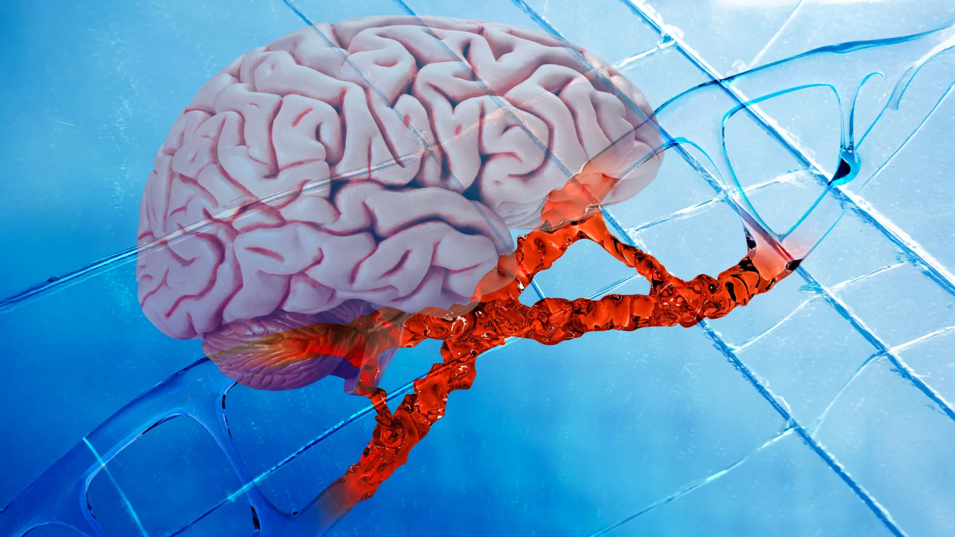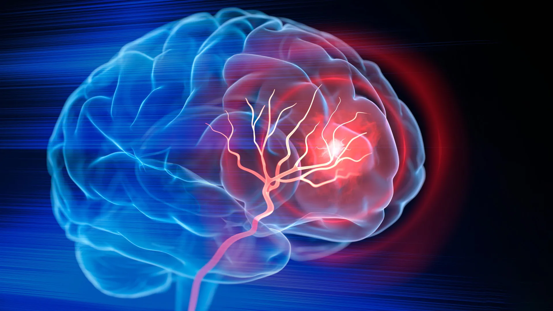
For decades, the standard of care for ischemic strokes has been limited to time-sensitive interventions. When a blood clot blocks an artery, the primary objective is reperfusion—the restoration of blood flow—using tissue plasminogen activator (tPA) or mechanical thrombectomy. While these procedures are life-saving, they are fundamentally reactive. Once the oxygen-deprived neural tissue dies, a necrotic cavity remains, serving as a permanent scar that hinders cognitive and motor function. Until now, the clinical focus has been almost exclusively on acute salvage rather than structural reconstruction.
The new research, published in the journal Cell Biomaterials, suggests a paradigm shift: instead of attempting to replace lost brain matter directly, engineers are creating a specialized "niche" that co-opts the body’s inherent biological machinery to rebuild the void.
The Engineering of a Healing Microenvironment
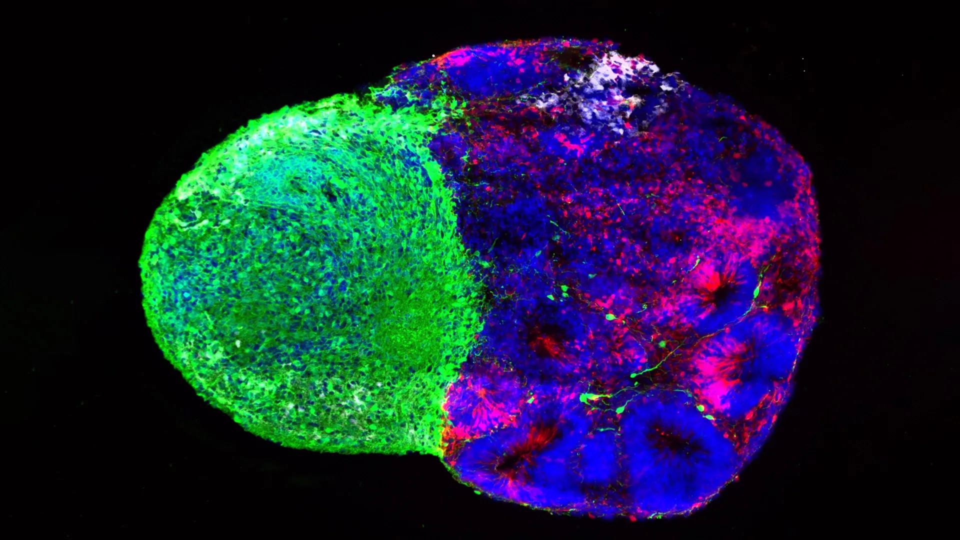
The Duke team, led by Tatiana Segura, the Robert Plonsey Distinguished Professor of Biomedical Engineering, utilized a proprietary technology known as MAPS, or microporous annealed particle scaffolds. These scaffolds are composed of hydrogel microparticles that, when injected, self-assemble into a porous, lattice-like structure.
The architecture is crucial. Unlike a solid gel, which might act as a barrier to cellular infiltration, the MAPS structure is intentionally open. This porosity provides a physical framework that allows endogenous cells—the body’s own immune, vascular, and neural components—to migrate into the injury site.
However, the scaffold itself is only the chassis. To drive the biological engine of repair, the team integrated extracellular vesicles (EVs). EVs are essentially biological nanoparticles, secreted by cells to communicate with their environment by ferrying proteins, lipids, and genetic material. By chemically tethering these vesicles to the surface of the hydrogel microparticles, the researchers ensured that signaling molecules remained concentrated within the scaffold rather than diffusing away into the surrounding healthy tissue.
The Role of Astrocytes and the Immune Response
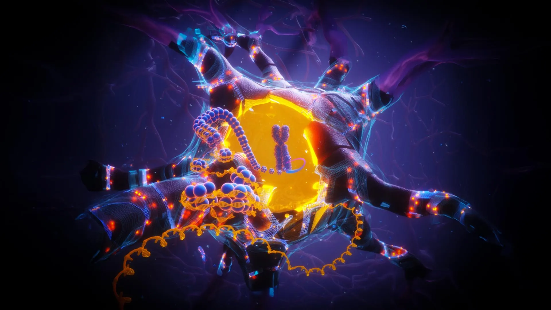
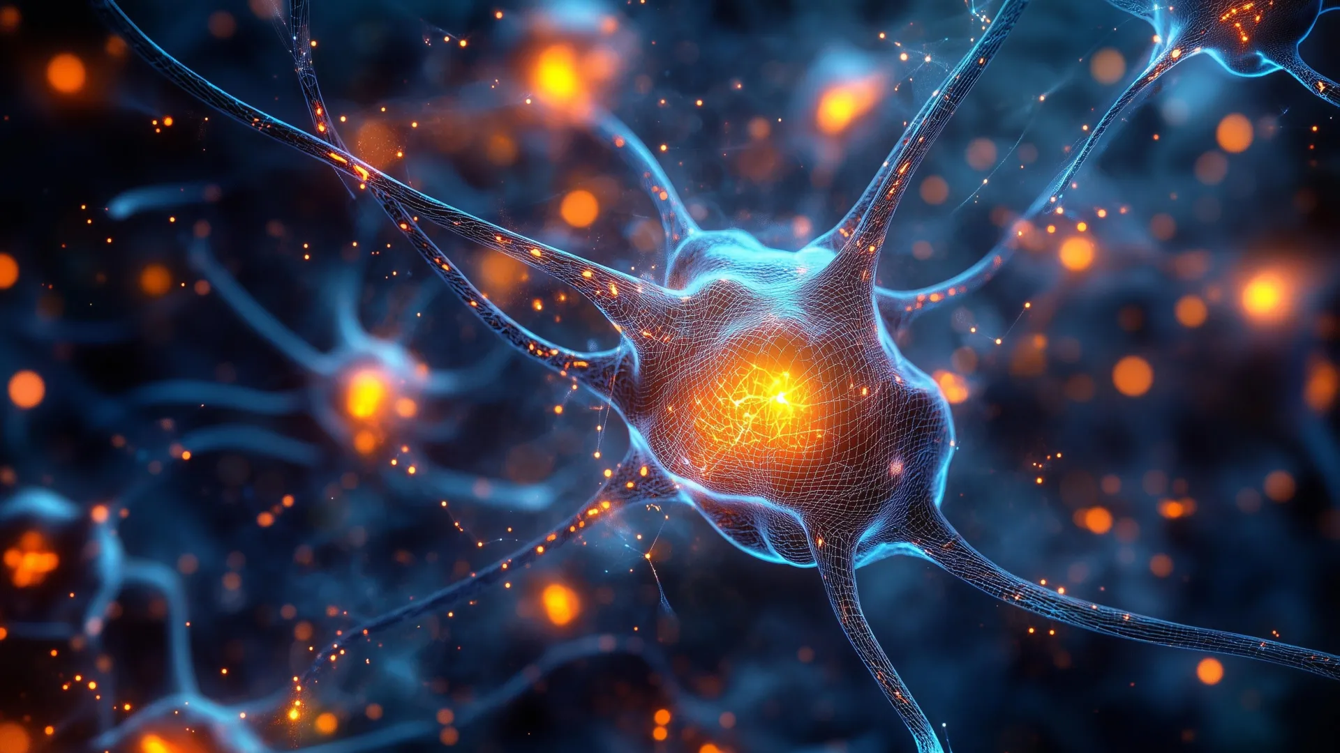
The study focused on astrocytes, the star-shaped glial cells that are among the first responders to central nervous system injury. Traditionally, astrocytes have been viewed through the lens of scar formation; they wall off the injury site to protect the brain, but in doing so, they often create a physical and chemical barrier to axonal regrowth.
In this experiment, the researchers harvested EVs from lab-grown astrocytes and loaded them with specific signaling molecules—namely IL-4 and C1q. These molecules were chosen for their ability to modulate the local immune response.
The strategy was highly successful in recruiting macrophages and, unexpectedly, a significant population of neutrophils. Neutrophils are often regarded as detrimental in the acute phase of a stroke because they exacerbate inflammation and contribute to collateral tissue damage. However, the Duke study reveals that this perspective may be incomplete. When present at the right time and in the right chemical environment—in this case, the optimized scaffold—neutrophils appear to shift their behavior, transitioning from destructive agents to facilitators of angiogenesis (the formation of new blood vessels) and tissue remodeling.
Chronology and Experimental Outcomes
In the murine models used for this study, the therapeutic intervention followed a rigorous timeline:
- Initial Phase: Following the induction of an ischemic injury, the MAPS scaffold containing IL-4 and C1q-loaded EVs was injected directly into the stroke cavity.
- Mid-Phase (Weeks 1–4): The scaffold acted as a bridge. Researchers observed a significant influx of immune cells, which effectively "primed" the area for vascularization. The presence of the scaffold prevented the collapse of the cavity and provided a surface for new blood vessels to traverse the previously empty space.
- Late Phase (Weeks 4–8): The integration of the scaffold was evidenced by the growth of axonal fibers, the long extensions of neurons that transmit electrical impulses. By the eight-week mark, the mice underwent grid-walking tests to assess motor coordination.
The results were statistically significant. Treated mice displayed forelimb placement precision that was indistinguishable from healthy control subjects, a level of functional recovery that persisted for the duration of the study. Crucially, when the researchers attempted to inject the EVs without the porous scaffold, the repair response was negligible. This confirmed that the scaffold was not merely a delivery vehicle, but an essential structural and environmental component of the healing process.
Implications for Clinical Neurology
The implications of this research are substantial, though the medical community remains cautious. Currently, there are no FDA-approved therapies that successfully reconstruct the brain cavity after a stroke. If this approach can be successfully translated to humans, it could fundamentally alter the prognosis for millions of patients who live with permanent motor or cognitive deficits post-stroke.
"The goal is not to force the brain to become what it was, but to provide it with the environmental conditions necessary to self-repair," said lead study author Shangjing Xin. "By managing the immune response and providing a physical template, we are essentially ‘reprogramming’ the injury site to accept new growth."
However, significant hurdles remain before this technology can enter clinical trials. The current study utilized rat-derived astrocytes for the EVs; for human application, the researchers are investigating the use of human induced pluripotent stem cell (iPSC)-derived astrocytes. These cells offer a more scalable and ethically straightforward source, while also providing a higher degree of control over the specific signaling profile of the EVs.
Furthermore, the transition from murine models to human anatomy presents a massive scale-up challenge. The human brain is exponentially larger and more complex than that of a mouse, and the long-term safety profile of the hydrogel material—including how it degrades and whether it triggers late-stage inflammatory responses in humans—remains to be fully established.
A New Paradigm in Regenerative Medicine
The work conducted at Duke represents a shift in the field of regenerative medicine, moving away from simple drug-delivery models toward the creation of "living" therapeutic environments. By acknowledging that the brain is an ecosystem rather than a collection of independent cells, the researchers have demonstrated that the path to recovery lies in the coordination of multiple cellular processes.
The ability to recruit and retain immune cells at the injury site marks a departure from traditional anti-inflammatory treatments, which often aim to suppress the immune system entirely. Instead, this strategy suggests that the immune system, when guided by precise biochemical cues, is a powerful ally in tissue regeneration.
As the Segura laboratory moves toward investigating human-derived cells, the broader scientific community is watching closely. If the results can be replicated in larger, more complex models, this injectable scaffold could serve as a blueprint for treating other forms of neural injury, including traumatic brain injury (TBI) and neurodegenerative conditions where tissue loss is a primary factor.
For now, the study provides a robust validation of the concept that the "dead" space left by a stroke can be re-engineered. By turning the cavity into a site of active biological signaling and structural support, researchers are offering a new vision for recovery: a future where the brain is not merely left to struggle with the aftermath of an injury, but is provided with the internal infrastructure to rebuild itself from within. The path to clinical application is long, but the successful restoration of motor function in animal models suggests that the engineering of the brain’s microenvironment may finally be within reach.