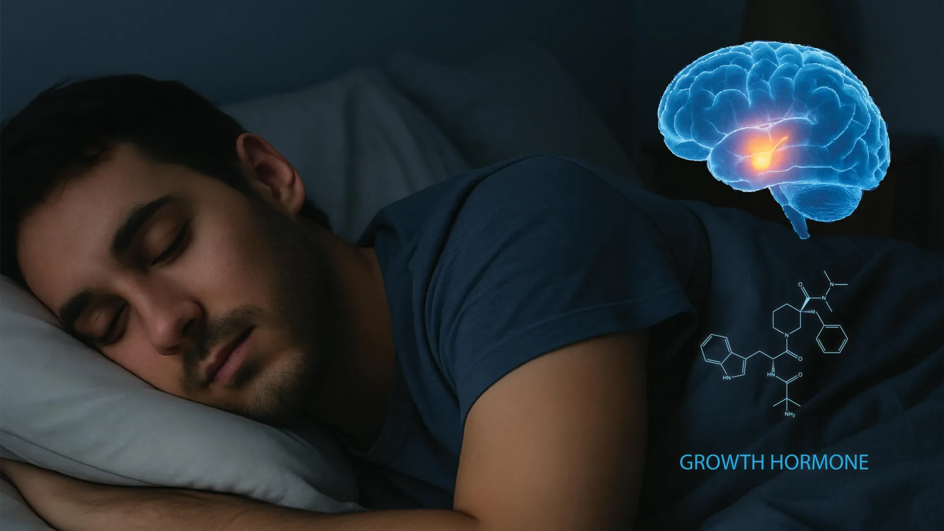
The intricate relationship between a restorative night’s rest and the physiological maintenance of the human body has long been a cornerstone of biological science, yet the specific mechanisms governing this link have remained largely shrouded in mystery. Researchers at the University of California, Berkeley, have recently achieved a significant breakthrough by identifying the specific neural circuitry responsible for regulating growth hormone (GH) release during sleep. This discovery, published in the prestigious journal Cell, not only clarifies how the brain coordinates the surge of hormones necessary for physical repair but also reveals a sophisticated feedback loop that balances sleep-wake cycles. By mapping these pathways, scientists have opened new avenues for treating a spectrum of conditions, ranging from metabolic syndromes like type 2 diabetes to neurodegenerative disorders such as Alzheimer’s and Parkinson’s disease.
Growth hormone is far more than a developmental catalyst for adolescents; it is a vital protein-based hormone produced by the pituitary gland that facilitates muscle protein synthesis, bone density maintenance, and the regulation of glucose and lipid metabolism throughout adulthood. It has been an established fact since the 1960s that the highest concentrations of GH are secreted during the deepest stages of non-rapid eye movement (NREM) sleep. However, the "black box" of the brain—specifically how neural signals translate sleep states into hormonal output—has eluded researchers until now. The UC Berkeley study provides the first direct recording of neural activity in live subjects to illustrate this process in real-time.
The Hypothalamic Command Center: Mapping the GHRH and Somatostatin Neurons
At the heart of this discovery lies the hypothalamus, a small but critical region located at the base of the brain that serves as the primary interface between the nervous system and the endocrine system. The research team, led by Yang Dan, a professor of neuroscience and molecular and cell biology, focused on two primary types of nerve cells within the hypothalamus: growth hormone-releasing hormone (GHRH) neurons and somatostatin neurons.
In a healthy biological system, GHRH acts as the "accelerator," signaling the pituitary gland to release growth hormone into the bloodstream. Conversely, somatostatin acts as the "brake," inhibiting GH release to prevent overproduction. Using advanced optogenetic techniques—a method that involves using light to control neurons that have been genetically sensitized—the researchers were able to observe how these two cell populations interact during different sleep stages.
The study found that during REM sleep—the stage associated with vivid dreaming—both GHRH and somatostatin neurons increase their activity. However, the specific timing and intensity of these signals during NREM sleep create the characteristic "pulse" of growth hormone that the body uses for cellular repair. This nuanced coordination ensures that the body receives a concentrated dose of GH when it is most capable of utilizing it for metabolic and structural maintenance.
A Novel Feedback Loop: The Role of the Locus Coeruleus
One of the most surprising findings of the UC Berkeley study is the identification of a previously unknown feedback system involving the locus coeruleus (LC). Located in the brainstem, the LC is the brain’s primary source of norepinephrine, a neurotransmitter responsible for alertness, vigilance, and the "fight or flight" response. Traditionally, the LC was thought to function primarily as a wakefulness promoter, keeping the brain alert during the day.
The researchers discovered that as growth hormone levels rise during sleep, the hormone travels back to the brain and activates neurons in the LC. Initially, this stimulation appears to encourage a transition toward wakefulness, acting as a natural alarm clock as the body completes its repair cycle. However, the feedback loop contains a self-regulating "fail-safe." If the activity in the LC becomes excessively high, it triggers a compensatory response that actually promotes sleepiness.
"This suggests that sleep and growth hormone form a tightly balanced system," explained Daniel Silverman, a UC Berkeley postdoctoral fellow and study co-author. "Too little sleep reduces growth hormone release, and too much growth hormone can in turn push the brain toward wakefulness. This balance is essential for growth, repair, and metabolic health."
This feedback loop explains why individuals with chronic sleep deprivation often suffer from metabolic imbalances. When sleep is cut short, the GH pulse is interrupted, which in turn fails to properly regulate the LC, leading to a cycle of daytime fatigue, reduced cognitive focus, and impaired metabolic recovery.
Methodology and Chronology of the Discovery
The breakthrough was the result of years of iterative research conducted at UC Berkeley’s Department of Neuroscience and the Helen Wills Neuroscience Institute. To achieve these results, the team utilized a mouse model, which is highly effective for sleep research due to the conserved nature of the hypothalamus across mammals.
- Phase One (Circuit Tracing): The team used viral tracers to map the physical connections between the hypothalamus and the pituitary gland, as well as the connections leading back to the locus coeruleus.
- Phase Two (Real-time Recording): Researchers implanted electrodes to monitor the firing patterns of specific neurons during the mice’s natural sleep cycles. Because mice sleep in short bursts throughout a 24-hour period, the team was able to collect data across hundreds of sleep-wake transitions.
- Phase Three (Optogenetic Manipulation): By stimulating GHRH neurons with light, the researchers could artificially trigger GH release and observe the subsequent effect on the LC and the animal’s arousal state.
- Phase Four (Data Integration): The final stage involved correlating hormonal blood levels with the recorded neural activity, proving that the neural firing they observed was directly responsible for the hormonal fluctuations.
This systematic approach allowed the researchers to move beyond correlation—simply knowing that GH and sleep happen together—to causation, identifying the exact "wiring" that makes it happen.
Supporting Data: The Impact of Sleep on Hormonal Health
The implications of this research are supported by a wealth of existing clinical data regarding sleep and human health. According to the Centers for Disease Control and Prevention (CDC), one in three adults in the United States does not get enough sleep. The UC Berkeley study provides a biological explanation for the statistical links between sleep debt and physical ailments:
- Metabolic Health: Growth hormone is essential for lipolysis (the breakdown of fats). Studies have shown that even one night of sleep deprivation can reduce the following day’s GH peak by up to 70%, contributing to insulin resistance and an increased risk of obesity.
- Athletic Recovery: In professional sports, "sleep hygiene" has become a primary metric for performance. GH released during sleep is responsible for repairing the micro-tears in muscle tissue caused by intense exercise.
- Aging and Longevity: Natural GH production declines with age, a phenomenon known as somatopause. This decline is often mirrored by a decrease in deep NREM sleep. The Berkeley findings suggest that by targeting the hypothalamic circuit, it may be possible to restore youthful GH levels in the elderly, potentially slowing the loss of muscle mass and bone density.
Clinical Implications: New Frontiers in Treatment
The identification of this circuit offers a "novel handle" for medical interventions. Current treatments for growth hormone deficiencies often involve direct injections of synthetic GH, which can have side effects and do not mimic the body’s natural pulsatile release.
"Understanding the neural circuit for growth hormone release could eventually point toward new hormonal therapies to improve sleep quality or restore normal growth hormone balance," said Daniel Silverman. He noted that experimental gene therapies could potentially target specific hypothalamic cells to "dial back" the excitability of the locus coeruleus in patients with insomnia or over-arousal disorders.
Furthermore, the connection to the locus coeruleus is particularly relevant for neurodegenerative disease research. The LC is one of the first areas of the brain to show signs of pathology in Alzheimer’s and Parkinson’s diseases. If growth hormone plays a role in maintaining the health of the LC, then maintaining healthy sleep-GH cycles could be a critical factor in delaying the onset of cognitive decline.
Analysis: A Paradigm Shift in Sleep Science
This study represents a shift in how scientists view the "purpose" of sleep. For decades, sleep was viewed primarily through the lens of cognitive restoration—the brain "cleaning" itself of metabolic waste. While that remains true, the UC Berkeley research emphasizes that sleep is also a period of intense endocrine activity that dictates the health of the entire body.
By proving that growth hormone feeds back into the brain to regulate wakefulness, the researchers have shown that the endocrine system is not just a passive recipient of brain signals; it is an active participant in the regulation of consciousness. This "cross-talk" between the body and the brain suggests that many conditions we currently classify as "mental" or "neurological" may have deep roots in hormonal imbalances triggered by poor sleep.
Institutional Support and Collaborative Effort
The research was a collaborative effort involving several prominent institutions and was supported by significant scientific grants. The study was funded by the Howard Hughes Medical Institute (HHMI) and the Pivotal Life Sciences Chancellor’s Chair fund.
In addition to lead author Xinlu Ding and co-author Daniel Silverman, the research involved a multi-disciplinary team including Peng Zhong, Bing Li, Chenyan Ma, Lihui Lu, Grace Jiang, Zhe Zhang, Xiaolin Huang, Xun Tu, and Zhiyu Melissa Tian from UC Berkeley. The team also collaborated with Fuu-Jiun Hwang and Jun Ding from Stanford University, highlighting the regional synergy in California’s premier research corridor.
As the scientific community digests these findings, the focus will likely shift toward human clinical trials. If the hypothalamic circuit identified in mice can be safely modulated in humans, the result could be a new generation of treatments that work with the body’s natural rhythms rather than against them, ushering in an era of precision medicine for sleep and metabolic health.