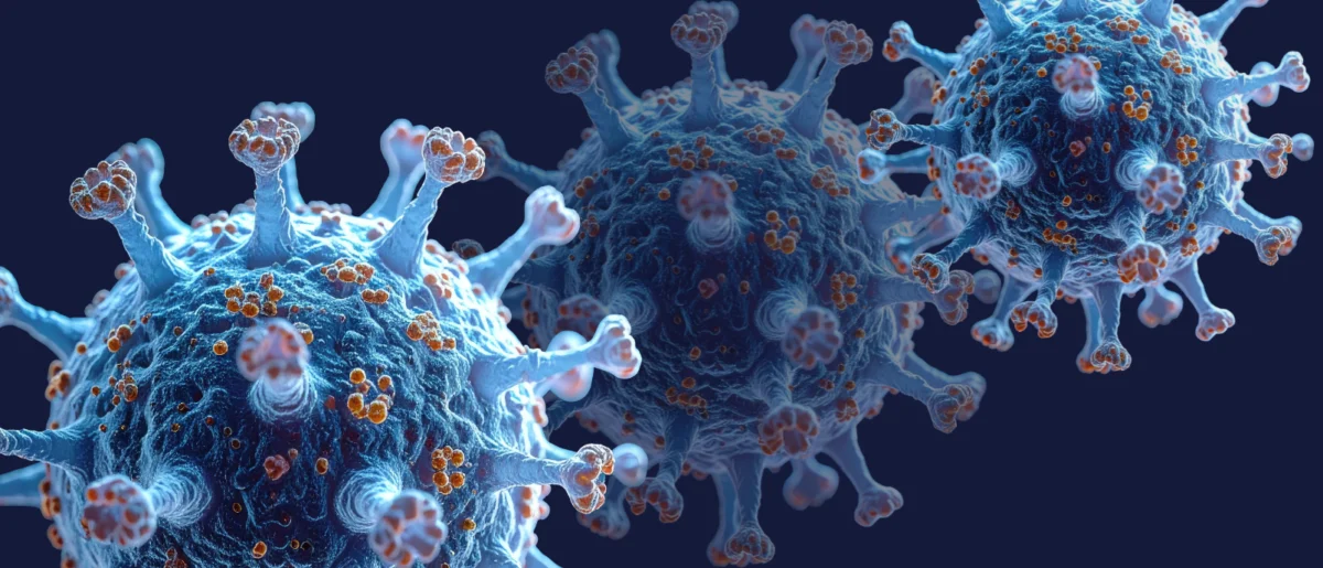
For decades, the mechanics of viral self-assembly have remained one of structural biology’s most elusive mysteries. Viruses—intricate packets of genetic material encased in protective protein shells known as capsids—manage to construct themselves with astonishing precision and reliability. Yet, how these complex biological architectures form spontaneously from individual molecular components without the aid of a cellular blueprint or directing machinery has puzzled scientists. Now, a pioneering study conducted by researchers at the University of Oxford has captured this step-by-step assembly process in real-time, providing an unprecedented molecular-level view of how viral capsids come together.
Published in September 2026, the breakthrough findings reveal how simple, reversible molecular interactions guide thousands of potential configuration pathways toward a single, successfully completed structure. By lifting the veil on how viruses solve this high-stakes geometric puzzle, the research not only illuminates fundamental biophysical principles but also paves the way for the design of novel antiviral therapeutics, next-generation vaccines, and advanced bio-engineered nanomaterials.
The Enigma of Viral Self-Assembly
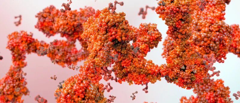
To understand the magnitude of the Oxford discovery, one must appreciate the sheer scale of the engineering challenge a virus faces. A typical viral capsid is composed of anywhere from tens to thousands of individual protein subunits. These building blocks must collide, interact, and lock into a specific, symmetrical geometry—often an icosahedron—to effectively shield the pathogen’s fragile genetic cargo from hostile cellular environments.
Historically, virologists and biophysicists have relied on bulk experiments, theoretical modeling, and static snapshots to infer how these structures form. While these methods successfully identified the starting components and the final assembled capsids, they routinely missed the fleeting, high-energy intermediate structures that bridge the gap. These critical milestones are typically scarce, short-lived, and statistically difficult to capture using conventional laboratory techniques. Consequently, the exact kinetic pathways and physical rules governing viral growth remained largely speculative.
To bypass these historical limitations, the interdisciplinary team at Oxford—led by prominent figures in chemistry and structural biology—turned to cutting-edge measurement technology combined with ingenious experimental design.
Weighing Molecules in Real Time: The Technological Leap
The cornerstone of the Oxford study lies in a powerful technique known as mass photometry, pioneered at the university. Mass photometry measures the mass of individual, unlabelled biomolecules in solution by precisely quantifying the amount of light they scatter. By coupling this technology with a novel method designed to confine individual molecules, the research team managed a scientific feat previously thought nearly impossible: they continuously "weighed" a growing virus-like particle as individual protein components attached to it one by one.
Using an engineered virus-like model consisting of precisely 60 protein subunits, co-lead authors Roi Asor and Professor Philipp Kukura, alongside colleague Jack Tan, were able to observe the step-by-step evolution of a single assembling particle.
"Until now, much of our understanding of how these structures assemble has had to be reconstructed from snapshots or theoretical models," explained Professor Kukura. "Being able to both quantify the underlying interactions and follow one particle as it grows changes that. We can see the important intermediate structures appear and deduce a detailed model of the construction process."
Navigating the Molecular Maze
The data captured by the Oxford team revealed that viral assembly does not rely on rigid, predetermined instructions. Instead, it operates through a remarkably forgiving system of trial and error, governed by statistical mechanics and thermodynamic stability.
When protein building blocks initially encounter one another, they form weak, highly reversible connections. This reversibility is vital to the process. If two proteins join in a non-productive arrangement or an unstable configuration, the weak bond allows them to simply fall apart, freeing the components to try a different orientation.
However, when these proteins happen to form specific closed structures, the dynamics shift dramatically. The simultaneous formation of multiple contact points exponentially increases the stability of the arrangement. These stable configurations function as critical molecular waypoints, or funnels, that progressively guide the sprawling maze of potential assembly routes down a streamlined pathway toward the finished capsid.

A pivotal turning point in this molecular journey occurs when five larger protein building blocks unite to form a closed pentagonal ring. This milestone represents the first major thermodynamic sink—a particularly stable structure in the otherwise volatile assembly pathway. Once this pentagonal cornerstone is established, the mathematics of the process change: fewer incoming protein units are required to reach subsequent stable stages, causing the overall rate of assembly to accelerate rapidly.
"A virus has to solve an extraordinary construction problem," noted Roi Asor. "Its components somehow have to find the right arrangement among a huge number of possibilities, without a blueprint or machinery directing the process. We can now watch what happens molecule by molecule and see the physical rules that make it possible."
Chronology and Context of the Breakthrough
The publication of this research builds upon years of incremental advancements in single-molecule biophysics at Oxford and international partner institutions. Over the past decade, mass photometry has steadily evolved from a novel optical method into an indispensable tool for structural biologists seeking to analyze protein interactions without the artifacts often introduced by fluorescent labeling or surface immobilization.
The integration of continuous single-particle monitoring represents the culmination of intense collaborative efforts between Oxford’s Department of Chemistry and its renowned medical and biological research divisions. By successfully applying this platform to a biological system as complex and dynamic as a self-assembling viral capsid, the team has established a new gold standard for studying transient macromolecular assemblies.
Furthermore, this discovery arrives amid a broader renaissance in the study of viral architecture and history. In parallel with modern structural studies, researchers have increasingly utilized advanced genomics and paleomicrobiology—such as extracting ancient viral DNA from historical artifacts and medieval manuscripts to map multi-millennial evolutionary timelines. Together, these complementary efforts highlight a comprehensive scientific push to understand viruses not just as static public health threats, but as dynamic, highly sophisticated biological machines whose fundamental rules of construction can eventually be exploited against them.
Implications for Antiviral Therapies and Vaccine Engineering
The broader applications of the Oxford study extend far beyond theoretical biophysics, holding profound implications for translational medicine and pharmacology.
For decades, pharmaceutical researchers have sought to develop broad-spectrum antiviral drugs that can disrupt the structural integrity of a virus. One promising strategy involves small-molecule therapeutics designed to bind to viral coat proteins, prematurely triggering their assembly or locking them into dysfunctional conformations before they can encapsulate viral genetic material. By decoding the exact sequence of molecular waypoints, the energetic barriers, and the critical nucleation steps required for capsid formation, the Oxford findings provide a precise roadmap for drug developers.
Armed with this mechanistic insight, pharmacologists can now rationally design targeted therapeutics that intentionally derail the assembly pathway of specific pathogens, neutralizing them before an infection can propagate within a host organism.
In addition to antiviral drug discovery, these insights offer substantial utility in the burgeoning field of bio-nanotechnology and vaccine engineering. Modern biotechnology frequently relies on virus-like particles (VLPs) to deliver targeted genetic instructions, therapeutic proteins, or antigenic payloads safely into human cells without causing disease. Understanding the precise rules governing how these protein shells spontaneously self-assemble will allow bioengineers to optimize production yields, enhance structural stability, and design customized delivery vectors with unprecedented precision.
Broader Biological Significance: Beyond Virology
While the immediate focus of the research centers on viral capsids, the underlying biophysical principles discovered by the Oxford team resonate throughout all of cellular biology.
Self-assembly is a foundational organizing principle of life. From the dynamic filaments of the cellular cytoskeleton and intracellular transport compartments to complex multienzyme machinery and membraneless organelles, biological systems constantly rely on spontaneous, macromolecular organization to maintain homeostasis and execute vital functions.
The methodological framework demonstrated in this study—combining mass photometry with continuous single-molecule observation—offers researchers a powerful new lens through which to investigate these diverse cellular processes. By shifting the scientific paradigm from static observation to real-time molecular tracking, the Oxford study opens an expansive new frontier in structural biology, promising to decode the assembly instructions of life itself, one molecule at a time.