
The fundamental process of cell division, the cornerstone of all life, has long presented a profound enigma to scientists, particularly concerning its earliest stages in embryonic development, especially within oviparous (egg-laying) species. Challenging long-held textbook paradigms, researchers from the esteemed Brugués group at the Cluster of Excellence Physics of Life (PoL) at TUD Dresden University of Technology have unveiled a previously unrecognized mechanism that enables large embryonic cells to divide without the absolute necessity of a fully formed contractile ring. Their groundbreaking findings, published in the prestigious scientific journal Nature, illuminate a sophisticated interplay between cytoskeletal components and the dynamic physical properties of the cell’s interior, orchestrated by what they term a ‘mechanical ratchet’ system. This discovery not only deepens our understanding of fundamental biological processes but also offers new perspectives on developmental biology and the evolution of cell division.
Unraveling the Paradox of Large Embryonic Cell Division
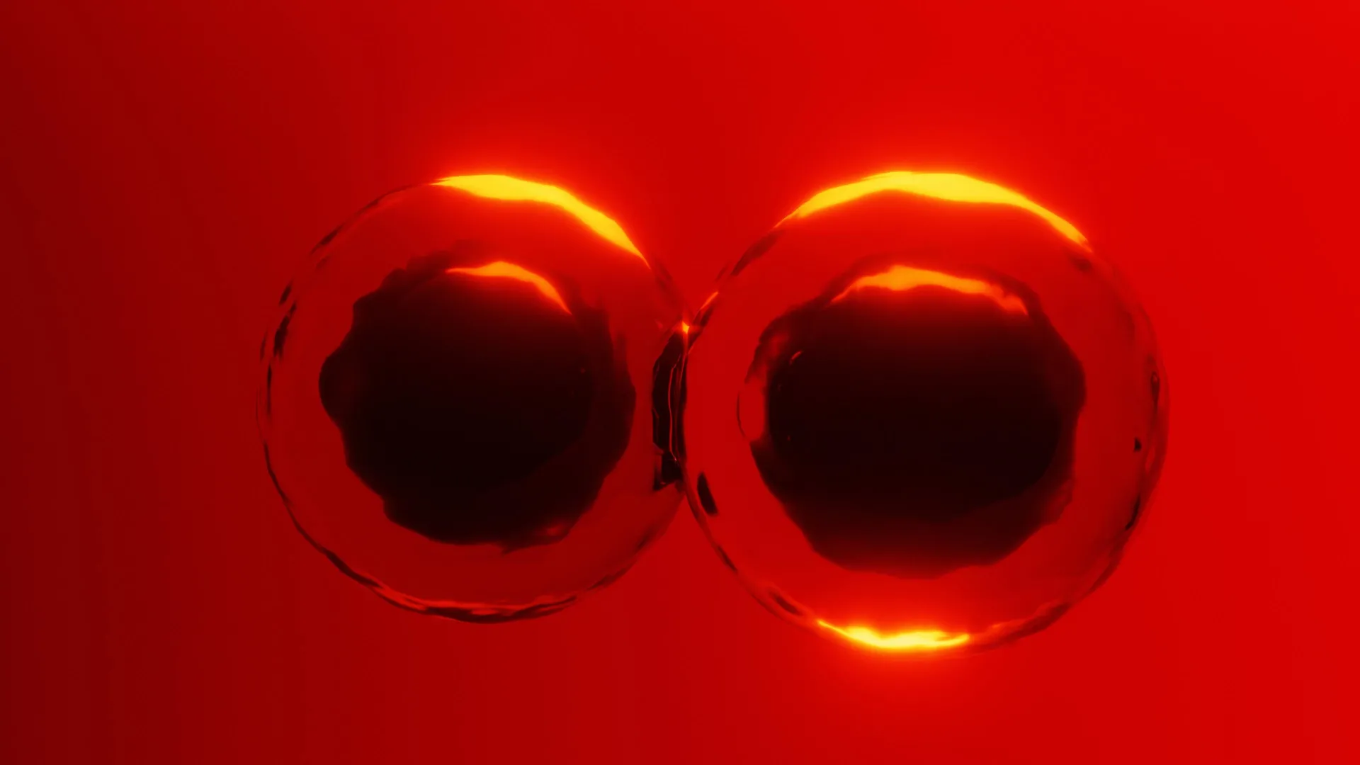
For decades, the prevailing model for cell division, known as cytokinesis, has centered on the formation of a contractile ring. This ring, primarily composed of the protein actin, assembles at the cell’s midpoint and constricts like a drawstring, effectively pinching the cell into two distinct daughter cells. This "purse string" model has proven remarkably effective in explaining division across a vast array of organisms. However, its limitations become starkly apparent when confronted with species characterized by exceptionally large embryonic cells, such as sharks, platypus, birds, and reptiles. In these organisms, the sheer volume of the cell, often occupied by a substantial yolk sac that nourishes the developing embryo, presents a significant geometric challenge. The immense size and the presence of the yolk sac impede the complete closure of an actin ring, leaving scientists to ponder the mechanisms by which these oversized cells successfully undergo division.
"With such a large yolk in the embryonic cell, there is a geometric constraint. How does a contractile band, with loose ends, remain stable and generate enough force to divide these huge cells?" posed Alison Kickuth, a recently graduated PhD student from the Brugués group and the lead author of the study. This question encapsulates the central puzzle that the research team set out to address. Their meticulous experimental work, detailed in the Nature publication, has now provided a compelling answer.
Zebrafish as a Model: Microtubules as Unexpected Stabilizers
To investigate this complex phenomenon, the researchers strategically selected zebrafish embryos. Zebrafish offer several advantages for developmental biology research: their embryos develop rapidly, and during their early stages, they possess large, yolk-rich cells that closely resemble those found in other oviparous species. This makes them an ideal model system for studying the unique challenges of large cell division.
The team employed advanced microscopy techniques and precise laser ablation to perturb the cellular machinery. In a pivotal experiment, Alison Kickuth used a laser to carefully sever the actin band. Counterintuitively, the severed band continued to move inward, suggesting that it was not solely anchored at its ends but was, in fact, supported along its entire length. This observation hinted at the involvement of other cellular components in maintaining the band’s integrity and propulsive force.
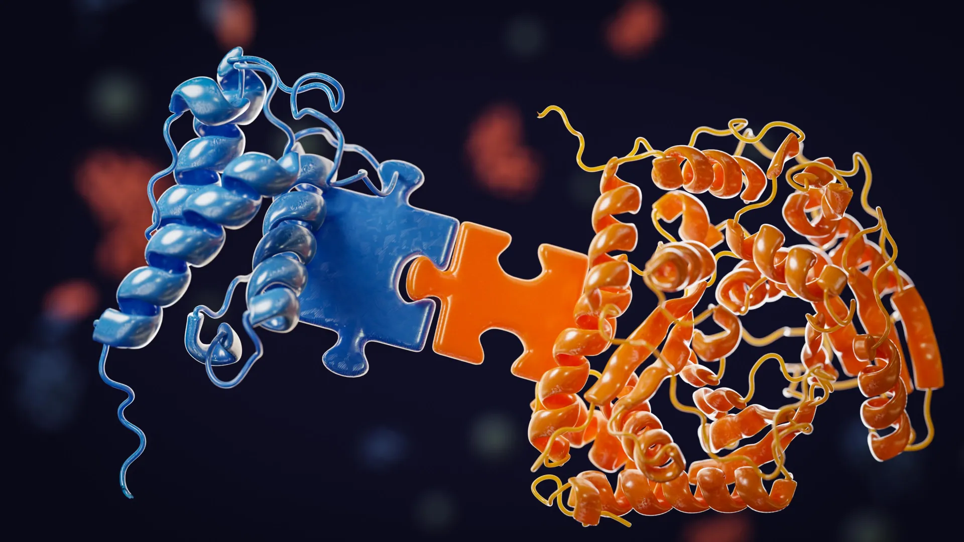
Further investigations revealed the critical role of microtubules, another essential component of the cytoskeleton. When the actin band was cut, microtubules in the vicinity were observed to bend and spread out. This behavior suggested that these fibrous structures were actively contributing to the stabilization of the actin band as it attempted to constrict. To rigorously test this hypothesis, the researchers employed two distinct methods to disrupt microtubule function. First, they used chemical agents to induce depolymerization, effectively halting the assembly of new microtubules. Second, they physically interfered with microtubules by introducing a tiny oil droplet as an obstacle within the cell. In both experimental scenarios, the absence or disruption of microtubules led to the collapse of the actin band. This provided unequivocal evidence that microtubules are not merely passive bystanders but play a crucial role in providing mechanical support and signaling pathways essential for both the formation and contraction of the actin ring.
Cytoplasmic Dynamics: A Shifting Landscape of Stiffness
The cytoskeleton is a dynamic network that undergoes continuous reorganization throughout the cell cycle. This cycle is broadly divided into interphase, a period of cell growth and DNA replication, and the mitotic phase (M-phase), during which chromosomes are segregated. A key event following DNA separation in M-phase is the expansion of large microtubule structures known as asters throughout the cytoplasm. These asters are known to play a role in determining the site where the actin band will subsequently form, effectively marking the future division furrow.
Recognizing that microtubules can influence the physical properties of the cytoplasm, the researchers hypothesized that asters might contribute to anchoring the actin band by stiffening the cellular interior. To investigate this, they devised an ingenious method to measure cytoplasmic stiffness. They introduced microscopic magnetic beads into the cells and then applied an external magnetic force to track their movement. By analyzing the displacement of these beads under magnetic influence, the researchers could precisely assess changes in cytoplasmic stiffness at different stages of the cell cycle.
Their findings revealed a remarkable temporal plasticity in the cytoplasm. During interphase, the cytoplasm becomes significantly stiffer, creating a supportive scaffold that helps to stabilize the actin band. Conversely, during M-phase, the cytoplasm transitions to a more fluid state, which allows the actin band to move inward, facilitating the physical separation of the cell. These cyclical shifts between stiffness and fluidity are not random occurrences but are precisely timed to enable efficient cell division.
The Mechanical Ratchet: Gradual Division Over Time
While the role of microtubules and cytoplasmic stiffness provided significant insights, a lingering puzzle remained. If the cytoplasm becomes more fluid during M-phase, how does the actin band avoid complete collapse during its contractile phase? The research team meticulously tracked the ends of the actin band over extended periods. Their observations revealed that while the band does exhibit a degree of instability during contraction in M-phase, it does not entirely fail. Instead, its partial retraction is effectively "rescued" by the rapid pace of early embryonic cell cycles.
This rescue mechanism operates in conjunction with the predictable cycle of cytoplasmic stiffness. As the cell re-enters interphase, the asters reform, and the cytoplasm stiffens once again, providing renewed stability to the actin band. During the subsequent fluid phase of M-phase, the band can then resume its inward movement. This cyclical process of temporary instability followed by robust stabilization repeats across multiple cell cycles. Rather than completing division in a single, decisive event, the cell achieves it incrementally, step by step, through a series of alternating physical states of its interior. This iterative process functions as a ‘mechanical ratchet,’ progressively advancing the division without demanding the formation of a completely closed contractile ring.
"The temporal ratchet mechanism fundamentally alters our view of how cytokinesis works," emphasized Jan Brugués, the corresponding author of the study. The researchers propose that this innovative mechanism offers an elegant and effective solution for very large embryonic cells that must divide rapidly and cannot rely on the conventional, single-cycle contractile ring model.
Alison Kickuth further elaborated on the significance of their findings, stating, "Zebrafish are a fascinating case, as cytoplasmic division in their embryonic cells is inherently unstable. To overcome this instability, their cells divide rapidly, allowing ingression of the band over several cell cycles by alternating between stability and fluidization until division is complete." This adaptive strategy highlights the remarkable ingenuity of biological systems in overcoming physical constraints.
Broader Implications and Future Directions
This groundbreaking research introduces a novel conceptual framework for understanding cell division in the context of large, yolk-rich embryos. The implications of these findings extend far beyond zebrafish, potentially applying to a wide array of oviparous species, from sharks and birds to reptiles. The study underscores the critical importance of precisely timed alterations in the material properties of the cytoplasm as a fundamental control mechanism for essential cellular processes.
The discovery of this mechanical ratchet mechanism could reshape how scientists approach the study of early development across diverse organisms. It suggests that variations in cytoplasmic rheology and cytoskeletal interactions may be key determinants of developmental success and evolutionary adaptation. Future research could explore whether similar mechanisms are at play in other challenging cell division scenarios, such as in large plant cells or during tissue morphogenesis.
Furthermore, the precise quantification of cytoplasmic stiffness using magnetic beads opens new avenues for investigating the physical basis of cellular processes. This methodology could be adapted to study cell migration, mechanotransduction, and the impact of disease on cellular mechanics. The interdisciplinary nature of this research, bridging physics and biology, exemplifies the power of a physics-informed approach to biological questions.
The research was generously supported by funding from the Deutsche Forschungsgemeinschaft (DFG, German Research Foundation) through Germany’s Excellence Strategy (EXC-2068-390729961 – Cluster of Excellence Physics of Life of TU Dresden). Additional support was provided by the Volkswagen ‘Life’ grant, number 96827, underscoring the collaborative and well-supported nature of this significant scientific endeavor. The insights gleaned from this study are poised to inspire a new generation of research into the fundamental mechanisms that drive life itself.


Leave a Reply