
The conventional understanding of disease progression, particularly in complex conditions like cancer and neurodevelopmental disorders, has long centered on genetic predispositions. While genetic risk factors are extensively identified and undeniably crucial, their mere presence often falls short of fully explaining why some individuals develop disease while others with similar genetic profiles do not. This fundamental gap in knowledge underscores a critical need for a more comprehensive approach, one that integrates a multitude of biological layers to unravel the intricate mechanisms driving pathogenesis across diverse human populations. This holistic perspective is precisely what leading researchers are championing, advocating for a multiomic strategy that considers genetics, epigenomics, cell-type-specific pathogenesis, and spatial context in conjunction. Such an integrated view allows for an unprecedented mapping of cellular identity, lineage, and interactions within intact tissues, providing a significantly more complete understanding of both normal development and the onset and progression of disease. At the forefront of this scientific revolution is Dr. Jasmine Plummer, the founding Director of the Center for Spatial Omics at St. Jude Children’s Research Hospital in Memphis, Tennessee, USA. Dr. Plummer’s groundbreaking work focuses on developing and deploying cutting-edge spatial genomics and multiomics technologies to meticulously map how cells communicate and organize within their native tissue environments. Her laboratory is dedicated to uncovering the cellular origins and molecular drivers of cancer and neurodevelopmental diseases by seamlessly integrating high-resolution spatial data with advanced single-cell systems biology. In a recent interview, Dr. Plummer shared profound insights into the innovative techniques her lab has pioneered, discussed the inherent limitations of current spatial technologies and data analysis tools, and shed light on the ambitious mission of the Global Alliance for Spatial Technologies initiative.
Unraveling Cellular Processes: The Plummer Lab’s Innovative Toolkit
Understanding the nuanced cellular processes that govern both normal physiological development and the aberrant pathways leading to oncogenesis requires an arsenal of sophisticated tools. The Plummer Lab has distinguished itself by combining established single-cell transcriptomics and epigenomics with a suite of cutting-edge, imaging-based multiomics approaches. These include in situ sequencing, which allows for the direct sequencing of RNA or DNA within tissue sections, thereby localizing genetic activity; multiplex in situ hybridization, enabling the simultaneous detection of multiple RNA targets; and cyclic immunofluorescence, a technique for visualizing numerous protein targets in a cyclical manner within the same tissue section. Complementing these imaging methods, the lab also employs advanced spatial transcriptomics, which provides a comprehensive map of gene expression across tissue sections, and develops scalable computational pipelines essential for managing and interpreting the immense datasets generated.
One of the lab’s notable innovations is STAMP (Spatial Transcriptomics Activated by Multiplexed Probes), a method developed in collaboration with Luciano Martelotto (formerly an Associate Professor at the University of Adelaide, Australia) and Holger Heyn (Single Cell Genomics Group Leader at the Centro Nacional de Análisis Genómico, Barcelona, Spain). STAMP cleverly transforms standard imaging platforms into powerful single-cell transcriptomic readers by projecting sequencing-based cell identities directly back onto the intricate architecture of the tissue. This allows researchers to pinpoint not just what genes are expressed, but where specific cell types expressing those genes are located within the tissue. This precision is critical for understanding localized disease mechanisms. Given the sheer size and complexity of spatial omics datasets, the Plummer Lab has also invested heavily in building advanced computational pipelines designed for faster, smarter analysis. Their commitment to standardization in this rapidly evolving field led to the creation of Spatial Touchstone, a robust toolkit specifically engineered to benchmark and validate samples across various platforms, ensuring data quality and comparability—a crucial step for broad adoption and reliable scientific progress.
The Indispensable Role of Spatial Context in Disease Research
A central tenet of modern biological research, powerfully articulated by Dr. Plummer, is the understanding that cells do not function in isolation; rather, they operate within highly structured and interactive environments. This fundamental principle underscores the critical importance of studying disease within its spatial context. Spatial approaches offer a profound advantage over single-cell methods alone by revealing intricate cell-cell interactions, preserving the vital tissue architecture, and identifying microenvironmental niches that are otherwise lost when tissues are dissociated into individual cells. These methods provide not only a catalog of which cells are present but crucially, where they are situated, how they are organized relative to one another, and which neighboring cells or structures might be influencing their behavior.
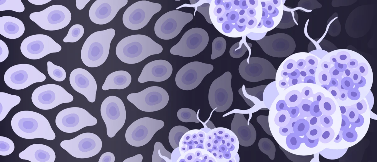
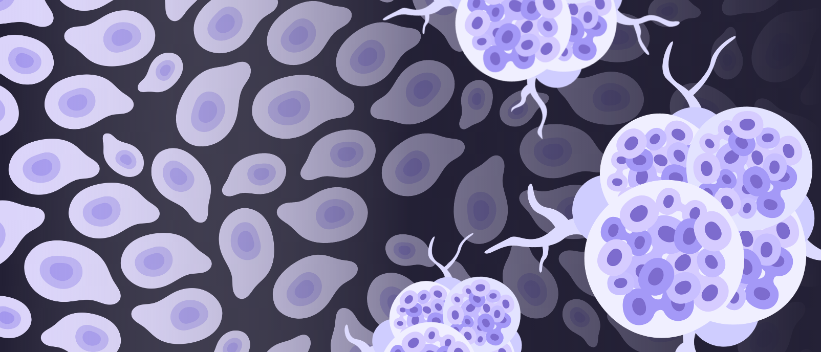
For instance, in a tumor microenvironment, the precise location of immune cells relative to cancer cells can dictate therapeutic response and patient prognosis. Similarly, in neurodegenerative diseases, the spatial distribution of amyloid plaques or tau tangles and their proximity to specific neuronal or glial cell types can illuminate disease progression and identify potential therapeutic targets. The key, Dr. Plummer emphasizes, lies in integration: single-cell data excels at defining cell states with high molecular resolution, providing a detailed molecular fingerprint of each cell, while spatial data masterfully places those precisely defined states back into their authentic biological context within the tissue. It is important to note that this is not about replacing single-cell approaches, which remain incredibly valuable for controlled perturbations and understanding intrinsic cellular programs. Instead, these perturbation datasets can be utilized to better inform and train spatial models, thereby improving the interpretation of spatial patterns and facilitating the construction of more predictive, biologically grounded insights into disease mechanisms. This synergistic approach ensures that researchers gain both a granular understanding of individual cell characteristics and a panoramic view of their collective function within a living system, leading to a more complete biological picture.
Analyzing the Spatial Omics Deluge: Leveraging AI for Deeper Insights
The collection of spatial omics data marks only the initial step; the subsequent analysis of these colossal and multifaceted datasets is where true biological insights are unearthed. The analytical journey typically commences with rigorous preprocessing, which includes alignment of data points to tissue images, accurate cell segmentation to delineate individual cells, and robust feature extraction to identify relevant molecular markers. Following these foundational steps, researchers proceed to identify distinct cell types or molecular patterns, meticulously mapping them back onto the preserved tissue structure. The analysis then delves into uncovering spatial relationships, such as the formation of cellular neighborhoods, the identification of specific cell-cell interactions, and the characterization of microenvironmental gradients that may influence cellular behavior.
However, the most profound and actionable insights emerge from the intelligent integration of spatial data with other crucial modalities, including single-cell data, proteomic profiles, high-resolution imaging, and invaluable clinical datasets. This is where Artificial Intelligence (AI) becomes an indispensable ally. AI algorithms are uniquely positioned to navigate the complexity of combining these diverse data types, enabling the discovery of hidden patterns, subtle correlations, and emergent properties that would be imperceptible to traditional analytical methods. By leveraging machine learning, researchers can build sophisticated predictive models that can forecast disease trajectories, identify individuals at risk, and even suggest optimal therapeutic strategies. This capability is particularly critical in fields like oncology, where early and accurate diagnosis can dramatically improve patient outcomes. Ultimately, this advanced integration of multiomic insights, powered by AI, promises to translate fundamental discoveries into faster, more accurate diagnostics. This will pave the way for clinically actionable tools, facilitating earlier disease detection, more precise patient stratification, and significantly more informed and personalized treatment decisions, marking a paradigm shift in clinical practice.
The Evolution of Omics and the Promise of Spatial Technologies
The journey towards spatial multiomics has been a remarkable progression in biological inquiry, reflecting an ever-deepening quest to understand life’s complexity. The late 20th century saw the dawn of genomics, focused on mapping and sequencing entire genomes. This was followed by transcriptomics, analyzing all RNA molecules; proteomics, studying proteins; and metabolomics, examining metabolites. Each ‘omic’ layer provided unprecedented detail, but initially, these were largely bulk analyses, averaging signals from millions of cells and thus obscuring crucial cellular heterogeneity. The early 2010s ushered in the single-cell revolution, allowing scientists to characterize individual cells, revealing distinct cellular populations and states previously masked by bulk approaches. This was a monumental step, but it came at a cost: the crucial spatial information—where these individual cells resided and interacted within a tissue—was lost during tissue dissociation.
The emergence of spatial omics, initially gaining significant traction with techniques like spatial transcriptomics in 2016, effectively bridged this critical gap. It allowed for the measurement of molecular profiles in situ, maintaining the invaluable tissue architecture. The rapid development of this field, driven by technological innovations in microscopy, advanced sequencing chemistry, and exponentially increasing computational power, reflects the scientific community’s profound recognition of the need for contextual biology. The global spatial omics market, for instance, is projected to grow from an estimated USD 300 million in 2022 to over USD 1.5 billion by 2027, with a compound annual growth rate (CAGR) exceeding 30%. This exponential growth underscores the perceived value and substantial investment flowing into these technologies, highlighting their potential to transform our understanding of disease and accelerate therapeutic development.
The urgency for these advancements is further underscored by the staggering global burden of diseases like cancer and neurological disorders. Cancer remains a leading cause of death worldwide, with an estimated 20 million new cases and 10 million deaths annually, according to the World Health Organization. Neurological disorders, including Alzheimer’s, Parkinson’s, multiple sclerosis, and various neurodevelopmental conditions, affect over 1 billion people globally, representing a significant cause of disability, morbidity, and immense healthcare expenditure. Despite decades of dedicated research, curative treatments remain elusive for many of these conditions, emphasizing the critical need for a deeper, spatially resolved understanding of their underlying pathology.
Navigating the Frontiers: Limitations and Solutions in Spatial Omics
Despite their immense promise, existing spatial omics technologies and data analysis tools are still in a phase of rapid evolution and inherently present certain limitations that researchers actively seek to overcome. Dr. Plummer candidly acknowledges these trade-offs: some platforms offer exceptionally high molecular depth, allowing for the quantification of thousands of genes or proteins, but at the expense of lower spatial resolution, meaning they cannot pinpoint molecular events to individual cells or subcellular compartments with precision. Conversely, other technologies excel at preserving exquisite spatial detail but are currently limited in the number of molecular targets they can simultaneously measure. This "resolution versus throughput" challenge is a common theme in developing new biotechnologies.
Further challenges encompass sensitivity, where detecting low-abundance molecules, crucial for early disease detection, can be difficult; issues with tissue quality and preservation, which can impact data integrity and experimental reproducibility; accuracy in cell segmentation, especially in densely packed or irregularly shaped tissues; scalability, as processing large numbers of samples remains resource-intensive and costly; and perhaps most critically, a lack of cross-platform standardization. This latter point means that comparing datasets generated using different technologies or in different labs can be exceedingly difficult, hindering collaborative efforts and the establishment of universally accepted benchmarks. Without standardization, validating findings across different research groups becomes a significant hurdle.

To overcome these multifaceted limitations, Dr. Plummer and her peers advocate for a multi-pronged strategy. Central to this is multimodal integration, combining complementary methods to leverage their individual strengths while mitigating their weaknesses. This involves meticulous experimental design, ensuring robust data generation from the outset. The development of powerful, flexible computational pipelines is also crucial for handling the immense scale and complexity of the data, allowing for efficient processing and analysis. Furthermore, fostering strong collaboration across diverse disciplines—from molecular biologists and pathologists to computational scientists and engineers—is essential for driving innovation and addressing complex challenges. As Dr. Plummer explains, "No single platform answers every question, so the most effective strategy is to combine complementary methods and build frameworks that allow results to be validated across datasets and biological systems." The ongoing push for standardization across the field, exemplified by initiatives like Spatial Touchstone and the Global Alliance for Spatial Technologies, is expected to significantly enhance the robustness and reproducibility of all platforms and data outputs over time, ultimately accelerating discovery and translation.
The Global Alliance for Spatial Technologies: Fostering a Collaborative Future
Recognizing the collective challenges and the unparalleled potential of spatial technologies, Dr. Jasmine Plummer has been instrumental in establishing the Global Alliance for Spatial Technologies (GAST). This initiative, born from a grassroots desire within the scientific community, embodies a crucial mission: to significantly accelerate the development, widespread adoption, and impactful application of spatially resolved technologies across the entire spectrum of biomedical research.
At its core, GAST seeks to forge a cohesive global community that transcends institutional and geographical boundaries. It aims to bring together a diverse array of stakeholders, including pioneering researchers, frontline clinicians, technology developers, and computational scientists. The alliance’s primary objectives are multifaceted: to build shared standards that ensure data comparability and reproducibility across different labs and platforms, to foster robust collaborative networks that facilitate knowledge exchange and resource sharing, and critically, to lower the barriers to entry for using these powerful tools. This democratic approach ensures that spatial technologies are not confined to a few specialist centers with extensive resources but become broadly accessible. By democratizing access and standardizing practices, GAST intends to empower a wider scientific community to apply these advanced methods to answer the most pressing biological and clinical questions, thereby accelerating discovery and translation on a global scale. This collaborative ethos is widely considered by experts across the scientific community to be indispensable for the maturation and widespread impact of any nascent scientific field, ensuring that the benefits of these revolutionary tools reach as many patients as possible.
Transformative Impact: Spatial Omics in Oncology and Neurology
The future impact of advanced spatial omics tools on both oncological and neurological research is envisioned as profoundly transformative, promising to reshape diagnostic and therapeutic paradigms. In oncology, these technologies are poised to revolutionize our understanding of cancer at an unprecedented level of detail. They will deepen our insights into tumor heterogeneity – the phenomenon where different cells within a single tumor exhibit distinct molecular characteristics – which is a major driver of treatment resistance and metastasis. Spatial omics will also illuminate the intricate organization of the immune microenvironment within and around tumors, identifying specific immune cell subsets and their precise locations, which can predict response to immunotherapies and other treatments. Furthermore, these tools will shed light on metastatic niches, revealing how cancer cells interact with distant tissue environments to establish secondary tumors, and unraveling the molecular mechanisms underlying treatment resistance, enabling the design of more effective therapeutic strategies. When paired with sophisticated AI tools, spatial omics will facilitate the discovery of more precise biomarkers, enabling better patient stratification for clinical trials, leading to higher accuracy diagnostics, and ultimately informing more rational and personalized therapeutic decisions. This could mean a future where a patient’s tumor is not just biopsied, but spatially mapped to inform a truly tailored treatment plan.
In neurology, where the nervous system is inherently organized into highly specialized circuits, distinct layers, and unique cellular neighborhoods, spatial tools will be nothing short of revolutionary. These technologies will provide an unparalleled capacity to understand how neurodegenerative, neuroinflammatory, or neurodevelopmental diseases unfold across specific brain regions, observing pathological changes in situ. They will enable researchers to meticulously track how glial cells (the brain’s crucial support cells) and neuronal interactions change over time in response to disease or injury, and how local microenvironments contribute to processes such as neurodegeneration, chronic inflammation, or attempts at neural repair. This contextual understanding is critical, as many neurological disorders are characterized by subtle, localized changes that propagate throughout complex neural networks. For example, understanding the precise spatial progression of amyloid plaques in Alzheimer’s disease and their impact on neighboring neurons could unlock new therapeutic avenues.
More broadly, Dr. Plummer believes that spatial omics will fundamentally shift both oncology and neurology from a descriptive cataloging of molecular events towards a far more integrated and functional view of disease. This paradigm shift will ensure that molecular changes are not interpreted in isolation but are intrinsically linked to tissue structure, physiological function, and ultimately, clinical outcome. This integration promises to accelerate the identification of novel therapeutic targets, refine existing drug development pipelines by providing a more accurate understanding of drug mechanisms of action, and significantly enhance our ability to predict patient response to therapies, thereby ushering in a new era of precision medicine for some of humanity’s most challenging diseases. The scientific community eagerly anticipates the breakthroughs that this powerful fusion of multiomic and spatial technologies will bring, holding immense promise for improved diagnostics, more effective treatments, and a deeper, more contextual understanding of life itself.


Leave a Reply