
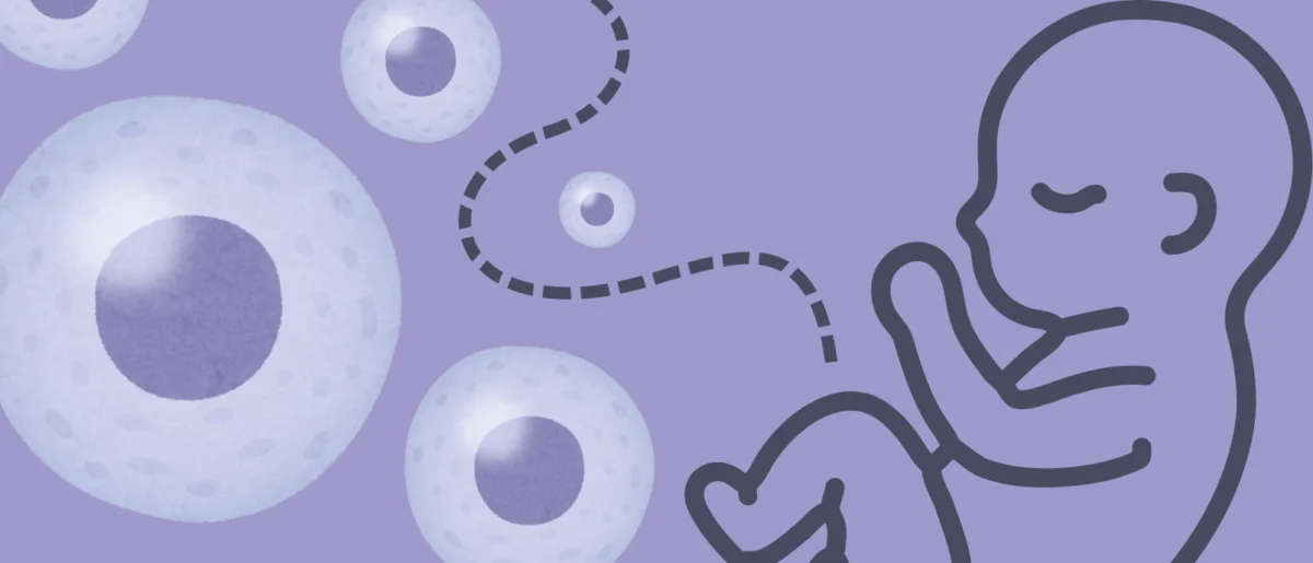
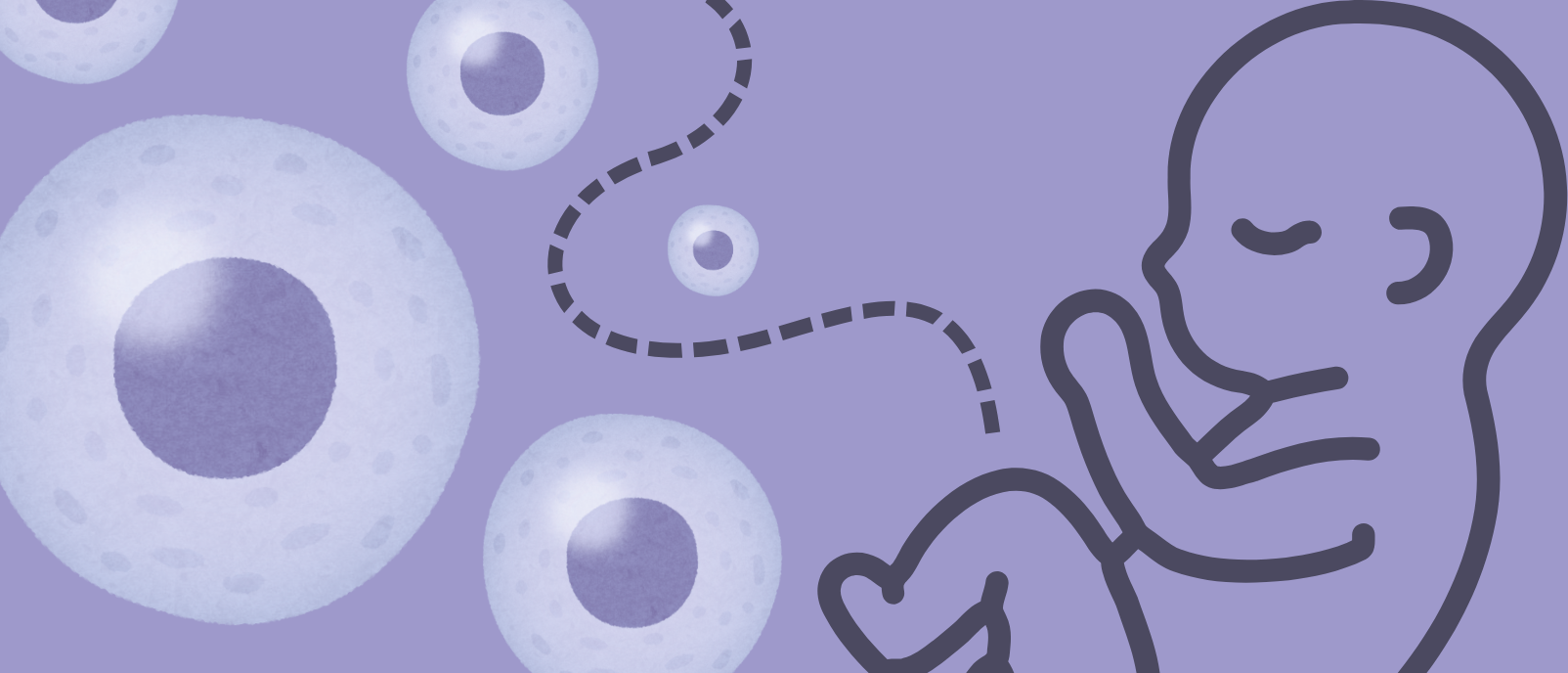
Researchers at the University of California San Francisco (UCSF) have achieved a significant breakthrough in understanding the intricate biological dialogue between a mother and her developing fetus. Their recent study, leveraging advanced single-cell and spatial omics technologies, has constructed the most comprehensive cellular atlas of the human maternal-fetal interface (MFI) to date. This monumental effort not only unveils previously unknown cell types and illuminates the complex process of healthy MFI development but also offers crucial insights into the cellular underpinnings of devastating pregnancy complications such as preeclampsia, miscarriage, and preterm birth.
Unlocking the Secrets of the Maternal-Fetal Interface
The maternal-fetal interface is a transient yet profoundly vital biological structure, forming the critical nexus where the maternal uterine tissues meet the fetal placental cells. This dynamic interaction is paramount for nutrient exchange, waste removal, and immune modulation, all essential for sustaining a healthy pregnancy and ensuring optimal fetal development. Dysfunction within this interface is a recognized precursor to a host of adverse pregnancy outcomes, which collectively affect millions of families globally each year. For instance, preeclampsia, a serious hypertensive disorder of pregnancy, complicates 2-8% of pregnancies worldwide, leading to significant maternal and fetal morbidity and mortality. Miscarriage, defined as pregnancy loss before 20 weeks of gestation, affects an estimated 10-20% of clinically recognized pregnancies, while preterm birth, occurring before 37 weeks, impacts about 10% of births globally and is the leading cause of death among children under five.
A Critical Yet Elusive Connection
Despite its indisputable importance, the MFI has historically presented substantial challenges to scientific investigation. Its transient nature, the complex interplay of diverse cell types from two genetically distinct individuals, and its deep-seated location within the uterus have made it notoriously difficult to study in high resolution. Traditional research methods often relied on bulk tissue analysis, which provides an averaged view of cell populations, masking the critical contributions and interactions of individual cell types. This limitation has hindered a comprehensive understanding of how healthy pregnancies develop and, critically, why complications arise, leaving many questions unanswered about the precise cellular and molecular mechanisms at play.
The Dawn of Advanced Omics Technologies
The UCSF team’s success in dissecting the MFI is a testament to the revolutionary capabilities of contemporary single-cell and spatial omics technologies. Over the past decade, these methods have transformed biological research by enabling scientists to analyze gene expression, protein profiles, and epigenetic modifications at the resolution of individual cells, while also preserving their spatial context within tissues. Single-cell sequencing, for example, allows researchers to identify rare cell populations, characterize cellular heterogeneity, and trace developmental trajectories that would be impossible to discern with bulk sequencing. Spatial transcriptomics takes this a step further, mapping gene expression profiles directly onto tissue sections, revealing where specific cell types and molecular activities occur relative to one another. These advancements provide an unprecedented "microscopic" view of biological processes, offering the detail necessary to untangle the MFI’s complexities.
Groundbreaking Methodology: A Multi-Omics Approach
To overcome the historical impediments, the UCSF researchers employed a sophisticated multi-omics strategy, combining several cutting-edge techniques. Their approach included:
- Single-nucleus multiome profiling: This technique simultaneously measures gene expression (transcriptomics) and chromatin accessibility (epigenomics) within individual cell nuclei. By analyzing both these layers of information, researchers can gain a deeper understanding of not only which genes are active in a cell but also how their expression is regulated, providing clues about cell identity and function.
- Spatial transcriptomics: This method allowed the team to map gene expression patterns directly onto tissue sections, revealing the precise anatomical locations of different cell types and their molecular interactions within the MFI. This spatial context is crucial for understanding how cells communicate and organize to form functional tissues.
- Multiplex protein imaging: By simultaneously visualizing multiple proteins within tissue samples, this technique provided detailed information about cell phenotypes, signaling pathways, and cell-cell interactions, complementing the genetic and transcriptomic data.
These methods were applied to snap-frozen MFI samples meticulously sourced from existing placenta tissue banks at Stanford University and UCSF. The samples represented healthy pregnancies that had been aborted during the first and second trimesters, providing a critical window into early and mid-gestation development. To ensure a comprehensive understanding across the entire pregnancy timeline, the study also incorporated analyses of fresh placental samples collected following healthy term deliveries. A stringent selection criterion was applied: all included tissues were confirmed to be free of any health abnormalities, ensuring that the resulting cellular atlas accurately reflected the processes of healthy MFI development, thereby serving as an invaluable reference point for future studies into pathological conditions.
Comprehensive Sample Collection and Ethical Considerations
The systematic collection and rigorous quality control of samples underscore the study’s commitment to robust scientific inquiry. The inclusion of samples from different gestational stages – early, mid, and late pregnancy – is particularly vital. The MFI undergoes dramatic transformations throughout gestation, adapting to the changing demands of the growing fetus. By mapping these changes at a cellular level, the UCSF team has provided a dynamic atlas, illustrating how cell populations emerge, differentiate, and interact over time.
It is important to acknowledge the ethical framework surrounding the use of fetal tissue in research. Research involving human fetal tissue, particularly from elective abortions, is subject to strict ethical guidelines and regulatory oversight in the United States and many other countries. These guidelines typically require informed consent from the donor, ensure the anonymity of the donor, and prohibit any financial incentive for donation. The use of such samples is often deemed ethically permissible when the research addresses significant health issues, there are no viable alternatives, and the research adheres to the highest scientific and ethical standards. In this context, the UCSF study, by shedding light on the mechanisms of pregnancy complications and potentially saving lives, exemplifies a pursuit of medical knowledge within established ethical boundaries.
Pioneering Discoveries: New Cell Types and Regulatory Mechanisms
The extensive cell atlas generated by this study represents a landmark achievement, meticulously documenting healthy MFI development from its nascent stages in early gestation through to term. Among its most striking revelations was the identification of a previously unknown maternal cell type. This novel cell, residing within the uterine tissue, plays a crucial regulatory role in the establishment and integration of placental cells. This process is fundamental for ensuring adequate blood flow from the mother to the fetus, a vital conduit for oxygen and nutrient delivery. Any disruption in this delicate balance can have severe consequences for fetal growth and overall pregnancy health.
The Unveiling of a Novel Maternal Cell Type
The discovery of this specific maternal cell type is significant because it adds a new piece to the puzzle of how the maternal immune system and uterine environment interact with the invading fetal trophoblast cells to form a stable placenta. Researchers speculate that understanding the precise signaling pathways and molecular machinery utilized by this cell type could open new avenues for interventions aimed at improving placental function in at-risk pregnancies. For instance, if this cell type is found to be deficient or dysfunctional in certain complicated pregnancies, it might become a target for future therapeutic strategies designed to enhance placental development and nutrient transfer.
Cannabinoid Receptors and Pregnancy Outcomes: A Crucial Link
Further investigation into this newly identified maternal cell type yielded another critical finding: the presence of a cannabinoid receptor on its surface. When these cells were exposed to cannabinoid molecules, a notable effect was observed – they restricted the establishment of placental cells. This discovery offers a compelling potential biological explanation for the poorer pregnancy outcomes that have been increasingly associated with cannabis use during pregnancy.
Cannabis use during pregnancy has become a growing public health concern, particularly with the increasing legalization of cannabis in various regions. Studies have linked prenatal cannabis exposure to adverse outcomes such as low birth weight, preterm birth, and neurodevelopmental issues in offspring. However, the precise biological mechanisms underlying these associations have remained largely unclear. The UCSF study now provides a direct cellular pathway through which cannabinoids could exert their detrimental effects on placental development and function. This finding reinforces existing public health recommendations from organizations like the American College of Obstetricians and Gynecologists (ACOG) and the Centers for Disease Control and Prevention (CDC), which strongly advise against cannabis use during pregnancy due to potential risks to both mother and fetus. The elucidation of this cellular mechanism provides powerful, scientific backing for these warnings, potentially strengthening public health campaigns and patient counseling efforts.
Deciphering Pregnancy Complications at Cellular Resolution
Beyond the discovery of new cell types and regulatory mechanisms, the UCSF team took a crucial step towards understanding pathological pregnancy states. By integrating the detailed cellular data from their healthy MFI atlas with genetic data from over 10,000 patients, they were able to identify specific cell types and states that demonstrated strong associations with common and severe conditions such as preeclampsia, miscarriage, and preterm birth. This data integration approach, combining single-cell resolution with large-scale patient genetic information, is a powerful paradigm for disease discovery.
Preeclampsia: New Insights into Uterine Vascular Remodeling
A particularly impactful finding emerged concerning preeclampsia. The study revealed that the cell types most significantly affected during preeclampsia were those involved in uterine blood vessel remodeling. Preeclampsia is a complex disorder characterized by the sudden onset of high blood pressure, often accompanied by protein in the urine or other signs of organ damage, typically after 20 weeks of gestation. It is a leading cause of maternal and fetal morbidity and mortality worldwide.
The pathogenesis of preeclampsia is understood to involve abnormal placental development and function, particularly a failure of the uterine spiral arteries to undergo proper remodeling. Normally, during early pregnancy, these maternal blood vessels are extensively modified by invading fetal trophoblast cells to become wide, low-resistance vessels, ensuring a robust and consistent blood supply to the placenta. In preeclampsia, this remodeling process is incomplete, leading to narrow, high-resistance vessels, placental ischemia, and the release of anti-angiogenic factors into the maternal circulation, which contribute to the systemic maternal symptoms.
The UCSF study’s pinpointing of specific cell types involved in this critical uterine blood vessel remodeling as the most affected in preeclampsia provides unprecedented clarity. This insight could guide future research toward identifying early biomarkers for preeclampsia, developing targeted therapies to correct the dysfunctional remodeling, or even predicting which pregnancies are at highest risk. For instance, if specific genetic variants are found to predispose these particular cell types to dysfunction, early genetic screening or monitoring of these cell populations could become part of routine prenatal care.
Broader Implications for Maternal and Fetal Health
The multimodal healthy MFI atlas presented by the UCSF team serves as a foundational resource, poised to accelerate research and innovation in maternal-fetal medicine. This comprehensive blueprint of cellular interactions and developmental trajectories offers an unparalleled reference for understanding normal physiological processes and, by extension, identifying deviations that lead to adverse outcomes.
Towards Enhanced Diagnostics and Targeted Therapies
The immediate implications of this research are vast. By pinpointing the specific cellular culprits or dysfunctions associated with conditions like preeclampsia and miscarriage, the study paves the way for the development of highly specific diagnostic tools. Imagine a future where early pregnancy screening could identify cellular biomarkers that indicate a heightened risk of preeclampsia, allowing for proactive interventions. Similarly, understanding the cellular basis of recurrent miscarriage could lead to personalized treatment strategies for affected individuals.
Moreover, the identification of novel cell types and their regulatory mechanisms opens exciting avenues for targeted therapeutic development. If a specific cell type is found to be underperforming or misbehaving, researchers could develop drugs or gene therapies designed to modulate its function, thereby restoring healthy MFI activity. For example, if the newly discovered maternal cell type’s function can be enhanced in situations where placental establishment is compromised, it could potentially prevent complications related to insufficient blood flow. The finding regarding cannabinoid receptors also suggests potential targets for pharmacological intervention, should it be deemed necessary to counteract the effects of cannabinoid exposure, or simply to understand the broader implications of receptor signaling in pregnancy.
Expert Perspectives and the Path Forward
Experts in the field are likely to view this study as a pivotal moment for maternal-fetal medicine. Dr. Tippi MacKenzie, a fetal surgeon and professor at UCSF not involved in this specific study but deeply familiar with the challenges of pregnancy complications, might comment on the transformative potential of such high-resolution data. "For too long, the maternal-fetal interface has been a black box," she might suggest. "This atlas is like shining a powerful light into that box, revealing the intricate machinery within. It fundamentally changes how we can approach diagnosing and treating pregnancy disorders." Similarly, Dr. Mary Norton, a professor of obstetrics and gynecology at UCSF, could emphasize the clinical relevance. "Understanding the specific cell types involved in conditions like preeclampsia is a game-changer. It moves us beyond treating symptoms to potentially addressing the root causes at a cellular level, offering hope for more effective interventions and, ultimately, healthier outcomes for mothers and babies."
From a public health standpoint, the clarity provided on the impact of cannabis use could strengthen public health messages. A representative from the CDC’s Division of Reproductive Health might reiterate, "This research provides further evidence supporting our recommendations against cannabis use during pregnancy. Understanding the specific cellular mechanisms helps us communicate the risks more effectively and underscores the importance of avoiding substances that can interfere with critical developmental processes."
Addressing the Gaps: Future Research Directions
While this study represents a monumental leap forward, the UCSF team readily acknowledges that the journey is far from over. They express confidence that "more cell states and subtypes associated with normal pregnancy and pathological deviations are to be discovered with further spatial and temporal investigations." The MFI is an incredibly dynamic environment, and even more granular temporal analyses, perhaps involving longitudinal studies or even more refined single-cell capture techniques, could uncover additional layers of complexity.
Future research will undoubtedly build upon this foundational atlas. This includes delving deeper into complicated pregnancies, not just healthy ones, to precisely map the cellular dysfunctions in various pathological states. Identifying the specific molecular pathways that are disrupted in these conditions will be critical for pinpointing potential therapeutic targets. Furthermore, the atlas could serve as a benchmark for evaluating new diagnostic markers or for testing the efficacy of novel therapeutic agents in preclinical models. The integration of even more diverse patient populations, including those from different ethnic backgrounds and geographical locations, will also be vital to ensure the generalizability of these findings and to address health disparities in maternal-fetal health.
The Transformative Potential for Global Health
In conclusion, the UCSF study on the maternal-fetal interface stands as a testament to the power of advanced biotechnologies in unraveling complex biological mysteries. By providing an unprecedented cellular and spatial map of this crucial connection, researchers have laid a robust foundation for a new era in maternal-fetal medicine. The identification of novel cell types, the elucidation of regulatory mechanisms, and the precise cellular associations with major pregnancy complications collectively offer a beacon of hope for improving pregnancy outcomes worldwide. This comprehensive atlas is not merely a collection of data; it is a springboard for future discoveries, promising to transform our understanding, diagnosis, and treatment of conditions that profoundly impact the health of mothers and their children across the globe.


Leave a Reply