
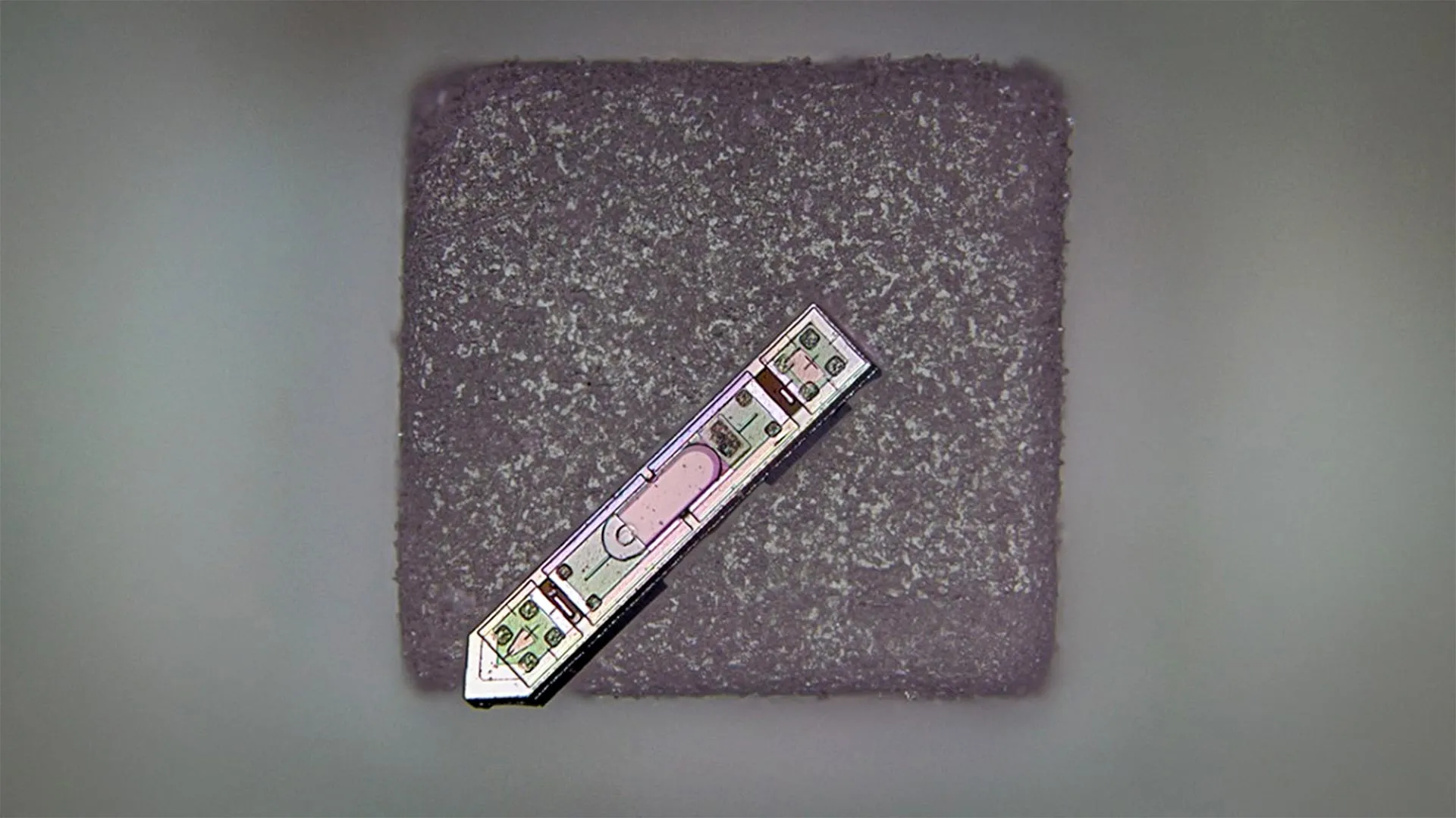
In a landmark achievement for the fields of bioengineering and microelectronics, a multidisciplinary team at Cornell University has announced the development of a neural implant so small it can rest atop a single grain of salt. This device, characterized as a microscale optoelectronic tetherless electrode (MOTE), represents a paradigm shift in how scientists and clinicians may eventually interface with the central nervous system. Despite its microscopic dimensions—measuring approximately 300 microns in length and 70 microns in width—the device is capable of recording and wirelessly transmitting high-fidelity brain activity data from a living subject for a duration exceeding 12 months. This breakthrough, recently detailed in the journal Nature Electronics, addresses several of the most persistent challenges in neurotechnology, including device longevity, tissue displacement, and the limitations of wired connections.
The development of the MOTE was spearheaded by Alyosha Molnar, a professor in the School of Electrical and Computer Engineering at Cornell University, in collaboration with Sunwoo Lee, an assistant professor at Nanyang Technological University in Singapore. The project’s roots trace back to Lee’s tenure as a postdoctoral researcher in Molnar’s laboratory, where the team sought to bridge the gap between the massive scale of modern microchips and the delicate, fluid environment of the mammalian brain. By leveraging advanced semiconductor materials and innovative optical communication methods, the researchers have created a platform that bypasses the need for bulky batteries or invasive wiring, potentially ushering in a new era of "invisible" medical monitoring.
Technical Specifications and the MOTE Architecture
At the heart of the MOTE device is a sophisticated integration of semiconductor physics and optical engineering. Unlike traditional neural probes that rely on radio-frequency (RF) waves or physical wires to transmit data, the MOTE utilizes light. The core component is a specialized semiconductor diode fabricated from aluminum gallium arsenide (AlGaAs). This material is chosen for its unique optoelectronic properties, which allow it to serve a dual purpose: it captures incoming light to power the device’s internal circuitry and simultaneously emits light to broadcast data back to an external receiver.
The internal architecture of the MOTE is a marvel of miniaturization. In addition to the AlGaAs diode, the 300-micron footprint houses a low-noise amplifier and an optical encoder. These components are manufactured using the same complementary metal-oxide-semiconductor (CMOS) technology that powers modern smartphones and computers, yet they have been refined to operate at a fraction of the power. To ensure the device can transmit data through the dense environment of the brain, the system employs red and infrared laser beams. These specific wavelengths are chosen because they can safely penetrate biological tissue with minimal absorption or scattering, allowing for a clear line of communication between the implant and an external optical sensor.
The data transmission itself is handled via Pulse Position Modulation (PPM). This is a highly efficient coding technique often utilized in deep-space satellite communications. In a PPM system, the timing of an optical pulse—rather than its intensity or frequency—carries the information. This method allows the MOTE to communicate complex electrical signals from the brain using an incredibly low power budget. According to Professor Molnar, this efficiency is what allows the device to remain operational and accurate over long periods without the heat generation that often plagues larger electronic implants.
A Chronology of Innovation: From Concept to Long-Term Success
The journey toward the MOTE began several years ago as researchers recognized the "size-power" paradox in neural engineering. Historically, increasing the number of recording sites in a neural implant required larger devices to handle the increased power and data demands. However, larger devices occupy more volume in the brain, leading to significant tissue damage and a robust immune response known as gliosis, where the body forms scar tissue around the implant, eventually insulating the electrodes and rendering them useless.
The Cornell team’s timeline involved several key stages of development:
- Phase I (Semiconductor Design): Early research focused on identifying a material that could handle both power harvesting and data transmission. AlGaAs was identified as the primary candidate due to its efficiency in the infrared spectrum.
- Phase II (Miniaturization and Fabrication): Utilizing Cornell’s nanofabrication facilities, the team shrunk the circuitry to the micron scale, ensuring that the final device would be smaller than a grain of table salt.
- Phase III (Wireless Powering Tests): Researchers had to prove that a laser could provide enough energy to the implant to run the amplifier and encoder without burning the surrounding brain tissue.
- Phase IV (In Vivo Validation): The most critical stage involved implanting the MOTE into a living animal model. The goal was not just to record data, but to do so over an extended period to prove the device’s stability.
The reporting of a one-year operational window is particularly significant. Most microscale implants fail within weeks or months due to moisture ingress or the biological rejection mentioned earlier. The MOTE’s ability to function for over a year suggests that its materials and "tetherless" nature—meaning it has no wires pulling on it as the brain moves within the skull—greatly enhance its biocompatibility.
Supporting Data: The Advantages of Optical Over Radio Frequency
One of the primary engineering decisions that sets the MOTE apart is the move away from Radio Frequency (RF) transmission. Most modern wireless implants, such as those developed by Neuralink or Paradromics, use RF to transmit data. While effective, RF transmission requires an antenna. The physics of electromagnetism dictates that an antenna must be of a certain size relative to the wavelength it is transmitting. This creates a "floor" for how small an RF-based device can be.
In contrast, optical transmission uses wavelengths in the hundreds of nanometers. This allows the "antenna"—in this case, the light-emitting diode—to be shrunk to the micron scale without losing efficiency. Furthermore, RF signals can interfere with other electronics and are susceptible to environmental noise. The MOTE’s optical system is inherently shielded from electromagnetic interference, which leads to a significantly higher signal-to-noise ratio.
Data collected during the study showed that the MOTE could successfully capture "local field potentials"—the summed electrical activity of thousands of neurons—with a level of clarity comparable to much larger, wired electrodes. The use of infrared light also ensures that the brain tissue is not subjected to harmful ionizing radiation, maintaining the safety profile of the device for chronic use.
Reactions and Perspectives from the Scientific Community
While the Cornell team has led the charge, the broader scientific community has responded to the Nature Electronics report with cautious optimism. Independent neuroscientists have noted that the "tetherless" aspect is perhaps the device’s greatest contribution to the field. Traditional electrodes are connected by wires to a connector on the skull; every time the subject moves, the wires tug on the brain, causing micro-traumas. By removing the tether, the MOTE moves in harmony with the brain tissue.
"As far as we know, this is the smallest neural implant that will measure electrical activity in the brain and then report it out wirelessly," Professor Molnar stated during the announcement. He emphasized that the goal was not just to make something small for the sake of size, but to solve the mechanical mismatch between rigid electronics and soft biological tissue.
Collaborator Sunwoo Lee highlighted the interdisciplinary nature of the work, noting that the transition of the technology from a lab setting to a functional, long-term implant required a convergence of electrical engineering and surgical precision. Academic peers have suggested that if this technology can be scaled to include multiple MOTEs distributed across the brain, it could provide a "distributed" map of neural activity that was previously impossible to obtain.
Future Horizons: MRI Compatibility and Beyond
The implications of the MOTE extend far beyond simple brain-machine interfaces. One of the most promising future applications involves the device’s compatibility with Magnetic Resonance Imaging (MRI). Current neural implants contain significant amounts of metal, which can heat up or cause massive distortions (artifacts) in MRI images, making it dangerous or impossible for patients with implants to receive necessary scans.
Because the MOTE is primarily composed of semiconductors and utilizes optical communication, it is theoretically transparent to the powerful magnets of an MRI machine. "The materials used in the MOTE could allow researchers to record brain activity during MRI scans, something that is largely not possible with current implants," Molnar noted. This would allow clinicians to correlate the microscopic electrical data from the MOTE with the macroscopic structural and functional data from the MRI, providing a dual-layered view of brain health.
Furthermore, the team is exploring the integration of these devices with "artificial skull plates." These would be transparent windows replaces a small portion of the skull, allowing lasers to reach the implants and data to be collected without any skin-penetrating wires. This "window to the brain" concept could revolutionize the treatment of chronic conditions like epilepsy, Parkinson’s disease, or even spinal cord injuries.
Broader Impact and Ethical Considerations
The successful deployment of the MOTE signals a shift toward less invasive, more durable bio-integrated sensors. Beyond the brain, the researchers suggest the technology could be adapted for use in the spinal cord or peripheral nerves, potentially helping to restore function in paralyzed patients. The extreme miniaturization also opens the door to "swarm" neural interfaces, where hundreds of tiny sensors are injected or placed throughout the cortex to provide a high-resolution, decentralized data network.
However, the rise of such capable, nearly invisible technology also brings ethical considerations to the forefront. As neural implants become smaller and more integrated into the body, the scientific community must address questions regarding data privacy, the long-term effects of laser exposure in the brain, and the permanence of such micro-devices.
For now, the Cornell-led breakthrough stands as a testament to the power of micro-scale engineering. By shrinking a wireless transmitter to the size of a grain of salt and proving its resilience over a year of continuous operation, Molnar, Lee, and their colleagues have provided a new toolkit for understanding the most complex organ in the known universe. The MOTE is not merely a piece of hardware; it is a bridge between the digital world of silicon and the biological world of the neuron, built at a scale that finally respects the delicacy of the human mind.


Leave a Reply