
In a landmark achievement for regenerative medicine and hematology, a multidisciplinary team of scientists at the University of Basel and University Hospital Basel has successfully engineered a fully functional, three-dimensional model of the human bone marrow niche using exclusively human cells. This breakthrough, recently detailed in the prestigious journal Cell Stem Cell, represents the first time that the intricate "blood factory" of the human body has been recreated in a laboratory setting with such anatomical and physiological accuracy. By integrating bone cells, blood vessels, nerves, and immune components into a single cohesive system, the researchers have provided the scientific community with a powerful new tool to study blood disorders, test pharmaceutical compounds, and move toward a future of personalized oncology.
The bone marrow is an exceptionally complex organ, acting as the primary site for hematopoiesis—the continuous production of all blood cells, including oxygen-carrying erythrocytes, infection-fighting leukocytes, and clot-forming platelets. Despite its critical importance, the marrow is often overlooked until pathological conditions, such as leukemia or myelodysplastic syndromes, disrupt its delicate balance. Historically, investigating these conditions has proven difficult because the marrow’s internal environment is notoriously hard to access and even harder to replicate outside the living body. For decades, researchers have relied on mouse models or simplified two-dimensional cell cultures, both of which frequently fail to capture the nuances of human biology and the specific interactions that drive disease progression.
The Architecture of the Endosteal Niche
At the heart of the Basel team’s research is the "endosteal niche," a specialized microenvironment located near the inner surface of the bone. This niche is far more than a simple storage space for cells; it is a sophisticated regulatory hub where various cell types communicate through chemical signals and physical contact to govern the behavior of hematopoietic stem cells. The endosteal niche is of particular interest to oncologists because it is often where cancer cells, such as those found in acute myeloid leukemia, seek refuge to survive intensive chemotherapy.
The complexity of this environment has long stymied bioengineering efforts. A functional niche requires a precise arrangement of osteoblasts (bone-forming cells), endothelial cells (which form blood vessels), mesenchymal stromal cells, and even nerve fibers. Until now, no laboratory model had successfully synthesized all these components into a unified human-derived system. Led by Professor Ivan Martin and Dr. Andrés García-García from the Department of Biomedicine, the Basel researchers overcame this hurdle by utilizing advanced stem cell technology and bio-synthetic scaffolds.
Chronology of Development: From Stem Cells to Tissue
The development of this model followed a rigorous, multi-stage chronological process. The project began with the selection of a suitable structural foundation. The team chose a scaffold made of hydroxyapatite, a naturally occurring mineral form of calcium apatite that serves as the primary inorganic constituent of human bone and tooth enamel. This scaffold provided the necessary mechanical rigidity and chemical cues to encourage cell attachment and growth.
Following the preparation of the scaffold, the researchers employed induced pluripotent stem cell (iPSC) technology. By taking human cells and "reprogramming" them into a pluripotent state using molecular biology techniques, the team created a blank slate of cells capable of becoming any tissue type. The next phase involved a highly controlled "orchestrated development." By introducing specific growth factors and signaling molecules at precise intervals, the scientists guided the stem cells to differentiate into the diverse array of cell types found in the bone marrow.
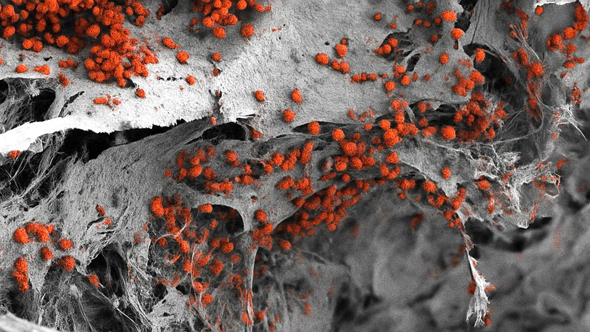
Over several weeks, these cells organized themselves within the hydroxyapatite framework, forming blood vessel networks, bone-like tissue, and rudimentary nervous structures. The result was a three-dimensional organoid measuring eight millimeters in diameter and four millimeters in thickness. While small to the human eye, this represents a significant scale in the world of micro-tissue engineering, providing enough volume to sustain biological processes that mimic those occurring in a patient’s body.
Validation and Supporting Data
To ensure the model’s validity, the researchers conducted extensive comparative analyses between their lab-grown tissue and actual human bone marrow samples. The data revealed a high degree of structural and functional similarity. Specifically, the model demonstrated the ability to maintain the "stemness" of human blood-forming cells. In traditional lab cultures, these stem cells often differentiate too quickly or die off, but within the engineered niche, the researchers successfully maintained human blood cell formation for several weeks.
Key data points from the study include:
- Spatial Distribution: Imaging confirmed that the different cell types organized themselves into distinct zones, mirroring the natural anatomy of the endosteal niche.
- Vascularization: The presence of functional endothelial networks allowed for the simulation of nutrient and waste exchange, a critical factor for long-term cell viability.
- Molecular Signaling: Transcriptomic analysis showed that the cells in the model were expressing the same sets of genes used by cells in a living human marrow to communicate and regulate blood production.
Official Responses and the Drive for the 3Rs
The success of the project aligns with a global movement in science known as the "3Rs"—the push to Replace, Reduce, and Refine animal experiments. Professor Ivan Martin emphasized that while animal models, particularly mice, have provided foundational knowledge, they are not perfect substitutes for human physiology.
"We have learned a great deal about how bone marrow works from mouse studies," Professor Martin noted. "However, our model brings us closer to the biology of the human organism. It could serve as a complement to many animal experiments in the study of blood formation in both healthy and diseased conditions."
This sentiment is echoed by the University of Basel’s institutional policy, which prioritizes the development of alternative methods to minimize the use of laboratory animals. By providing a human-cell-based alternative, the researchers hope to increase the "translatability" of laboratory findings—meaning that discoveries made in the lab are more likely to hold true when applied to human patients in clinical trials.
Implications for Oncology and Drug Development
The practical applications of this model are vast, particularly in the realm of drug discovery. Currently, the pharmaceutical industry faces a high "attrition rate," where drugs that appear successful in animal trials fail during human clinical trials due to unforeseen toxicity or lack of efficacy. A human-based bone marrow model could serve as a high-fidelity testing ground for new chemotherapy agents or immunotherapies, allowing researchers to see how drugs interact with human tissue before they ever reach a patient.
However, Dr. Andrés García-García pointed out a current technical hurdle regarding the model’s size. "For the specific purpose of drug development, the size of our bone marrow model might be too large," he explained. In industrial drug screening, where thousands of compounds must be tested simultaneously, smaller "organ-on-a-chip" versions of the model will be necessary to ensure cost-effectiveness and speed. The team is already looking into miniaturization techniques to adapt their system for high-throughput screening.
Beyond mass drug testing, the model holds immense promise for personalized medicine. In the future, a patient diagnosed with a complex blood cancer could have their own cells used to create a "digital twin" or "biological avatar" of their bone marrow. Doctors could then test various combinations of treatments on the patient’s specific marrow model to identify which therapy is most effective at eradicating the cancer while sparing healthy blood production. This would move oncology away from a "one-size-fits-all" approach toward a strategy tailored to the unique genetic and cellular profile of the individual.
Analyzing the Path Forward
The creation of the human bone marrow niche model is a significant milestone, yet it is part of a broader, ongoing evolution in bioengineering. The study demonstrates that we are moving past the era of viewing tissues as simple collections of cells and toward understanding them as integrated, multi-system environments.
One of the critical implications of this research is its potential to shed light on "dormant" cancer cells. In many leukemia patients, the disease appears to go into remission, only to return years later. It is widely believed that the bone marrow niche provides a protective environment that allows a small number of cancer cells to go into a sleep-like state, rendering them immune to drugs that target rapidly dividing cells. By using this new 3D model, scientists can now investigate the exact signals that keep these cells dormant and, more importantly, figure out how to "wake them up" or eliminate them in their hiding spots.
Furthermore, the integration of nerve cells into the model is a particularly sophisticated touch. Recent research has suggested that the nervous system plays a role in regulating the release of blood cells into the circulation, especially during times of stress or injury. By including these neural components, the Basel team has opened the door to studying the "neuro-hematopoietic" axis, an area of biology that is still poorly understood.
Conclusion
The work of Professor Martin, Dr. García-García, and their colleagues at the University of Basel represents a synthesis of biology, engineering, and medicine. By successfully recreating the human endosteal niche, they have bridged a gap that has existed in medical research for decades. While further refinements are needed to scale the technology for widespread clinical and industrial use, the foundation has been laid for a new era of hematological research—one that is more ethical, more accurate, and more focused on the unique complexities of human biology. As this technology matures, it may very well change the way we understand the "blood factory" within us, leading to more effective treatments and a deeper understanding of the very essence of human life.


Leave a Reply