
A groundbreaking development from Rice University has unveiled a simple yet powerful method to investigate pyroglutamate, a pervasive but historically overlooked posttranslational modification (PTM) of proteins. This innovation promises to unlock profound new insights into how proteins achieve their myriad forms and functions, potentially revolutionizing our understanding of cellular processes and disease mechanisms. The research, spearheaded by Rice University chemistry professor Zachary Ball, describes a novel photochemical technique that selectively targets pyroglutamate, a modification whose subtle nature has long rendered it impervious to detailed study.
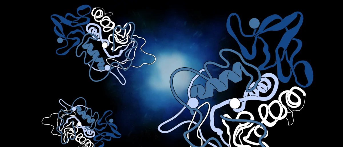
Proteins, often described as the workhorses of the cell, are complex macromolecules built from sequences of amino acids, much like intricate structures assembled from different types of Lego blocks. While there are only 20 standard amino acids, the sheer diversity of protein functions—from catalyzing biochemical reactions and transporting molecules to providing structural support and facilitating cell signaling—far exceeds the variety achievable by simple linear arrangements. This functional versatility is largely conferred by posttranslational modifications (PTMs), chemical alterations that occur to amino acids after a protein has been synthesized. PTMs dramatically expand the functional repertoire of proteins, influencing their stability, localization, interactions with other molecules, and overall biological activity. Common PTMs include phosphorylation, glycosylation, acetylation, and methylation, each playing critical roles in health and disease, and often serving as targets for drug development.
Among the vast array of known PTMs, pyroglutamate stands out as particularly enigmatic. It forms when a chemical reaction causes an amino acid called glutamate (or glutamine, which can cyclize to form pyroglutamate after deamidation) to cyclize, creating a five-membered lactam ring structure and expelling a molecule of water. This modification typically occurs at the N-terminus of a protein or peptide. Despite its ubiquitous presence across various biological systems and its known involvement in a range of physiological and pathological processes, pyroglutamate has remained largely uncharacterized. The primary reason for this scientific blind spot lies in its chemical nature: the modification introduces a relatively small change to the protein’s mass and does not significantly alter its overall charge or hydrophobicity, making it exceedingly difficult to detect and study using conventional biochemical methods. This subtlety has meant that while researchers have frequently observed pyroglutamate, they have lacked the tools to systematically investigate its distribution, the enzymes responsible for its formation or removal, and its precise impact on protein structure and function.
"We don’t know a lot about pyroglutamate beyond that we see it frequently," Professor Ball, the corresponding author of the paper, shared. This statement underscores the prevailing challenge that has long frustrated biochemists and molecular biologists. The lack of specific, easy-to-use tools has prevented researchers from asking fundamental questions about pyroglutamate’s role in vital biological processes, from protein folding and degradation to cellular signaling pathways. The new technique developed by Ball’s team is poised to change this by providing an unprecedented window into the world of this "hidden" protein mystery.
The innovation lies in its elegant simplicity and its harnessing of photochemistry—the use of light to drive chemical reactions. The method involves exposing a mixture containing the protein of interest, a specific tagging reagent, and two different chemical catalysts to blueish light, typically at wavelengths of either 350 or 400 nanometers. Under these precise conditions, one of the catalysts, which contains nickel, selectively binds to the pyroglutamate region on the protein. This initial binding event is crucial, as the nickel complex then acts as a secondary catalyst, facilitating the attachment of the tagging reagent specifically to the pyroglutamate site.
Professor Ball elaborated on the mechanism: "We had been experimenting with photochemistry, which is using light to initiate new reactions. Light can excite molecules enough to allow them to form new bonds. In this case, shining a light with a wavelength of either 350 or 400 nanometers allows us to initiate remarkable chemistry that is otherwise impossible." This controlled application of light provides the energy needed to overcome reaction barriers, enabling a highly selective and efficient labeling process that was previously unattainable. The precise reasons behind the nickel catalyst’s remarkable selectivity for pyroglutamate are still under investigation, but the practical outcome is a robust and repeatable method for targeting this elusive modification. With this new technique, Ball and other biochemists can now begin to ask and answer critical questions that have long lingered in the field.
The immediate implications of this breakthrough are far-reaching within basic scientific research. For the first time, scientists can systematically map the distribution patterns of pyroglutamate across the proteome of different cells, tissues, and organisms. This will allow them to identify which proteins are modified by pyroglutamate, and under what physiological or pathological conditions. Furthermore, the tool enables detailed studies into the kinetics of pyroglutamate formation and turnover, shedding light on the enzymes and cellular machinery involved. Critically, researchers can now investigate how pyroglutamate influences protein folding, stability, and intermolecular interactions. Does it stabilize certain protein conformations? Does it promote or prevent protein aggregation? Does it alter the binding affinity of a protein for its ligands or partners? These are fundamental questions that can now be systematically addressed, potentially revealing novel regulatory mechanisms within cells.
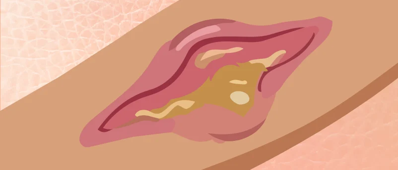
Beyond basic science, the broader impact of this tool extends into clinical and disease research, particularly in the realm of neurodegenerative disorders. Pyroglutamate has long been implicated in the pathogenesis of Alzheimer’s disease (AD), one of the most devastating neurological conditions globally. Specifically, N-terminally modified pyroglutamate amyloid-beta (Aβ) peptides (AβpE3-42 and AβpE11-42) are known to be abundant in amyloid plaques, a hallmark pathological feature of AD brains. These pyroglutamate-modified Aβ peptides are more prone to aggregation, are more resistant to degradation, and exhibit enhanced neurotoxicity compared to their unmodified counterparts. The formation of these modified Aβ species is believed to accelerate plaque formation and disease progression. The original article linking to a piece titled "Illuminating the link between amyloid beta and tau in Alzheimer’s disease" subtly hints at the potential for this new tool to deepen our understanding of such critical connections.
With Professor Ball’s new method, researchers can now precisely identify and quantify pyroglutamate modifications on Aβ peptides and other proteins implicated in AD. This capability could lead to a clearer understanding of how these modifications contribute to protein misfolding and aggregation, key events in neurodegeneration. Such insights could pave the way for the development of novel diagnostic biomarkers for early disease detection or even new therapeutic strategies aimed at preventing or reversing the formation of pathogenic pyroglutamate-modified proteins. For instance, if specific enzymes responsible for pyroglutamate formation in the context of AD can be identified, they could become promising drug targets. The implications also extend to other protein misfolding diseases, such as Parkinson’s disease or Huntington’s disease, where similar PTMs might play an underappreciated role.
The development of this tool also highlights the critical role of chemical biology in advancing our understanding of life sciences. By creating precise chemical probes and reaction methodologies, chemists enable biologists to observe and manipulate biological systems with unprecedented resolution. This iterative relationship between chemistry and biology is a cornerstone of modern scientific discovery, constantly pushing the boundaries of what is knowable. Rice University, with its strong traditions in both chemistry and engineering, continues to foster an environment ripe for such interdisciplinary breakthroughs. The university’s commitment to fundamental research provides the fertile ground for innovations that, while seemingly niche at first glance, possess the potential to ripple across multiple scientific disciplines and ultimately improve human health.
In conclusion, the development of a simple, light-activated method to study pyroglutamate represents a significant leap forward in our quest to understand the intricate world of proteins. By illuminating this previously hidden posttranslational modification, Professor Zachary Ball and his team at Rice University have not only provided a powerful new instrument for basic scientific inquiry but have also opened new avenues for exploring the molecular underpinnings of complex diseases. The ability to precisely target and investigate pyroglutamate promises to redefine our understanding of protein function, folding, and pathology, paving the way for a new era of discoveries in proteomics and biomedicine. This breakthrough underscores the enduring power of fundamental research to unravel the mysteries of life and translate them into tangible benefits for society.


Leave a Reply