New research conducted by neuroscientists at the Fralin Biomedical Research Institute at VTC has fundamentally questioned a decades-old paradigm regarding how the brain coordinates movement and how chronic neurological disorders are studied. For years, the scientific community has operated under the assumption that monitoring the activity of Purkinje cells—the large, intricate neurons located in the outer layer of the cerebellum—provides a direct and reliable window into the behavior of the deep cerebellar nuclei, which serve as the brain’s primary output hub for motor coordination. However, a study led by Meike van der Heijden, an assistant professor at the institute and the Virginia Tech School of Neuroscience, suggests that this linear relationship is far more complex and less predictable than previously believed, particularly in the context of disease states such as dystonia, ataxia, and tremor.
The cerebellum, often referred to as the "little brain," is a densely packed region at the base of the skull responsible for fine-tuning motor activity, maintaining balance, and ensuring that movements are fluid and precise. When the internal communication of the cerebellum is disrupted, the results are often debilitating. Patients may suffer from dystonia, characterized by painful, involuntary muscle contractions and abnormal postures; ataxia, which manifests as a lack of muscle coordination and "drunken" gait; or tremors, involving rhythmic, uncontrollable shaking of the limbs or head.
The Traditional Model of Cerebellar Communication
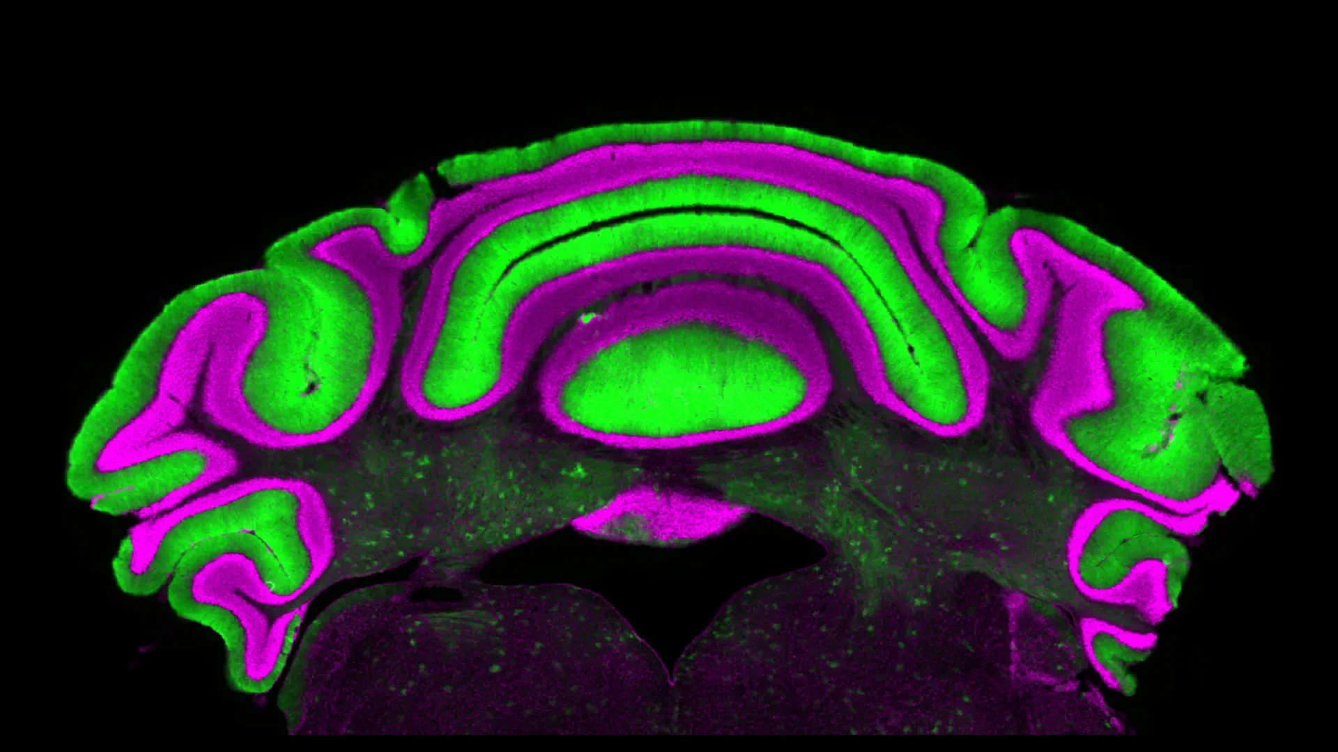
To understand the significance of this new research, one must first look at the established anatomical hierarchy of the cerebellum. The traditional model centers on the relationship between two primary cell populations. The Purkinje cells are the sole output of the cerebellar cortex. They are inhibitory neurons, meaning they release neurotransmitters—primarily gamma-aminobutyric acid (GABA)—that suppress the activity of their target cells. Those targets are the deep cerebellar nuclei (DCN), which sit deep within the white matter of the cerebellum.
Because the Purkinje cells directly inhibit the DCN, researchers have long utilized a "mirror" logic: if Purkinje cell activity increases, DCN activity should decrease, and vice versa. This assumption made the Purkinje cells an attractive target for study. Located in the cerebellar cortex, they are relatively accessible to modern recording technologies and imaging techniques. In contrast, the deep nuclei are buried under layers of tissue, making them significantly harder to reach and monitor in living subjects. Consequently, the Purkinje cell became the de facto biomarker for cerebellar health. If a researcher observed "noisy" or irregular firing in a Purkinje cell, they concluded that the same irregularity was being passed down to the deep nuclei, and subsequently to the rest of the motor system.
A Paradigm Shift in Neurological Observation
The new study, published in the Journal of Physiology, provides empirical evidence that this predictive relationship is fundamentally flawed. By analyzing an extensive database of electrophysiology recordings from pre-clinical models of cerebellar disease, Van der Heijden and her team discovered that activity levels in Purkinje cells did not correlate with the activity levels in the deep nuclei cells in a statistically significant way.
"We see that there’s not a clear linear relationship between activity in the Purkinje cells and in the deep nuclei cells," said Van der Heijden. "So there’s very limited predictive power in monitoring one to understand what’s going on in the other."
This finding suggests that the deep cerebellar nuclei are not merely passive recipients of Purkinje cell inhibition. Instead, they likely integrate a much wider array of inputs from other regions of the brain and spinal cord, or they possess intrinsic firing properties that allow them to maintain independent activity patterns even when their primary inhibitory input is compromised. This revelation is a "cautionary tale," as Van der Heijden describes it, for the entire field of neurobiology. It suggests that years of data derived solely from Purkinje cell observations may not accurately reflect the physiological reality of the cerebellar output that actually reaches the muscles.
Detailed Analysis of Dystonia, Ataxia, and Tremor
The implications of this research are most profound for the millions of people worldwide living with cerebellar disorders. Understanding the specific mechanics of these conditions is essential for developing effective therapies.
-
Dystonia: This condition affects an estimated 250,000 people in the United States alone. It causes muscles to contract involuntarily, leading to repetitive or twisting movements. While the exact cause is often unknown, it is widely recognized as a failure in the brain’s ability to inhibit unnecessary muscle movements. If researchers have been focusing on Purkinje cells to solve dystonia while the actual "broken" signal resides or originates independently in the deep nuclei, current treatment paths may be misaligned.
-
Ataxia: Often resulting from damage to the cerebellum or its pathways, ataxia leads to a lack of voluntary coordination of muscle movements. It can affect speech, eye movement, and swallowing. The "lack of correlation" found in the Virginia Tech study suggests that the irregular movements seen in ataxia may be the result of complex interactions within the deep nuclei that Purkinje cell monitoring simply cannot capture.
-
Tremor: Essential tremor is one of the most common neurological diseases, affecting nearly 10 million Americans. It involves a rhythmic shaking that occurs during voluntary movements. Deep brain stimulation (DBS) is a common treatment for severe cases, often targeting the thalamus, which receives direct input from the deep cerebellar nuclei. Van der Heijden’s research suggests that if we want to optimize DBS or pharmacological treatments, we must focus on the DCN as the primary source of the tremor’s rhythm.
Methodology and Data Collection
The research team, led by first author Alyssa Lyon, a doctoral candidate in Virginia Tech’s Translational Biology, Medicine, and Health Graduate Program, utilized a meta-analytical approach to electrophysiology. Electrophysiology involves the study of the electrical properties of biological cells and tissues, specifically the "firing rates" of neurons.
The team examined data from various disease models, comparing the firing frequency and regularity of Purkinje cells against those of deep nuclei cells. In a healthy system, the inhibitory "brakes" applied by Purkinje cells are finely tuned. In diseased models, these brakes are often described as "erratic." However, the team found that even when Purkinje cells fired in highly abnormal patterns, the deep nuclei did not always respond with a corresponding abnormal pattern. In some cases, the deep nuclei appeared to "filter out" the noise from the Purkinje cells, while in others, they generated their own unique forms of dysfunction.
"Purkinje and cerebellar deep nuclei cell activity is disrupted in a disease state, and a better understanding of the relationship between these neuron types will ultimately help optimize treatments," Lyon stated. Her work emphasizes that the cerebellum acts more like a complex network than a simple relay station.
Timeline of Cerebellar Research and Future Directions
The study of the cerebellum has evolved through several distinct phases over the last century:
- Early 1900s: Anatomists like Santiago Ramón y Cajal mapped the unique structure of Purkinje cells, noting their massive dendritic trees.
- Mid-20th Century: The "Marr-Albus-Ito" theory established the cerebellum as a site of motor learning, focusing heavily on the plasticity of the Purkinje cell synapses.
- Late 20th Century: The development of electrophysiology allowed researchers to record from live neurons, leading to the assumption that Purkinje cells were the primary drivers of cerebellar function.
- 2024: The Virginia Tech study marks a pivotal moment where the focus shifts from the "input/processing" layer (Purkinje) to the "output" layer (Deep Nuclei) as the key to understanding disease.
Looking forward, the Fralin Biomedical Research Institute plans to expand this research by utilizing more advanced recording techniques that can reach the deep nuclei in awake, behaving models. By observing how these cells behave during actual movement—rather than just recording their baseline firing rates—researchers hope to decode the "language" of the deep nuclei.
Broader Impact and Clinical Implications
The discovery that Purkinje cells are not reliable biomarkers for the deep nuclei has immediate consequences for the pharmaceutical industry and clinical neurology. Many drug trials for movement disorders target receptors specifically located on Purkinje cells, with the hope that stabilizing these cells will "fix" the output of the cerebellum. Van der Heijden’s findings suggest that such treatments might fail if the deep nuclei have already moved into a state of independent dysfunction.
Furthermore, in the realm of neurosurgery and medical device development, this research supports a shift toward targeting the deep cerebellar nuclei directly. "We suggest that if you want to know how the cerebellum is behaving in a disease state, you have to look at the deep nuclei neurons, not just the Purkinje cells," Van der Heijden noted.
This research also serves as a reminder of the dangers of scientific reductionism. While it is tempting to simplify the brain’s circuitry into linear "if-then" pathways, the reality is a multi-layered system of checks and balances. The deep nuclei receive inputs from mossy fibers and climbing fibers, which carry sensory and motor information from the rest of the body, completely bypassing the Purkinje cells. It is likely that these alternative pathways play a much larger role in disease pathology than previously credited.
As the scientific community digests these findings, the focus of cerebellar research is expected to shift. The "cautionary tale" provided by the Van der Heijden lab highlights a fundamental truth in neuroscience: the most accessible part of the brain to study is not necessarily the most important part for understanding a disease. By looking deeper—both literally and figuratively—researchers at Virginia Tech are paving the way for a new era of precision medicine in the treatment of chronic movement disorders. The goal remains clear: to move beyond assumptions and develop therapies based on the actual, recorded behavior of the brain’s most critical output centers.