Every year, millions of individuals worldwide suffer from ischemic strokes, medical emergencies triggered by a sudden blockage of blood vessels supplying oxygen and nutrients to the brain. While modern medicine has made tremendous strides in acute stroke intervention—such as utilizing clot-dissolving pharmaceutical agents like tissue plasminogen activator (tPA) and performing mechanical thrombectomies to physically extract obstructions—these immediate therapies possess a critical limitation. They can successfully restore blood flow and salvage threatened, yet still viable, peri-infarct brain tissue, but they are entirely powerless to replace the neural architecture that has already been irreversibly destroyed.
When a severe stroke occurs, the resulting localized cell death frequently leaves behind a physical cavity or fluid-filled void at the primary site of injury. Historically, clinical management following the acute phase has relied heavily on neurorehabilitation. Through physical, occupational, and speech therapy, surviving neural networks can adapt and compensate for lost functions, but the biological reality remains stark: the brain itself does not inherently regenerate lost tissue on its own.
Seeking to rewrite this biological narrative, a multidisciplinary team of biomedical engineers at Duke University has developed an innovative, injectable biomaterial designed to fundamentally alter the internal landscape of the stroke cavity. Delivered more than 24 hours after the initial ischemic event in preclinical models, this advanced biomaterial acts as a regenerative scaffold. By intelligently recruiting and orchestrating the body’s innate immune system, the therapy actively stimulates the growth of new blood vessels, encourages neural remodeling, and restores sophisticated motor performance in subjects.
The Science of MAPS: Engineering a Regenerative Microenvironment
Led by Tatiana Segura, the Robert Plonsey Distinguished Professor of Biomedical Engineering at Duke University, the research team focused on creating a supportive structural and biochemical framework that encourages tissue repair rather than scar formation. The foundational technology behind this breakthrough centers on MAPS, or microporous annealed particle scaffolds.
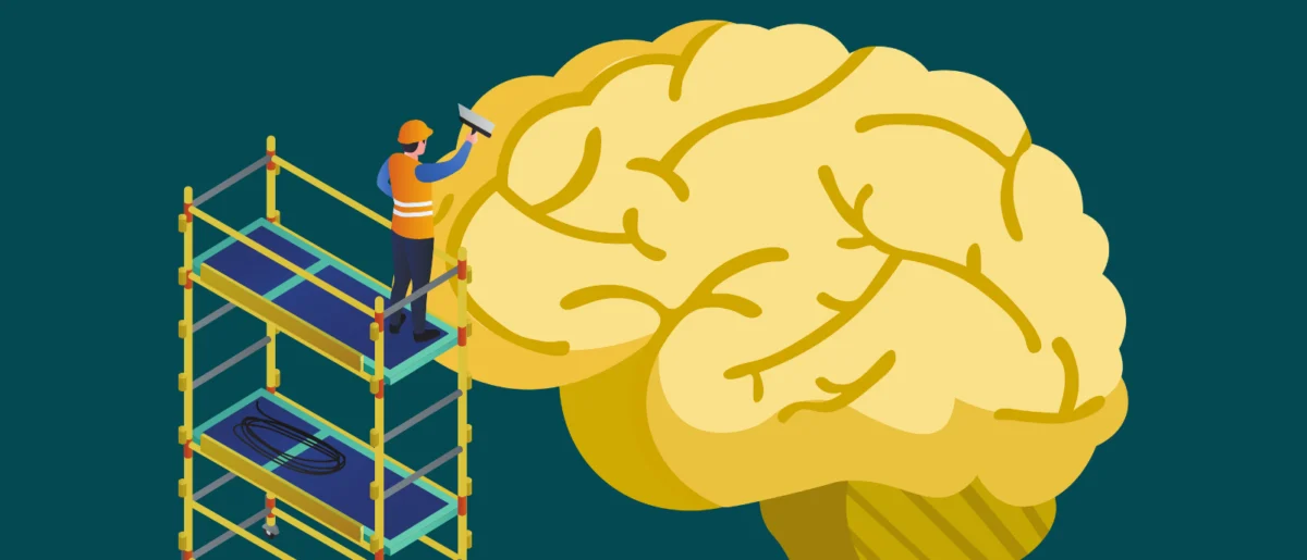
Unlike traditional monolithic hydrogels that form a solid, impenetrable barrier, MAPS are composed of microscopic hydrogel particles that assemble together to form a highly interconnected, porous network. This porous microstructure serves as a welcoming architectural scaffold, acting much like a cellular scaffolding system at a construction site, allowing host cells to migrate inward, organize, and build new tissue matrices.
However, providing a physical home for cells is only half the battle. In the complex biochemical environment of the brain, introducing a foreign material can often trigger defensive inflammatory responses that result in chronic scarring rather than healing. To overcome this obstacle, Segura and her colleagues looked to the brain’s own cellular communicators for inspiration: astrocytes.
Astrocytes are star-shaped glial cells that perform critical housekeeping functions in the normal, healthy central nervous system, maintaining the blood-brain barrier, providing nutritional support to neurons, and reacting swiftly to injury. When trauma occurs, astrocytes communicate with other biological systems by releasing extracellular vesicles (EVs). These nanoscale cellular packages function as biological delivery vans, loaded with vital proteins, lipids, and genetic material capable of influencing nearby cellular behavior.
Harnessing Cellular Messengers via Chemical Anchoring
To leverage these natural signaling properties, the Duke researchers collected extracellular vesicles harvested from lab-grown astrocytes. Rather than simply injecting these vesicles blindly into the bloodstream or the brain—where they would likely degrade rapidly or diffuse away from the target site—the team engineered a precise local delivery mechanism.
Utilizing a specialized chemical reaction, the researchers permanently anchored the astrocyte-derived extracellular vesicles directly onto the exterior surfaces of the hydrogel microparticles before injection. This crucial methodological step ensured that the biological signals remained strictly localized within the porous MAPS scaffold, establishing a persistent biochemical beacon for incoming cells.
"We are not simply placing a material into the brain," explained Segura, emphasizing the sophistication of the approach. "We are engineering a local environment that can coordinate several parts of the repair response."
Screening various combinations of signaling molecules loaded onto the scaffold, the team discovered that a specific pairing of interleukin-4 (IL-4) and complement component C1q was uniquely potent. This biochemical combination excelled at recruiting beneficial immune cells into the otherwise isolated and deteriorating stroke cavity.
Re-evaluating the Role of Neutrophils in Neural Repair
Among the immune cells recruited to the biomaterial scaffold, one specific population surprised the research team: neutrophils. Conventionally, neutrophils are viewed by immunologists and neurologists as detrimental actors during the immediate aftermath of a stroke. As a primary component of the acute inflammatory response, they rush to the site of injury and are frequently associated with exacerbating tissue damage and oxidative stress.
However, the Duke study challenges this dogma. The findings indicate that when neutrophils encounter the specialized microenvironment created by the IL-4 and C1q-loaded MAPS scaffold at a slightly later stage—more than 24 hours post-stroke—their biological function shifts dramatically from destructive inflammation to active tissue repair and vascular remodeling.

When the researchers experimentally depleted this neutrophil-rich immune cell population in the test subjects, the subsequent growth of new blood vessels and the overall remodeling of the scaffold dropped precipitously. This empirical proof demonstrated that neutrophils were not merely bystanders, but essential contributors to the regenerative cascade.
"This result changes how we think about neutrophils after stroke," noted Shangjing Xin, a postdoctoral fellow in the Segura laboratory and lead author of the study. "Their role appears to depend on when they arrive, where they are located, and the signals they receive from their surroundings. Our study demonstrates a potential engineering strategy to recruit and retain these cells at the right time."
Preclinical Success and Functional Recovery
The downstream consequences of this precisely orchestrated immune response extended far beyond initial cellular recruitment. Following the administration of the optimized MAPS scaffold, histological analyses revealed robust neoangiogenesis—the formation of an extensive network of new blood vessels permeating the previously barren stroke cavity. Furthermore, researchers observed a significant proliferation of axonal fibers, the foundational wiring of the central nervous system, winding through and around the repaired injury zone.
To determine whether these structural and cellular improvements translated into actual functional recovery, the researchers subjected the animal models to rigorous behavioral testing. Using a standardized grid-walking test that evaluates precise forelimb placement and motor coordination, subjects treated with the biomaterial demonstrated marked improvements over time.
By eight weeks post-treatment, the motor performance of the mice receiving the optimized scaffold was statistically indistinguishable from that of healthy, uninjured control animals. Crucially, these functional gains were sustained throughout the duration of the study.
Control experiments further validated the necessity of the biomaterial design. When extracellular vesicles were administered alone without the structural support of the MAPS scaffold, they failed to yield comparable vascular repair or functional restoration. This proved that the therapeutic efficacy relied synergistically on both the biochemical cargo and the physical, porous architecture of the hydrogel system.
Pathways to Clinical Translation: Challenges and Next Steps
Despite the profound promise of these preclinical findings, the research team emphasizes that the therapy remains in the preclinical phase of development. The current iterations of the MAPS scaffold were evaluated in rodent models via direct intracranial injection into the damaged tissue site.
Translating this complex regenerative strategy to human patients will require overcoming substantial regulatory, technical, and physiological hurdles. Future investigations must rigorously evaluate the long-term safety profiles of the biomaterial, map out the precise mechanistic contributions of various immune-cell subsets throughout the healing timeline, and scale the testing up to larger, gyrencephalic animal models whose cerebral anatomy more closely mirrors that of human patients.
Moreover, the current study relied on extracellular vesicles harvested from primary rat astrocytes. To advance toward human clinical trials, the Segura laboratory is actively exploring the use of EVs generated by human induced pluripotent stem cell (iPSC)-derived astrocytes. This cutting-edge cell source could provide a scalable, ethically viable, and clinically standardized supply chain for the therapy, while affording researchers finer control over the specific therapeutic cargo loaded onto the vesicles.
Broader Implications for Regenerative Medicine
The paradigm established by the Duke University research team marks a philosophical and practical shift in how neuroscientists view central nervous system trauma. For decades, therapeutic strategies focused heavily on neuroprotection—attempting to shield vulnerable cells from dying immediately following an ischemic insult. While vital, neuroprotection alone leaves clinicians empty-handed once tissue necrosis has occurred.
By treating the stroke cavity not as an inert scar to be managed, but as a dynamic biological ecosystem capable of regeneration, this biomaterial-based approach opens new frontiers across neurology and tissue engineering.
"You do not restore an ecosystem simply by containing the initial damage," Segura concluded. "You have to create the conditions that allow life to return. That is how we think about the stroke cavity. The material is not intended to reproduce the brain itself, but to create an environment where the body’s own cells can enter, communicate, and participate in rebuilding vascularized tissue."
As research progresses toward human applicability, this marriage of biomaterials science and immunology offers a renewed sense of optimism for millions of stroke survivors worldwide, pointing toward a future where the brain’s lost territory is actively reclaimed and repaired.