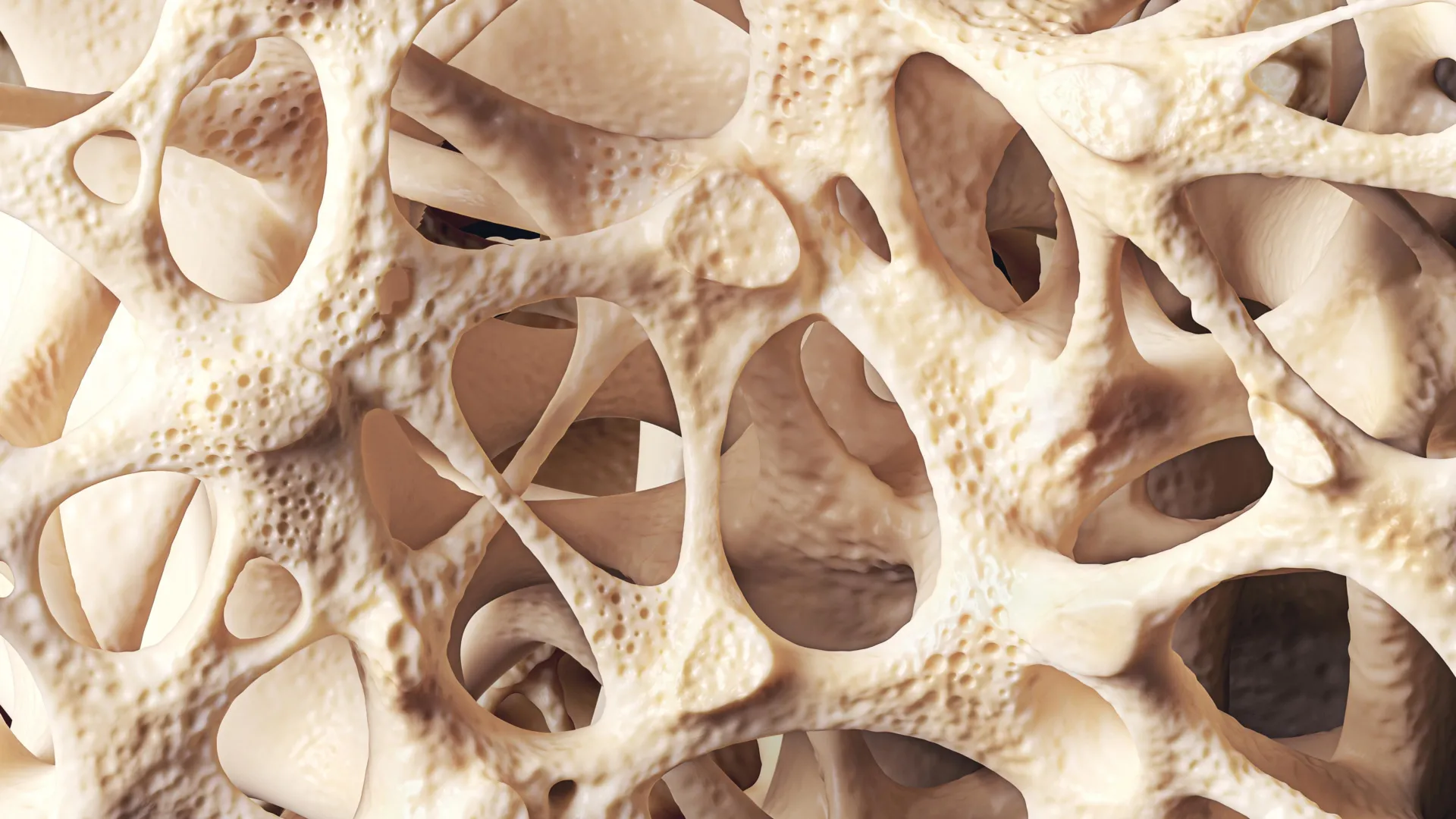
Osteopenia—a widespread medical condition characterized by a subclinical reduction in bone mineral density that falls short of the diagnostic threshold for osteoporosis—affects approximately 40% of the global adult population. Despite its staggering prevalence, particularly among elderly individuals and postmenopausal women, the condition frequently develops in absolute silence. It rarely presents with overt symptoms, leaving millions of individuals unaware that their skeletal framework is weakening until a minor trauma triggers an unexpected fragility fracture. In the United Kingdom alone, public health data indicates that low bone density accounts for upwards of 500,000 fractures annually, generating substantial strain on acute healthcare services, orthopedic wards, and long-term rehabilitation systems.
As global populations continue to age, health authorities are increasingly casting a spotlight on osteopenia, reframing it not merely as a benign consequence of growing older, but as a critical window of clinical opportunity. Early detection, paired with proactive lifestyle adjustments and targeted medical interventions, can halt or even reverse bone degradation, preventing the cascade of morbidity and mortality associated with severe osteoporotic fractures.
The Biological Mechanism: The Lifelong Balance of Bone Remodeling
To understand why osteopenia occurs, it is essential to examine the highly dynamic, continuous biological process known as bone remodeling. Far from being inert scaffolding, bone is living tissue that undergoes unceasing renewal throughout an individual’s lifetime. This ongoing cycle relies on a finely tuned equilibrium between two distinct cellular actions: bone resorption, wherein specialized cells called osteoclasts break down and remove old or micro-damaged bone tissue, and bone formation, wherein cells called osteoblasts synthesize new extracellular matrix and mineralize it to create fresh bone.
During childhood, adolescence, and early adulthood, the formation of new bone vastly outpaces its resorption, resulting in a net accumulation of skeletal mass. Peak bone mass—the maximum strength and density an individual’s bones will ever achieve—is typically attained between a person’s mid-20s and early 30s.
Once this biological zenith is reached, the physiological paradigm shifts. Gradually, the rate of bone resorption begins to outstrip bone formation. This subtle, age-related deficit means that every remodeling cycle results in a tiny, net loss of bone mass. Over decades, this cumulative deficit leads to declining bone mineral density, transitioning an individual from healthy bone density into the realm of osteopenia, and potentially further down the spectrum toward full-scale osteoporosis if the rate of loss accelerates or is left unchecked.
Chronology and Risk Factors: From Peak Bone Mass to Accelerated Loss
While chronological aging serves as the primary and universal driver of bone density decline, a complex matrix of biological, hormonal, lifestyle, and environmental factors can profoundly accelerate the process.
The most pronounced acceleration of bone loss occurs during midlife, specifically surrounding the menopausal transition in women. The sharp, natural decline in estrogen levels following menopause significantly impairs skeletal protection. Estrogen plays a vital, regulatory role in bone metabolism by promoting the apoptosis (programmed cell death) of bone-resorbing osteoclasts while safeguarding osteoblasts. When estrogen levels plummet, this protective brake is removed, unleashing unchecked bone resorption. Epidemiological data underscores the severity of this hormonal shift, revealing that approximately one in two women over the age of 50 will ultimately suffer a fragility fracture in their remaining lifetime.
Beyond hormonal shifts, daily lifestyle choices accumulated across decades exert a heavy toll on skeletal integrity. Chronic physical inactivity denies the skeleton the mechanical loading it requires to stimulate bone-forming cells. Nutritional deficiencies further compromise the body’s structural raw materials; insufficient dietary calcium intake starves the mineralization process, while inadequate vitamin D levels drastically impair the gut’s ability to absorb whatever calcium is consumed. Furthermore, noxious habits such as tobacco smoking and excessive alcohol consumption directly inhibit osteoblast activity and disrupt endocrine pathways vital for bone health.
Medical history and pharmacotherapy also play a pivotal role in the chronological progression of bone loss. Individuals managing chronic inflammatory conditions—such as Crohn’s disease, ulcerative colitis, or coeliac disease—frequently suffer from malabsorption syndromes, depriving their bones of essential nutrients even when dietary intake appears adequate. Additionally, long-term therapeutic use of specific medications, most notably systemic corticosteroids (glucocorticoids) prescribed for autoimmune and respiratory disorders, aggressively suppresses bone formation while accelerating resorption, placing patients at an exceptionally high risk of rapid skeletal deterioration.
Diagnostic Precision: Unmasking the Silent Threat
Because osteopenia produces no localized pain, swelling, or physical deformity in its early stages, clinical discovery relies almost exclusively on screening protocols, incidental findings during imaging for other conditions, or investigations triggered after a low-trauma fracture has already occurred.
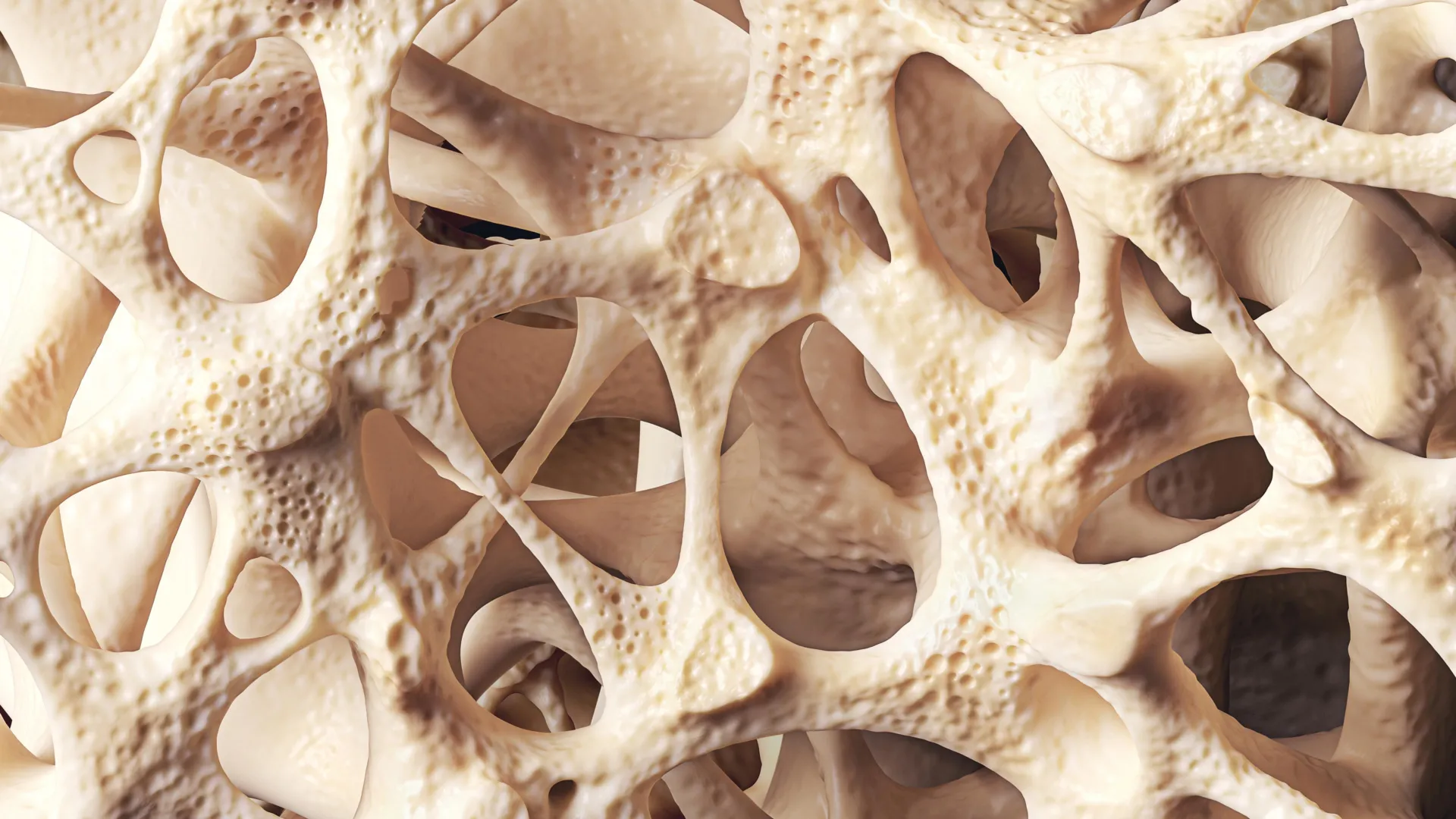
The gold standard for quantifying skeletal health and diagnosing osteopenia is the dual-energy X-ray absorptiometry (DXA) scan. This non-invasive, low-dose radiological examination measures bone mineral density at critical fracture-prone sites, most commonly the lumbar spine and the proximal femur (hip).
The results of a DXA scan are reported using a metric known as a T-score, which statistically compares the patient’s measured bone density to the expected mean bone density of a healthy young adult reference population of the same sex at their peak bone mass.
- A T-score between 0 and −1.0 is considered normal bone density.
- A T-score ranging between −1.0 and −2.5 establishes the clinical diagnosis of osteopenia.
- A T-score of −2.5 or lower meets the diagnostic threshold for osteoporosis, signifying an advanced reduction in bone mass and a sharply elevated risk of fracture.
Clinical Management and Strategic Intervention
The primary objective in managing osteopenia is not necessarily to restore the skeletal density of a twenty-year-old, but rather to stabilize bone mass, prevent further deterioration, and structurally reinforce the skeleton to dramatically lower the 10-year probability of a debilitating fracture.
Clinicians utilize validated risk assessment calculators—such as the FRAX tool—which integrate a patient’s DXA T-score with clinical risk variables, including age, body mass index, personal and family fracture history, smoking status, and systemic corticosteroid use. This calculation helps determine whether an individual requires lifestyle modifications alone or if they would benefit from pharmacological intervention.
For the majority of individuals diagnosed with osteopenia, management begins with foundational lifestyle modifications:
Nutritional Optimization
Sustaining structural strength requires an adequate daily supply of elemental calcium, sourced primarily from dairy products, dark leafy green vegetables, and fortified plant-based alternatives. However, calcium cannot be effectively utilized without sufficient vitamin D. Because vitamin D deficiency remains remarkably common across temperate regions like the UK due to limited year-round sunlight exposure, health authorities frequently recommend daily vitamin D supplementation.
Targeted Physical Activity
Mechanical loading is a powerful stimulus for skeletal adaptation. Weight-bearing aerobic exercises—such as brisk walking, dancing, and jogging—force the skeleton to work against gravity, signaling osteoblasts to lay down new bone tissue. Resistance training, utilizing free weights or resistance bands, provides even greater mechanical tension, concurrently enhancing muscle mass. Furthermore, specialized movement practices like Tai Chi have been clinically proven to improve dynamic balance, proprioception, and core stability, thereby directly reducing the incidence of accidental falls that frequently precipitate fractures in vulnerable populations.
Pharmacological Considerations
While lifestyle interventions form the cornerstone of care, high-risk patients—particularly those with concurrent clinical risk factors or those who have already sustained a fragility fracture—may be candidates for pharmacological therapies. Antiresorptive medications, such as bisphosphonates, are occasionally prescribed off-label for severe osteopenia when fracture risk models indicate a high probability of future skeletal failure. These drugs work by binding to the surface of bones, inhibiting the action of osteoclasts, and effectively halting the relentless breakdown of bone matrix.
Broader Public Health Implications and Future Outlook
The rising prevalence of osteopenia represents a formidable challenge for global healthcare infrastructure. As life expectancies lengthen, the cumulative burden of chronic musculoskeletal conditions threatens to overwhelm clinical resources unless public health strategies undergo a fundamental shift.
Health economists and epidemiologists emphasize that viewing osteopenia merely as a normal, untreatable aspect of aging is a costly miscalculation. Reframing the condition as a critical window of vulnerability allows healthcare providers to intercept the trajectory of bone loss long before irreversible architectural damage occurs.
Current clinical consensus stresses that the progression from osteopenia to osteoporosis is far from inevitable. With early, systematic screening using DXA technology, combined with widespread public health messaging encouraging lifelong physical activity, balanced nutrition, and the early mitigation of modifiable risk factors, millions of individuals can successfully preserve their skeletal integrity. Ultimately, safeguarding bone health demands a life-course perspective—recognizing that the strength of our skeleton in later life is the direct, cumulative reflection of our health, habits, and physiological changes across every preceding decade.