Around 40% of adults worldwide are affected by osteopenia, a prevalent yet frequently overlooked condition characterized by a decline in bone mineral density. While it rarely makes headline news, this silent musculoskeletal issue serves as a critical bridge between healthy aging and severe skeletal degradation. Particularly common among postmenopausal women and elderly populations, low bone density is responsible for an estimated staggering volume of health complications globally. In the United Kingdom alone, health authorities attribute more than 500,000 fractures annually to compromised bone strength, placing an immense, recurring burden on emergency departments, orthopedic services, and long-term care systems.
Despite its high prevalence, osteopenia rarely announces itself with overt symptoms. Unlike acute conditions that trigger immediate pain or distress, bone loss develops insidiously and silently over many years. Millions of individuals walk, work, and exercise entirely unaware that their internal scaffolding is weakening. Frequently, a patient only discovers they have the condition after suffering a sudden fragility fracture from a minor fall, or following a routine bone density scan recommended by a general practitioner due to age or menopausal status. This silent nature transforms osteopenia from a simple physiological decline into a significant, under-recognized public health crisis requiring urgent medical attention and public awareness.
The Biological Timeline of the Human Skeleton
To understand why bones weaken, medical science looks closely at the dynamic lifecycle of skeletal tissue. Bones are not static, lifeless pillars; they are metabolically active organs that undergo continuous renewal through a highly regulated physiological cycle known as bone remodeling. This lifelong process consists of two opposing phases: bone resorption, where specialized cells break down and remove old or damaged bone tissue, and bone formation, where other cells deposit new tissue to replace it.
During childhood and adolescence, bone formation significantly outpaces resorption, allowing the skeleton to grow in size, density, and strength. This biological momentum continues into early adulthood, culminating in what researchers term peak bone mass. For most individuals, peak bone density is achieved somewhere between their mid-20s and early-30s.
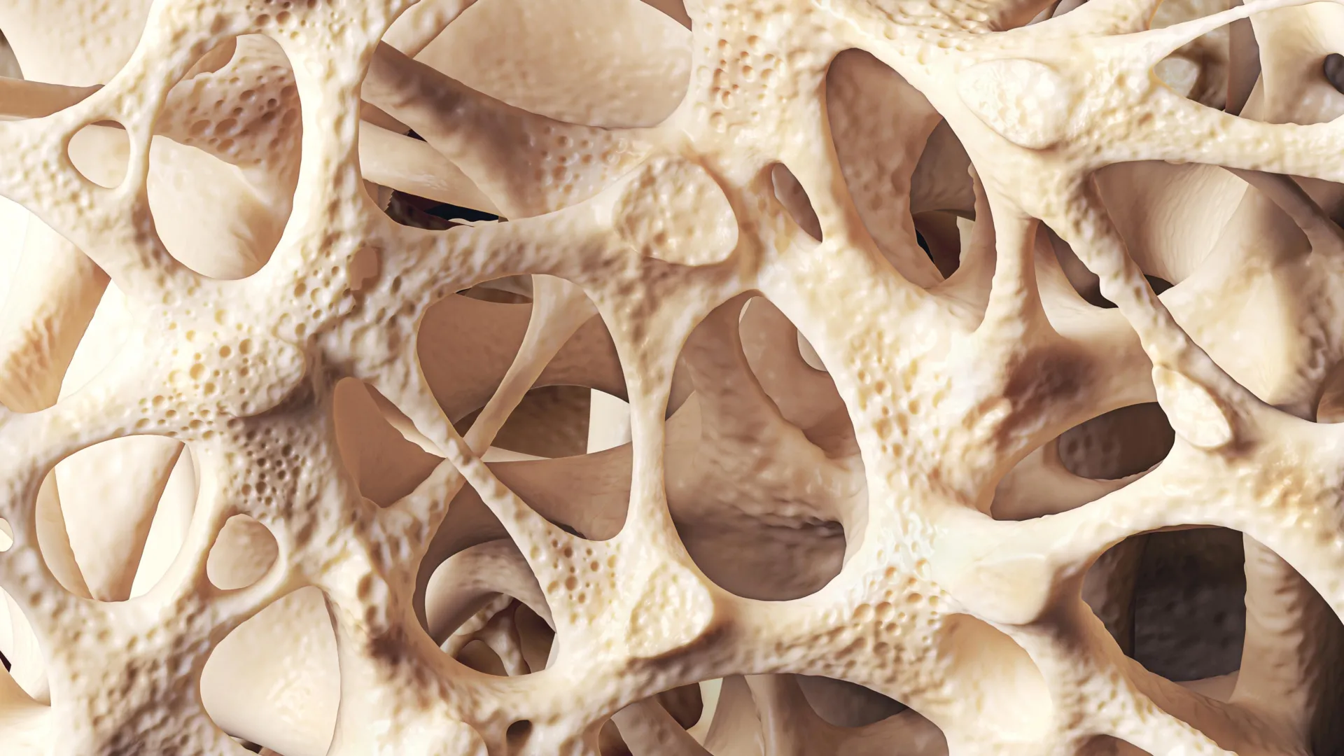
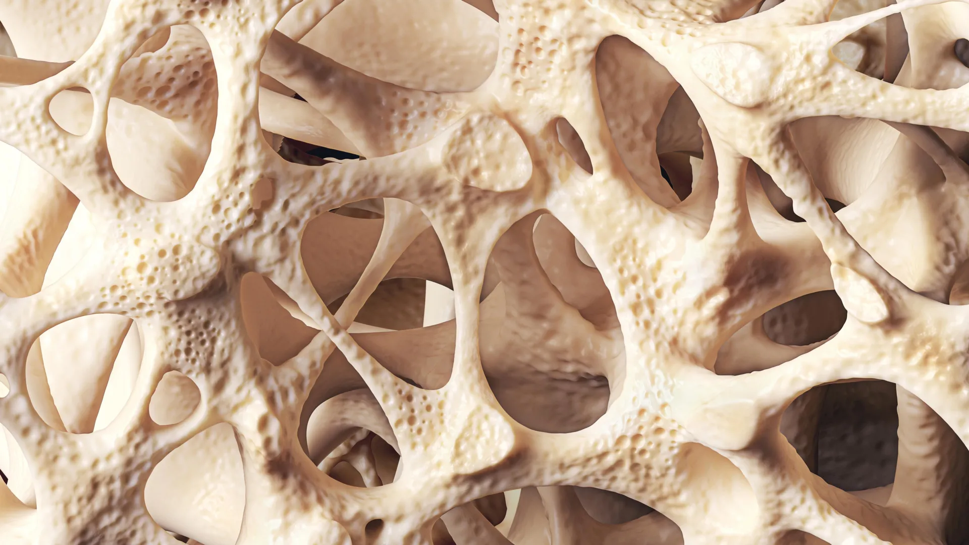
Once this biological peak is reached, the trajectory of skeletal health shifts. As aging progresses into middle age and beyond, the delicate balance of bone remodeling begins to falter. Slowly and progressively, the rate of bone resorption starts to exceed the rate of bone formation. Year by year, the microscopic architecture of the bone thins, trabecular struts become fragile and sparse, and overall bone mineral density declines. While this age-related reduction is a natural part of the human lifecycle, several external and internal catalysts can dramatically accelerate the process, turning a natural aging trajectory into a pathological vulnerability.
Risk Factors and Physiological Triggers
Ageing remains the primary and most unyielding risk factor for bone loss, but a constellation of hormonal, genetic, and environmental factors can profoundly impact skeletal integrity. Among these, hormonal shifts play a starring role.
In women, the sudden and steep decline in estrogen levels following menopause acts as a major accelerator of bone breakdown. Estrogen serves a protective function for the skeleton by suppressing the activity of osteoclasts—the cells responsible for bone resorption. When estrogen levels plummet during and after menopause, this natural brake is released, leading to rapid bone loss. Epidemiological data indicates that roughly one in every two women over the age of 50 will experience a fragility fracture in their remaining lifetime, heavily driven by this postmenopausal hormonal shift.
Beyond endocrinology, modern lifestyle choices heavily dictate long-term bone health. Chronic behaviors such as cigarette smoking, excessive alcohol consumption, and prolonged physical inactivity undermine the body’s ability to maintain bone strength. Concurrently, nutrition is foundational. Diets chronically lacking in adequate calcium intake—the primary mineral structural component of bone—or deficient in vitamin D, which facilitates intestinal calcium absorption, starve the skeleton of the raw materials it needs to repair and fortify itself.
Furthermore, medical interventions and underlying health conditions can exacerbate bone deterioration. Patients requiring long-term corticosteroid therapy for inflammatory conditions face a markedly elevated risk of drug-induced bone loss. Similarly, gastrointestinal disorders that impair nutrient absorption, such as Crohn’s disease or coeliac disease, frequently leave patients malnourished of vital minerals, indirectly triggering secondary osteopenia or osteoporosis.
Diagnostic Pathways and Clinical Assessment
Detecting osteopenia before it escalates into severe skeletal disease is the cornerstone of modern preventive orthopedics. Clinicians emphasize that identifying low bone density early provides a critical window of opportunity to intervene, halting or even reversing the trajectory of bone loss before a catastrophic fracture occurs.
The gold standard for diagnosing osteopenia is a dual-energy X-ray absorptiometry, commonly known as a DXA scan. This specialized, low-dose radiographic imaging procedure evaluates bone strength at key fracture sites, such as the hip, spine, and forearm. The resulting output is quantified as a T-score, which statistically compares the patient’s measured bone mineral density against the average peak bone mass of a healthy young adult of the same sex.
According to World Health Organization diagnostic criteria, a T-score between −1.0 and −2.5 classifies as osteopenia. A score dropping to −2.5 or lower crosses the diagnostic threshold into osteoporosis, where the risk of spontaneous or low-impact fracture rises exponentially.
To refine treatment strategies, physicians rarely rely on T-scores alone. Because not every patient with osteopenia requires immediate pharmacological intervention, clinicians frequently deploy advanced fracture risk assessment tools. These digital algorithms calculate a patient’s ten-year probability of suffering a major osteoporotic fracture by synthesizing multiple variables, including age, exact bone mineral density, history of steroid usage, body mass index, and familial history of hip fractures.
Comprehensive Management and Therapeutic Strategies
When an individual receives a diagnosis of osteopenia, management protocols generally focus on a multi-pronged approach: slowing down further bone degradation, optimizing nutritional status, enhancing muscular and skeletal resilience through physical activity, and, where risk profiles demand it, introducing targeted pharmaceutical therapies.
Lifestyle modifications form the bedrock of non-pharmacological management. Stopping smoking, moderating alcohol intake, and maintaining a healthy body weight reduce unnecessary physiological stress on the skeleton. Additionally, targeted physical activity is profoundly osteoprotective. Weight-bearing exercises—such as brisk walking, dancing, and light jogging—place mechanical strain on the skeleton, signaling bone-forming cells to lay down new mineral matrix. Resistance training further amplifies this effect by pulling on bone structures while simultaneously building the musculature needed for dynamic stability.
Clinical research underscores that regular physical movement not only preserves bone mineral density but also enhances overall neuromuscular coordination. Programs like Tai Chi, for instance, have been shown to significantly improve balance and core strength in older adults, thereby reducing the frequency of accidental falls that frequently precipitate fractures in weakened bones.
Nutritional optimization runs parallel to physical rehabilitation. Adequate daily calcium intake—sourced from dairy products, fortified plant milks, and dark leafy greens—provides the essential building blocks for bone tissue. However, calcium cannot be utilized without sufficient vitamin D. Because natural synthesis via sunlight is limited in many northern latitudes, health authorities in regions like the UK routinely recommend vitamin D supplementation for adults, particularly during autumn and winter months, to ensure optimal mineral metabolism.
In cases where fracture risk assessment tools indicate an elevated danger of skeletal failure, or if a patient has already suffered a fragility fracture, physicians may turn to prescription medications. Antiresorptive drugs, such as bisphosphonates, are designed to slow down the bone breakdown process, helping patients preserve existing density. While these therapeutics are widely utilized in established cases of osteoporosis, growing clinical consensus supports their selective use in high-risk osteopenic patients to preemptively thwart further deterioration.
Implications and the Paradigm Shift in Public Health
Medical professionals increasingly urge society to reframe how it perceives osteopenia. Rather than dismissing it as a benign side effect of aging or a harmless precursor, healthcare leaders view osteopenia as a critical warning sign and an invaluable point of intervention. The transition from osteopenia to full-scale osteoporosis is by no means an inevitability.
Emerging clinical data highlights that early detection, coupled with targeted lifestyle adjustments and proactive medical oversight, can successfully stabilize bone health, slow down resorption rates, and drastically lower the lifetime risk of debilitating fractures. In some favorable clinical scenarios, appropriate interventions can even yield modest improvements in bone density measurements.
Ultimately, protecting skeletal health requires a long-term, preventative perspective. Bone strength is not determined by a single life stage; rather, it is the cumulative reflection of decades of nutritional habits, physical activity levels, hormonal milestones, and overall health status. Cultivating and maintaining supportive lifestyle habits across the entire lifespan remains the single most effective strategy for safeguarding human mobility, independence, and long-term vitality.