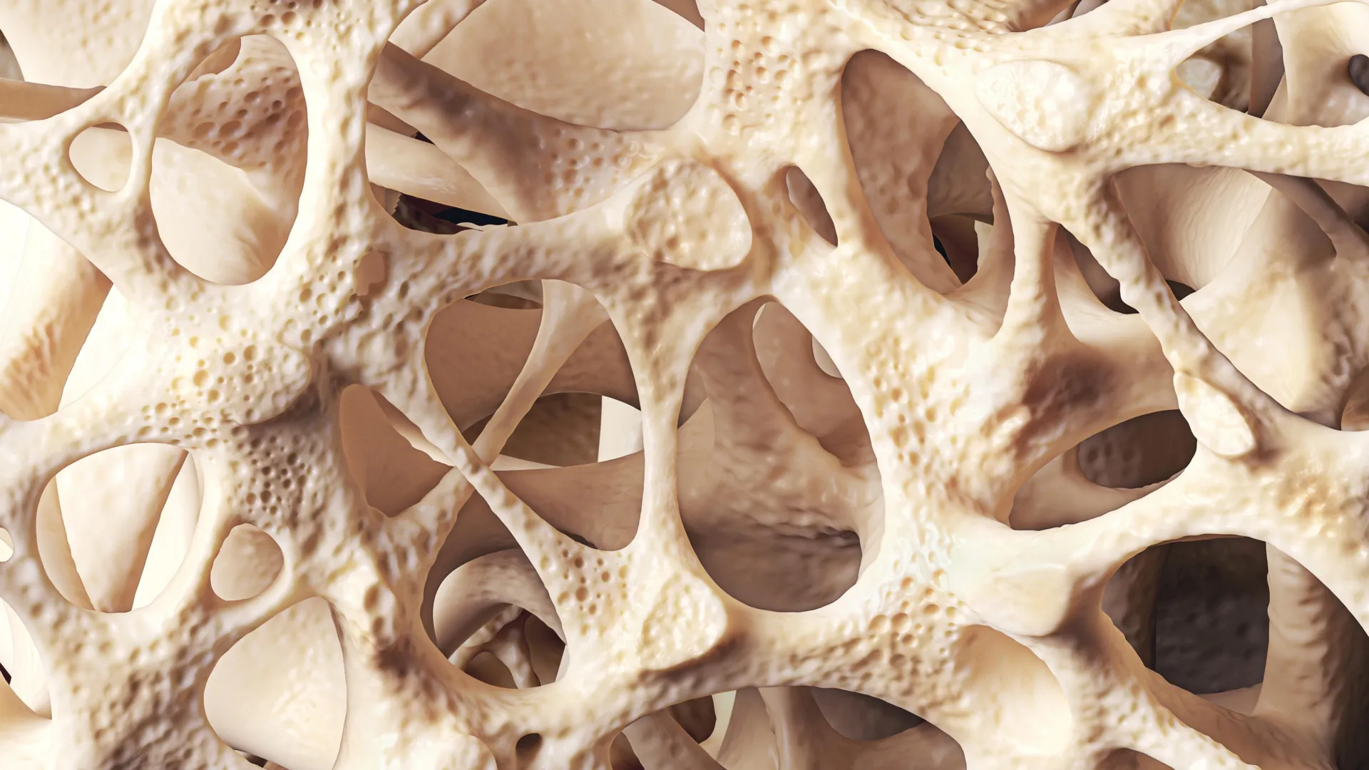
Around 40% of adults worldwide are affected by osteopenia, a condition characterized by a progressive loss of bone mineral density that falls short of the diagnostic threshold for osteoporosis. While it rarely makes headlines, this silent condition represents a massive, under-recognized public health challenge. It is particularly prevalent among postmenopausal women and elderly adults, laying the groundwork for severe skeletal vulnerabilities later in life. In the United Kingdom alone, health authorities estimate that more than 500,000 fractures occur annually due to low bone density, placing an immense operational and financial burden on acute healthcare systems, emergency departments, and social care infrastructure.
Unlike acute injuries or systemic illnesses that manifest with immediate pain, osteopenia develops quietly and without overt symptoms. Millions of individuals live with diminishing bone mass entirely unaware of their physiological status until a minor fall or impact triggers a fragility fracture, or until a routine bone density scan uncovers the deterioration. Because the condition masquerades as a normal part of aging, clinicians and public health agencies are increasingly urging a paradigm shift: viewing osteopenia not merely as a precursor to osteoporosis, but as a critical window of opportunity for early intervention and preventive medicine.
The Biological Timeline of Bone Remodeling and Peak Mass
To understand how osteopenia develops, researchers examine the dynamic biology of the human skeleton. Bone is not an inert scaffolding; it is living tissue that undergoes continuous renewal through a highly regulated, lifelong process known as bone remodeling. This mechanism relies on a delicate biological equilibrium between two distinct cellular activities: bone resorption, where specialized cells called osteoclasts break down and remove old bone tissue, and bone formation, where cells called osteoblasts build new matrix.
During childhood, adolescence, and early adulthood, the formation of new bone outpaces the resorption of old tissue, allowing individuals to steadily build skeletal mass. This trajectory reaches a crucial milestone known as peak bone mass, which typically occurs during a person’s mid-20s to early-30s. Peak bone mass serves as a physiological "savings account" for the rest of life. Individuals who achieve a higher peak bone mass during their developmental years possess greater resilience against age-related bone decline.
However, once this biological peak is achieved, the balance shifts. Gradually, bone resorption begins to outpace bone formation. As people enter their fourth and fifth decades of life, a slow, continuous net loss of bone tissue becomes the norm. When this cumulative loss lowers bone mineral density below optimal levels—yet stops short of the severe depletion seen in osteoporosis—clinicians diagnose osteopenia.
Diagnostic Criteria and the DXA Scan
Detecting this gradual loss requires specialized medical imaging, as standard X-rays are generally insensitive to minor reductions in bone density. The gold standard for assessment is the dual-energy X-ray absorptiometry (DXA) scan. This non-invasive, low-dose imaging procedure measures bone mineral density at key weight-bearing and fracture-prone sites, such as the hip, spine, and forearm.
DXA scan results are quantified using a T-score, which compares a patient’s measured bone density to the normative peak bone density of a healthy young adult of the same sex. According to World Health Organization criteria:
- A T-score of –1.0 or higher is considered normal.
- A T-score between –1.0 and –2.5 indicates osteopenia.
- A T-score of –2.5 or lower defines osteoporosis.
Public health guidelines generally recommend DXA screening for individuals presenting with specific risk factors, such as women aged 65 and older, postmenopausal women with elevated risk profiles, men aged 70 and older, or anyone who has experienced a low-impact fragility fracture. Early identification through these scans allows medical professionals to stratify patients by risk and implement strategies before bones become dangerously fragile.
Key Risk Factors and Accelerating Triggers
While natural aging is the primary driver of bone loss, a confluence of genetic, hormonal, lifestyle, and medical factors can significantly accelerate the rate of deterioration.
Hormonal fluctuations play a disproportionate role, particularly the sharp decline in estrogen levels that occurs during and after menopause. Estrogen acts as a protective shield for the skeletal system by promoting the apoptosis (programmed cell death) of osteoclasts, thereby slowing bone breakdown. When estrogen levels plummet, this regulatory brake is removed, leading to accelerated bone resorption. Epidemiological data indicates that approximately one in two women over the age of 50 will experience a fragility fracture in their lifetime, largely driven by postmenopausal bone loss.
Lifestyle factors compound these hormonal vulnerabilities. Chronic physical inactivity denies the skeleton the mechanical loading necessary to stimulate osteoblast activity. Furthermore, modifiable behaviors such as cigarette smoking and excessive alcohol consumption exert direct toxic effects on bone-building cells and disrupt calcium regulation.
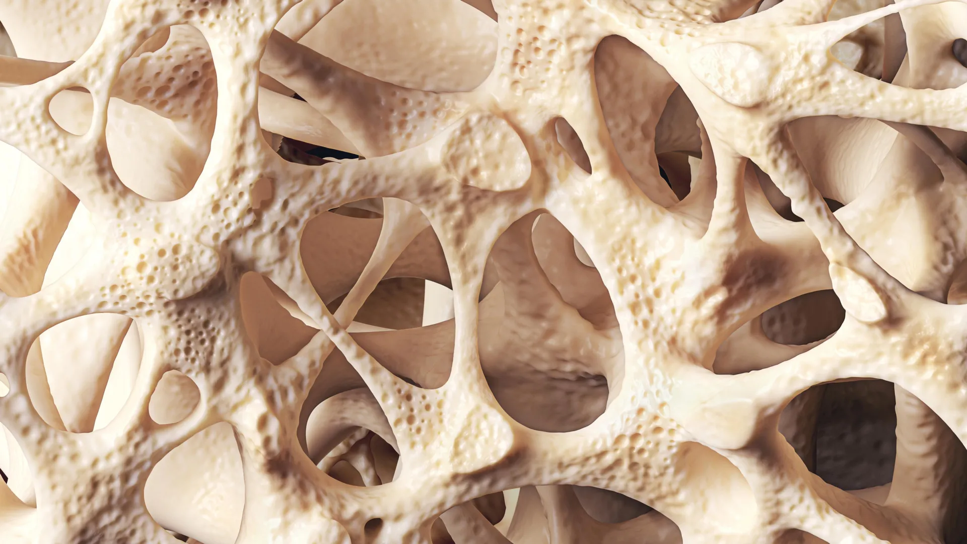
Nutritional deficiencies also undermine structural integrity. Inadequate dietary intake of calcium starves the body of the primary mineral required for rigid bone matrix, while low vitamin D levels impair the intestinal tract’s ability to absorb calcium efficiently. Public health data underscores that vitamin D insufficiency is widespread in northern latitudes, including the UK, making seasonal or year-round supplementation a frequent recommendation by health authorities.
Finally, underlying medical conditions and pharmacological interventions can heavily influence bone density. Chronic inflammatory and gastrointestinal disorders that impair nutrient absorption—such as Crohn’s disease and coeliac disease—frequently compromise bone health. Additionally, long-term systemic corticosteroid therapy, widely prescribed for autoimmune and inflammatory diseases, significantly suppresses bone formation while enhancing resorption, placing patients at high risk of rapid bone mass depletion.
Clinical Management and Strategic Intervention
When a patient is diagnosed with osteopenia, the primary clinical objective is to halt or slow the progression of bone loss and minimize the lifetime risk of fractures. Because not every patient requires pharmaceutical intervention, management strategies are tailored to individual risk assessments.
Clinicians frequently utilize sophisticated evaluation tools, such as the Fracture Risk Assessment Tool (FRAX), to calculate a patient’s ten-year probability of suffering a major osteoporotic or hip fracture. This calculation integrates age, bone mineral density T-scores, body mass index, smoking status, alcohol use, glucocorticoid history, and secondary causes of osteoporosis.
For individuals with low to moderate risk, management centers heavily on non-pharmacological lifestyle modifications:
-
Targeted Exercise Programs: Weight-bearing aerobic activities—such as brisk walking, dancing, and jogging—force the skeletal system to work against gravity, triggering osteoblasts to synthesize new bone tissue. Resistance training further strengthens both bone and surrounding muscle tissue, improving overall stability. Specialized movement practices like Tai Chi have also been shown in clinical research to enhance balance and proprioception, thereby lowering the incidence of falls that cause fractures.
-
Nutritional Optimization: Ensuring a balanced intake of calcium-rich foods (such as dairy products, fortified plant-based milks, and leafy greens) combined with adequate vitamin D supports the ongoing mineralization of bone tissue. Where dietary intake falls short, targeted supplementation is often prescribed under medical supervision.
-
Behavioral Adjustments: Cessation of smoking, moderation of alcohol consumption, and the maintenance of a healthy body mass index help preserve bone strength and reduce systemic inflammation.
In cases where a patient’s FRAX score reveals a high risk of fracture, or if they have already sustained a fragility fracture, clinicians may introduce pharmacological treatments. Antiresorptive medications, most notably bisphosphonates, are frequently deployed to slow down the action of osteoclasts, protecting existing bone mass and reducing fracture incidence. While these drugs are more commonly associated with osteoporosis treatment, growing clinical consensus supports their selective use in high-risk osteopenia patients to prevent further decline.
Broader Economic and Public Health Implications
The rising prevalence of osteopenia against the backdrop of an aging global demographic poses a profound challenge to healthcare systems. Fragility fractures resulting from compromised bone density often lead to hospitalization, prolonged rehabilitation, loss of independence, and increased mortality rates among older adults.
Public health economists emphasize that investing in early detection and proactive management of osteopenia yields significant long-term savings. By identifying bone loss at the osteopenic stage, healthcare providers can prevent the costly cascade of events associated with advanced osteoporosis and severe fractures.
Crucially, experts stress that the progression from osteopenia to osteoporosis is not an inevitable fate. Because bone health reflects the cumulative impact of nutrition, physical activity, and hormonal health across a person’s lifespan, adopting protective habits early—and maintaining them consistently—offers the most reliable shield against skeletal decline. Through a combination of widespread public awareness, accessible diagnostic screening, and targeted lifestyle and medical interventions, modern medicine possesses the tools to turn the tide against this silent epidemic, preserving mobility, independence, and quality of life for millions of aging adults.