
Researchers at Oregon Health & Science University (OHSU) have unveiled a groundbreaking discovery: a previously unrecognized internal system within cells that functions akin to "trade winds," enabling the rapid and directed transport of vital proteins to the cell’s leading edge. This revelation fundamentally reshapes scientific comprehension of cellular locomotion, the metastasis of cancer, and the intricate processes of wound healing. The findings, published in the esteemed journal Nature Communications, challenge long-standing paradigms in cell biology, suggesting a far more active and orchestrated internal environment than previously theorized.
A Paradigm Shift from Passive Diffusion to Active Transport
For decades, the prevailing model of protein movement within cells largely attributed it to diffusion—a passive, random process where molecules gradually spread out from areas of high concentration to low concentration. This "textbook" view depicted proteins drifting until they serendipitously reached their intended destinations. However, the OHSU study provides compelling evidence that cells possess a sophisticated mechanism for directed transport, generating internal fluid currents that actively propel proteins toward the cellular frontier. This leading edge is the dynamic region where cells extend, migrate, and engage in tissue repair. The implications of this shift are profound, suggesting that cellular organization and function are not solely governed by stochastic events but by actively regulated internal dynamics.
Serendipitous Discovery in a Classroom Setting
The genesis of this significant scientific breakthrough can be traced back to an unexpected observation made during a neurobiology course at the Marine Biological Laboratory in Massachusetts. Co-corresponding authors Catherine (Cathy) Galbraith, Ph.D., and James (Jim) Galbraith, Ph.D., were overseeing a routine experiment with students when they noticed an anomaly.
"It actually started out as an unexpected finding," Cathy Galbraith recounted. "We were just conducting an experiment with students in class." The experiment involved using a laser to temporarily render proteins invisible within a specific strip at the rear of a living cell, a standard technique for visualizing intracellular transport. During this observation, the researchers noted the unexpected appearance of a distinct, darker band at the front edge of the cell—the very area that elongates and advances during cell migration.
"We kind of did it for fun and then realized this gave us a way of measuring something that wasn’t able to be measured before," she explained, highlighting the serendipitous nature of the discovery. This observation provided a novel method for quantifying an previously unobserved cellular phenomenon.
Unveiling the "Dark Band": Soluble Actin Under Pressure
Further investigation into the mysterious "dark band" revealed it to be a wave of soluble actin, a critical protein responsible for cellular shape, movement, and internal organization. Previously, the scientific consensus held that actin primarily reached the cell’s leading edge through the slow process of diffusion. The new findings, however, demonstrated a fundamentally different mechanism at play.
"We realized the cartoon models in textbooks were missing a huge piece," Jim Galbraith stated, emphasizing the inadequacy of existing theoretical frameworks. "There had to be some kind of flow in the cell pushing things forward. Cells really do ‘go with the flow.’" This statement encapsulates the core of their discovery: an active, directed flow system within the cell.
The "Atmospheric Rivers" of the Cell: Directed Fluid Flows
Building on their initial classroom observation, Cathy and Jim Galbraith, who joined OHSU in 2013 after their tenure at the National Institutes of Health, collaborated with Nobel Laureate Eric Betzig, Ph.D., at the Howard Hughes Medical Institute’s Janelia Research Campus. This collaboration leveraged advanced imaging techniques, enabling the team to visualize and analyze these internal cellular currents with unprecedented clarity.
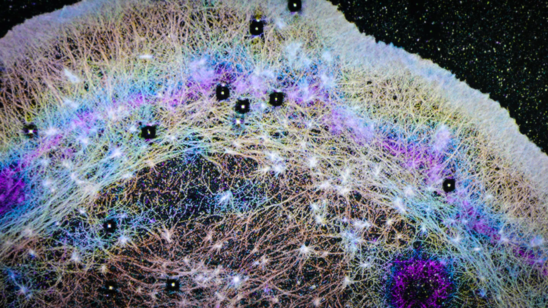
Utilizing specialized imaging tools, the researchers identified that cells actively generate directional fluid flows. They aptly compared these flows to "atmospheric rivers," powerful currents that transport vast quantities of water vapor across continents. These internal cellular currents effectively transport actin and other essential proteins towards the cell’s front, a process significantly faster and more efficient than diffusion alone.
"We found that the cell can actually squeeze at the back and target where it sends that material," Jim Galbraith elaborated, drawing an analogy to a sponge. "If you squeeze half a sponge, the water only goes on that half. That’s basically what the cell is doing." This analogy underscores the cell’s ability to actively manipulate internal pressure and fluid dynamics to direct the movement of its components.
These discovered flows are characterized as "nonspecific," meaning they can simultaneously carry a diverse array of proteins. This attribute makes the system remarkably efficient, supporting a multitude of cellular processes including protrusion (extending outward), adhesion (sticking to surfaces), and rapid shape changes. These capabilities are fundamental to cell migration, the intricate workings of the immune system, and the critical process of tissue repair.
A key aspect of this newly identified system is its localization within a specialized region at the cell’s anterior. This area is demarcated by an actin-myosin condensate barrier, a dynamic boundary that effectively segregates this protein-rich zone and channels the directed flows towards the advancing edge. This structural organization further highlights the cell’s sophisticated internal architecture for managing protein distribution.
Precision Imaging Reveals Cellular Currents: The FLOP Experiment
To visualize these elusive internal flows, the OHSU team developed a modified fluorescence microscopy technique. Instead of the conventional method of photobleaching (dimming) fluorescent molecules with a laser, they chose to activate fluorescent molecules at a single point and meticulously track their subsequent movement. This innovative approach allowed for precise measurement of the speed and direction of internal cellular transport.
One of their pivotal experiments was aptly named FLOP, standing for Fluorescence Leaving the Original Point. "It wasn’t a flop at all," Cathy Galbraith humorously stated. "It was the opposite. It is anything but a flop, because it worked." The success of FLOP provided the empirical data necessary to confirm the existence and characteristics of these internal cellular currents.
Implications for Cancer Metastasis and Therapeutic Development
The implications of this discovery extend significantly to our understanding of cancer cell migration and metastasis. The aggressive and rapid movement observed in some highly invasive cancer cells may be directly attributable to the enhanced efficiency of this internal transport system.
"We know these highly invasive cells have this really cool mechanism to push proteins really fast, really rapidly where they need them at the front of the cell," Jim Galbraith explained. He further elaborated on the comparative biology of cells: "All cells have basically the same components inside, much like a Porsche and a Volkswagen have many of the same parts, but when those parts are assembled into the final machine, they behave and function very differently." This suggests that while the fundamental machinery of the transport system may be conserved across cell types, its regulation and efficiency can vary dramatically, leading to the aggressive phenotypes observed in malignant cells.
By elucidating how cancer cells exploit this internal flow system in ways that differ from normal cells, scientists may be able to develop novel therapeutic strategies. The goal would be to target these aberrant transport mechanisms to impede or halt the spread of cancer. "If you can understand the differences, you can target future therapies based on how cancer cells and normal cells work differently," he emphasized, highlighting the potential for precision oncology.
A Symphony of Disciplines: Advanced Imaging and Interdisciplinary Collaboration
The successful identification and characterization of these cellular "trade winds" were the result of a highly interdisciplinary effort, bringing together experts in engineering, physics, microscopy, and cell biology. Crucial contributions were made by collaborators at the Janelia Research Campus in Virginia, including specialists in fluorescence correlation spectroscopy and 3D super-resolution imaging.
"The instrumentation we needed doesn’t exist in most places," Cathy Galbraith noted, underscoring the unique capabilities of the Janelia facility. "Janelia had a one-of-a-kind setup that let us test and confirm what we were seeing." This access to state-of-the-art imaging technology was indispensable for visualizing structures and processes at the nanoscale.
The study heavily relied on advanced imaging tools developed at Janelia, particularly iPALM (interferometric photoactivated localization microscopy). iPALM’s remarkable ability to resolve structures at the nanometer scale was instrumental in physically visualizing the compartmentalized nature of these internal flows. "iPALM allowed us to physically see the compartments," Jim Galbraith stated. "There’s no other light-based technique that could do that." This technological synergy was critical in translating theoretical possibilities into observable biological realities.
The "Pseudo-Organelle": A Novel Functional Compartment
The researchers have characterized this newly identified system as a "pseudo-organelle." Unlike traditional organelles that are enclosed by a membrane, this functional compartment operates through regulated fluid dynamics and molecular interactions, playing a pivotal role in orchestrating cellular behavior.
"Just as small shifts in the jet stream can change the weather, small changes in these cellular winds could change how diseases begin or progress," Cathy Galbraith remarked, drawing a compelling parallel to large-scale atmospheric phenomena. This analogy effectively conveys the potential for subtle alterations in these internal cellular currents to have significant downstream effects on health and disease.
The research team posits that their discovery has the potential to influence a broad spectrum of scientific fields. These include, but are not limited to, cancer research, where it could lead to new diagnostic and therapeutic targets; drug delivery, by understanding how cells internalize and transport molecules; tissue engineering and regenerative medicine, by better comprehending and manipulating cell-mediated repair processes; and synthetic biology, by providing new blueprints for designing artificial cellular systems.
"All you had to do was look," Cathy Galbraith concluded with a sense of awe. "The flows were there all along. Now we know how cells use them." This sentiment reflects the profound impact of observing the seemingly obvious with new eyes and advanced tools, unlocking fundamental secrets of cellular life.
The study’s coauthors include Brian English, Ph.D., from Janelia Research Campus, and Ulrike Boehm, Ph.D., formerly with Janelia and now affiliated with Carl Zeiss AG in Germany. The research was generously supported by grants from the National Institute of General Medical Sciences (part of the National Institutes of Health), the U.S. National Science Foundation, the W. M. Keck Foundation, and the Howard Hughes Medical Institute, including its Janelia Visiting Scientist Program. Funding for the advanced imaging techniques, such as iPALM and SIM (structured illumination microscopy), was also provided by dedicated grants and core facility support from Janelia and OHSU School of Medicine.


Leave a Reply