
In a revelation that fundamentally alters the established understanding of neural architecture, neuroscientists at the Massachusetts Institute of Technology (MIT) have identified millions of "silent synapses" within the adult brain. These immature, inactive connections between neurons represent a massive, previously hidden reserve of potential connectivity, remaining dormant until they are recruited to facilitate the formation of new memories. The discovery challenges the long-standing neuroscientific dogma that silent synapses are a transient feature of early development, suggesting instead that the adult brain retains a high degree of "plasticity" through these specialized structures.
For decades, the consensus in the field of neuroscience was that silent synapses—connections that lack the necessary machinery to transmit electrical signals under normal conditions—were exclusive to the neonatal stage. During these early windows of development, the brain is highly malleable, absorbing vast amounts of sensory information to map the environment. Previous studies in mice indicated that these connections typically matured or disappeared by approximately 12 days of age, an interval roughly equivalent to the first few months of human life. However, the MIT study, published in the journal Nature, demonstrates that roughly 30 percent of the synapses in the adult mouse cortex remain silent, acting as a "backup" system for lifelong learning.
The Stability-Plasticity Dilemma in Neural Networks
The existence of these silent synapses provides a biological solution to a fundamental problem in both neuroscience and artificial intelligence: the stability-plasticity dilemma. For a memory system to be effective, it must be flexible enough to acquire new information (plasticity) but stable enough to prevent new data from overwriting or corrupting established long-term memories (stability).
If every synapse in the adult brain were equally easy to modify, the introduction of new information would likely interfere with existing knowledge. Conversely, if all synapses were "fixed" and difficult to change, the brain would lose its ability to learn. The MIT team’s findings suggest that the brain manages this balance by maintaining two distinct populations of synapses. Mature, active synapses serve as the stable repository for existing knowledge, while the silent synapses—primarily located on tiny dendritic protrusions called filopodia—provide the flexibility needed for new acquisitions.
"These silent synapses are looking for new connections, and when important new information is presented, connections between the relevant neurons are strengthened," explains Dimitra Vardalaki, an MIT graduate student and lead author of the study. "This lets the brain create new memories without overwriting the important memories stored in mature synapses, which are harder to change."
A Chronology of Synaptic Discovery and Theoretical Clues
The concept of silent synapses first emerged in the 1990s, when researchers identified neural connections that possessed NMDA (N-methyl-D-aspartate) receptors but lacked AMPA (α-amino-3-hydroxy-5-methyl-4-isoxazolepropionic acid) receptors. In the decades that followed, these structures were viewed primarily as a developmental tool. However, certain anomalies in later research suggested the story was incomplete.
Studies into the neurobiology of addiction—often characterized as a form of maladaptive learning—hinted that silent synapses might reappear in the brains of adults. Furthermore, theoretical models proposed by neuroscientists Stefano Fusi and Larry Abbott argued that a mixture of "hard" and "soft" synapses was mathematically necessary for a memory system to remain robust over time. Despite these theoretical hints, physical evidence of widespread silent synapses in the healthy adult brain remained elusive until the MIT team applied advanced imaging techniques to the problem.
Advanced Imaging: The eMAP Breakthrough
The discovery was not the result of a direct search for silent synapses but rather a consequence of an investigation into dendrites—the branch-like extensions of neurons that receive input from other cells. Mark Harnett, an associate professor of brain and cognitive sciences at MIT and the study’s senior author, was investigating how dendrites process signals differently depending on their location within the brain’s architecture.
To visualize these processes at a molecular level, the researchers utilized a technique known as eMAP (epitope-preserving Magnified Analysis of the Proteome). Developed by co-author Kwanghun Chung, an associate professor of chemical engineering at MIT, eMAP involves physically expanding brain tissue using a hydrogel and then labeling specific proteins with fluorescent markers. This allows researchers to view the internal components of neurons with a resolution that far exceeds traditional light microscopy.
"The first thing we saw, which was super bizarre and we didn’t expect, was that there were filopodia everywhere," Harnett says. Filopodia are extremely thin, hair-like protrusions from dendrites. While they had been observed in the past, their diminutive size made them difficult to study, and many researchers assumed they were merely vestigial structures or precursors to functional synapses that had failed to mature.
Identifying the Molecular Signature of Silence
By applying eMAP to the adult mouse visual cortex and other brain regions, the team discovered that these filopodia were present in numbers far exceeding previous estimates. More importantly, they were able to map the receptors within these structures. They found that while filopodia contained NMDA receptors, they were almost entirely devoid of AMPA receptors.
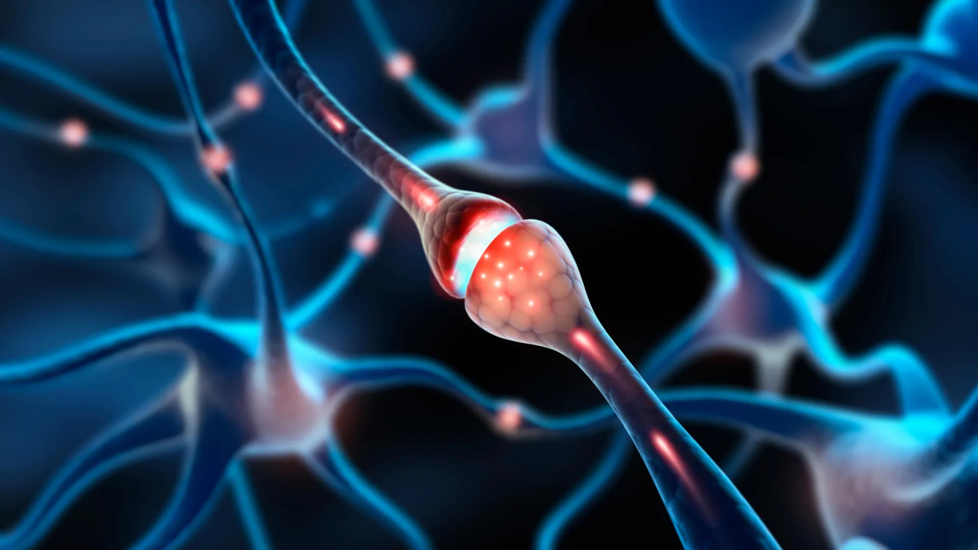
In the mechanics of neural transmission, glutamate is the primary neurotransmitter. For a signal to pass between neurons, it must activate both AMPA and NMDA receptors. AMPA receptors act as the "gatekeepers" that initiate the electrical signal. NMDA receptors, meanwhile, are typically blocked by magnesium ions and only open when the neuron is already partially depolarized. Without AMPA receptors, the filopodia cannot transmit an electrical signal in response to glutamate, rendering the synapse "silent."
The Mechanics of Unsilencing: Forming New Memories
To confirm that these filopodia were indeed functional silent synapses, the researchers employed a modified patch-clamping technique. This allowed them to monitor the electrical activity of individual filopodia in response to the simulated release of glutamate from neighboring neurons.
The experiments confirmed that glutamate alone failed to trigger a response in the filopodia. However, when the researchers experimentally removed the magnesium block from the NMDA receptors, they were able to record electrical currents. This proved the filopodia were "wired" and ready to receive signals, despite their current state of inactivity.
The team then demonstrated that these connections could be "unsilenced" through a process of synaptic plasticity. By pairing the release of glutamate with a strong electrical pulse from the neuron’s cell body, the researchers induced the rapid accumulation of AMPA receptors at the filopodia. Within minutes, the previously silent connection transformed into a functional, active synapse.
Crucially, the study found that it was significantly easier to "unsilence" a filopodium than it was to modify a synapse that was already active. This suggests that the brain’s reserve of filopodia is specifically tuned for high-speed learning, while mature synapses are optimized for long-term data retention.
Supporting Data and Statistical Context
The quantitative data provided by the study offers a new perspective on the brain’s storage capacity:
- Prevalence: Approximately 30% of all synaptic connections in the adult mouse cortex were found to be silent.
- Distribution: Filopodia were found across various layers of the cortex and in multiple brain regions, including the visual cortex and the hippocampus, suggesting a universal mechanism for mammalian learning.
- Comparison: The threshold for inducing plasticity in filopodia was found to be significantly lower than in mature synapses, which require much more intensive or repetitive signaling to change their "weight" or strength.
Implications for Aging and Neurological Health
The discovery of a hidden reserve of silent synapses has profound implications for the study of aging and neurodegenerative diseases. As the brain ages, cognitive flexibility often declines, making it more difficult to learn new skills or adapt to new environments. The MIT team is now investigating whether the number or function of these silent synapses decreases with age.
If the "pool" of silent synapses is depleted over time, it could explain why the elderly often find it more difficult to form new memories while maintaining vivid recollections of the distant past. Furthermore, in conditions such as Alzheimer’s disease, the disruption of synapse formation and maintenance is a primary driver of cognitive decline. Understanding the molecular players that keep filopodia in a "ready" state could lead to new therapeutic targets aimed at restoring memory flexibility.
"It’s entirely possible that by changing the amount of flexibility you’ve got in a memory system, it could become much harder to change your behaviors and habits or incorporate new information," Harnett notes. "You could also imagine finding some of the molecular players that are involved in filopodia and trying to manipulate some of those things to try to restore flexible memory as we age."
A New Paradigm for Neuroscience
The MIT study represents a shift in how the scientific community views the adult brain. Rather than being a relatively static organ that slowly loses its capacity for change, the brain appears to be a dynamic system that maintains a massive "surplus" of potential connections. This hidden infrastructure ensures that even in adulthood, the mammalian brain remains a high-performance learning machine, capable of integrating new experiences without jeopardizing the stability of a lifetime of gathered knowledge.
The research was supported by several prestigious organizations, including the Boehringer Ingelheim Fonds, the National Institutes of Health, the James W. and Patricia T. Poitras Fund at MIT, the Klingenstein-Simons Fellowship, the Vallee Foundation, and the McKnight Foundation. As researchers move forward to verify these findings in human brain tissue, the "silent synapse" may become a central pillar in our understanding of human intelligence and cognitive resilience.


Leave a Reply