
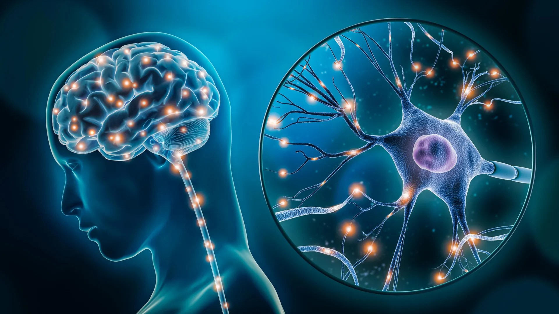
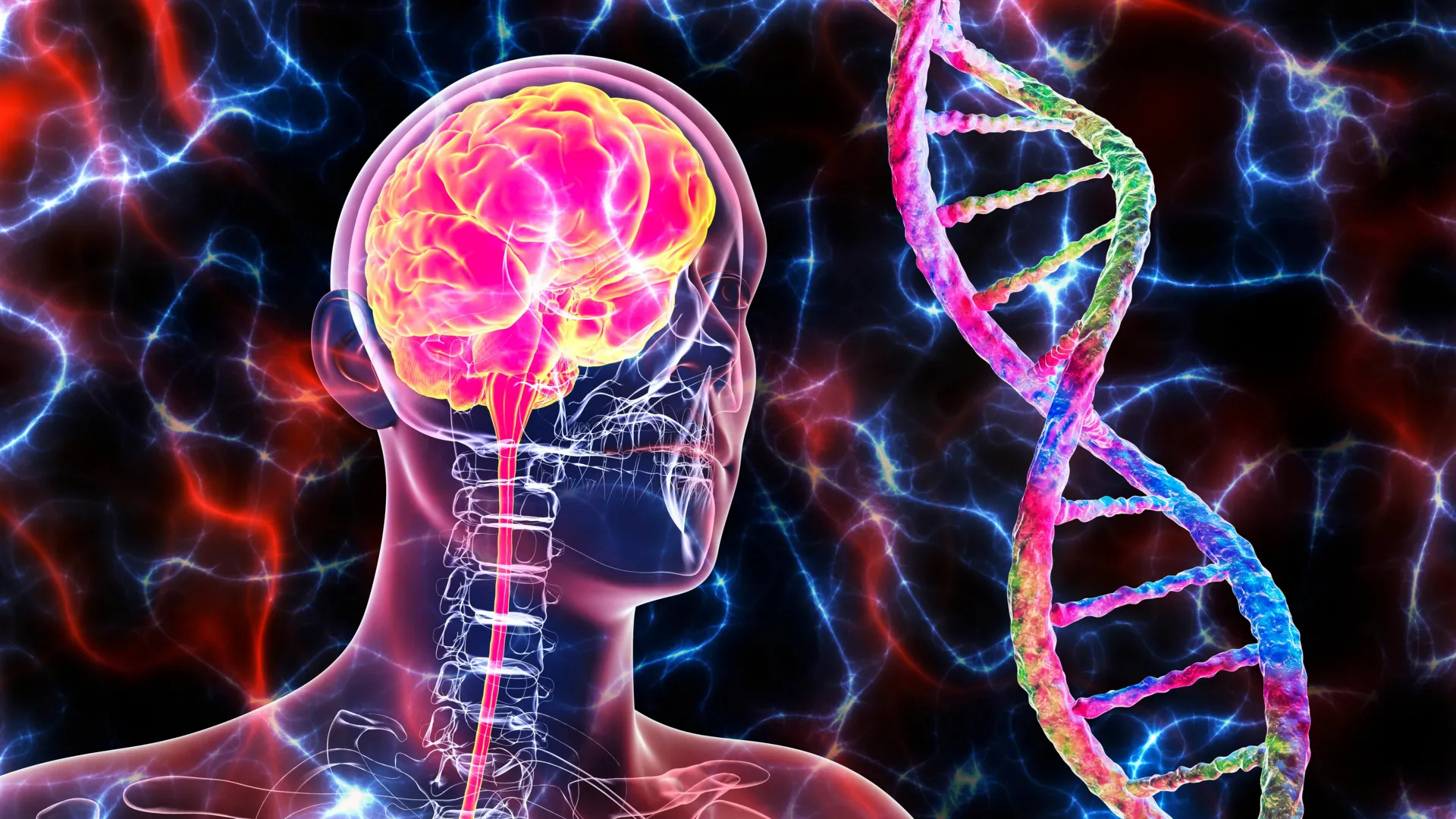
A groundbreaking study led by researchers at McGill University and the Douglas Research Centre has unveiled specific cellular abnormalities in the brains of individuals diagnosed with major depressive disorder, marking a significant shift in the biological understanding of mental health. The research, published in the prestigious journal Nature Genetics, identifies two distinct types of brain cells—excitatory neurons and microglia—that exhibit significantly altered genetic activity in those suffering from depression. This discovery provides the most detailed map to date of the molecular disruptions occurring within the human brain during depressive episodes, offering a new frontier for the development of targeted pharmacological interventions.
By utilizing advanced single-cell genomic techniques on rare post-mortem brain tissue, the team was able to move beyond generalized observations of brain activity to pinpoint the exact cellular drivers of the condition. The findings suggest that depression is not merely a "chemical imbalance" in the traditional sense, but a complex disruption of specific cellular ecosystems responsible for mood regulation and immune response. As depression continues to be a leading cause of disability worldwide, affecting an estimated 264 million people, these insights arrive at a critical juncture for psychiatric medicine.
The Scientific Breakthrough: Mapping the Cellular Landscape
The study, titled "Single-nucleus chromatin accessibility profiling identifies cell types and functional variants contributing to major depression," was spearheaded by senior author Dr. Gustavo Turecki. Dr. Turecki, a professor at McGill University and the Canada Research Chair in Major Depressive Disorder and Suicide, emphasized that this research represents a paradigm shift in neurobiology. For decades, researchers have struggled to reconcile the psychological symptoms of depression with its physical manifestations in the brain.
"This is the first time we’ve been able to identify what specific brain cell types are affected in depression by mapping gene activity together with mechanisms that regulate the DNA code," Dr. Turecki stated. "It gives us a much clearer picture of where disruptions are happening, and which cells are involved."
The team’s approach involved examining the "epigenetic landscape" of the brain. While every cell in the body contains the same DNA, the way that DNA is packaged—known as chromatin—determines which genes are accessible and active in a given cell. By profiling the chromatin accessibility of individual nuclei, the researchers could see which parts of the genetic code were "turned on" or "turned off" in depressed individuals compared to a control group.
Methodology and the Role of the Douglas-Bell Canada Brain Bank
The success of the study was predicated on the availability of high-quality human brain tissue. The researchers analyzed samples from 59 individuals who had been diagnosed with major depressive disorder and 41 individuals who served as a control group. These samples were provided by the Douglas-Bell Canada Brain Bank, one of the few institutions globally equipped to facilitate such granular biological research into psychiatric conditions.
The Brain Bank, founded in 1980, houses over 3,000 brains, many of which come from donors with well-documented psychiatric histories. This resource is invaluable because animal models, while useful, often fail to capture the unique complexity of the human prefrontal cortex—the area of the brain responsible for complex cognitive behavior and emotional regulation.
To process these samples, the McGill team employed single-nucleus RNA sequencing and ATAC-seq (Assay for Transposase-Accessible Chromatin using sequencing). These high-throughput technologies allow scientists to isolate individual cells from a piece of tissue and analyze their genetic output. In this study, thousands of individual cells were categorized and compared, allowing the team to identify specific "clusters" of cells that behaved abnormally in the depressed cohort.
Identifying the Key Cellular Players: Neurons and Microglia
The analysis highlighted two primary cell types that showed the most significant divergence in activity:
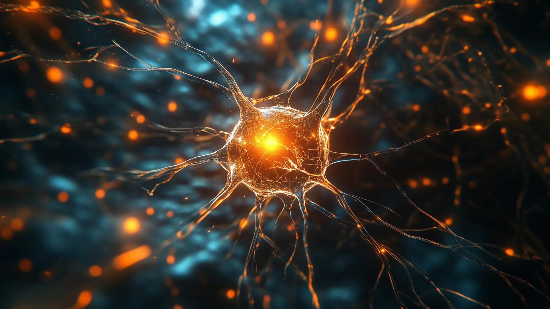
1. Excitatory Neurons
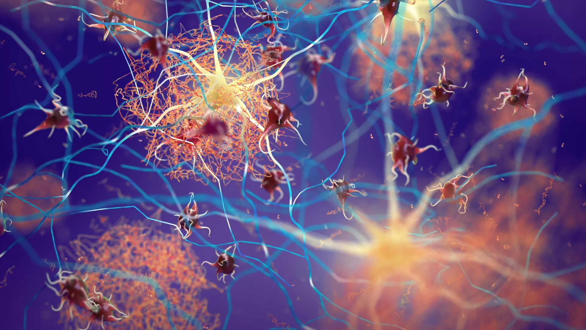
Excitatory neurons are the workhorses of the brain’s communication network, using the neurotransmitter glutamate to send signals between different regions. In the depressed brain, these neurons showed altered activity in genes related to synaptic plasticity—the brain’s ability to form new connections and adapt to new information.
When these neurons fail to function correctly, the brain’s "circuitry" for mood regulation becomes sluggish or disconnected. This finding aligns with recent clinical successes in using ketamine-based treatments, which target glutamate receptors, suggesting that the McGill study has identified the cellular foundation for why such treatments are effective.
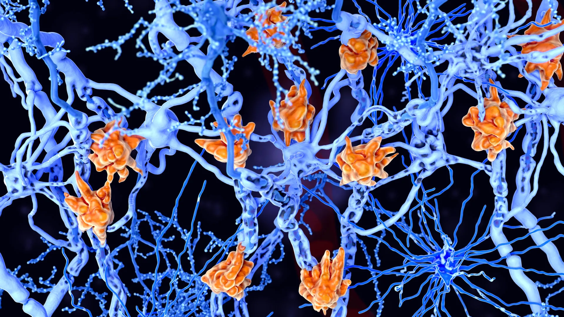
2. Microglia and the Immune Response
Perhaps more surprising was the involvement of microglia. These are the brain’s resident immune cells, responsible for cleaning up cellular debris and defending against pathogens. However, the study found that a specific subtype of microglia in depressed individuals showed genetic signatures associated with inflammation.
This supports the "inflammatory hypothesis" of depression, which suggests that a hyperactive immune system in the brain can lead to neurotoxicity and mood disturbances. By identifying the specific microglia involved, researchers can now look for ways to "calm" these immune cells without affecting the rest of the brain’s protective systems.
Chronology of Discovery in Depression Research
To understand the significance of this study, it is necessary to view it within the timeline of psychiatric evolution:
- 1950s–1960s: The "Monoamine Hypothesis" emerges, suggesting depression is caused by a deficiency in neurotransmitters like serotonin and norepinephrine. This led to the development of the first antidepressants.
- 1980s–1990s: The rise of SSRIs (Selective Serotonin Reuptake Inhibitors) dominates the market. However, researchers begin to notice that these drugs do not work for roughly one-third of patients.
- 2000s: Focus shifts to the "Neuroplasticity Theory," suggesting that depression involves the shrinking of neurons in the hippocampus and prefrontal cortex.
- 2010s: Genomic studies begin identifying hundreds of small genetic variations associated with depression risk, but they lack "cellular resolution"—they know the genes, but not which cells they are in.
- 2024: The McGill/Douglas study provides that missing resolution, mapping the genetic variations to specific excitatory neurons and microglia.
Global Context and the Socioeconomic Burden of Depression
The implications of this research extend far beyond the laboratory. Major Depressive Disorder (MDD) is a global health crisis. According to the World Health Organization (WHO), depression is a leading cause of disability and a major contributor to the overall global burden of disease.
The economic impact is equally staggering. In the United States alone, the economic burden of MDD was estimated at over $210 billion annually, a figure that includes direct healthcare costs, suicide-related costs, and workplace productivity losses. Despite this, the development of new psychiatric drugs has lagged behind other fields like oncology or cardiology, largely because the underlying biology of the brain remained a "black box."
By proving that depression has a measurable biological foundation at the cellular level, the McGill study helps to dismantle the stigma that mental health issues are purely psychological or a matter of "willpower."
"This research reinforces what neuroscience has been telling us for years," Dr. Turecki said. "Depression isn’t just emotional, it reflects real, measurable changes in the brain."
Official Responses and Scientific Peer Review
The publication in Nature Genetics followed a rigorous peer-review process, during which independent experts evaluated the team’s methodology. Academic reactions have been overwhelmingly positive, with many noting that the study’s use of single-cell genomics sets a new standard for psychiatric research.
The study was a collaborative effort involving several prominent institutions. Funding was provided by the Canadian Institutes of Health Research (CIHR), the Brain Canada Foundation, and the Fonds de recherche du Québec – Santé (FRQS). Additionally, the "Healthy Brains, Healthy Lives" (HBHL) initiative at McGill University played a pivotal role in supporting the computational analysis required to handle the massive datasets generated by single-cell sequencing.
Dr. Anjali Chawla, the study’s first author, noted that the complexity of the data required new ways of thinking about genetic risk. By identifying "functional variants"—genetic changes that actually alter how a cell works—the team has provided a roadmap for future drug developers.
Future Implications: Toward Precision Psychiatry
The next phase for Dr. Turecki and his team involves investigating how these cellular differences manifest in living patients. While post-mortem tissue provides a "snapshot" of the brain at the end of life, the researchers hope to use this data to develop biomarkers—biological signs that can be detected through blood tests or brain imaging.
If clinicians can identify which cell types are most affected in an individual patient, they could move toward a model of "precision psychiatry." Instead of a trial-and-error approach with standard antidepressants, a patient with high microglial inflammation might receive an anti-inflammatory neurological treatment, while a patient with excitatory neuron dysfunction might receive a glutamate-modulating therapy.
Furthermore, this research opens the door for gene-silencing technologies and other advanced molecular therapies. By targeting the specific mechanisms that regulate DNA in microglia or neurons, it may one day be possible to "reset" the brain’s genetic activity to a healthy state.
Conclusion
The findings from McGill University and the Douglas Institute represent a cornerstone in the modern era of neuroscience. By bridging the gap between genetics and cellular biology, the research provides a definitive answer to the question of what happens in the brain during depression. As the scientific community continues to digest these findings, the focus will undoubtedly shift from understanding the "what" of depression to mastering the "how" of its cure. For the millions of people worldwide living under the shadow of major depressive disorder, this cellular map offers more than just data; it offers a tangible path toward more effective, biology-based hope.


Leave a Reply