
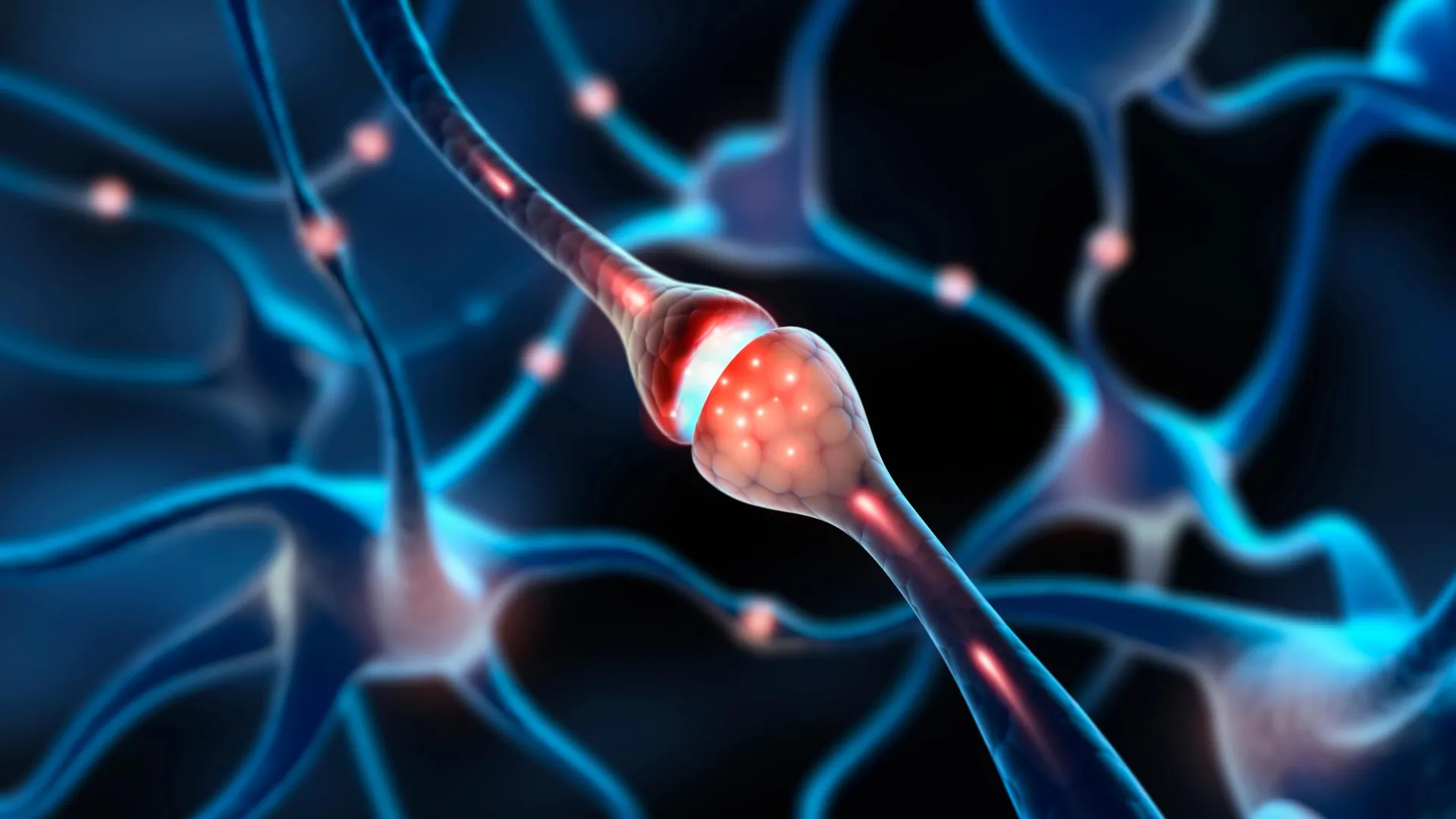
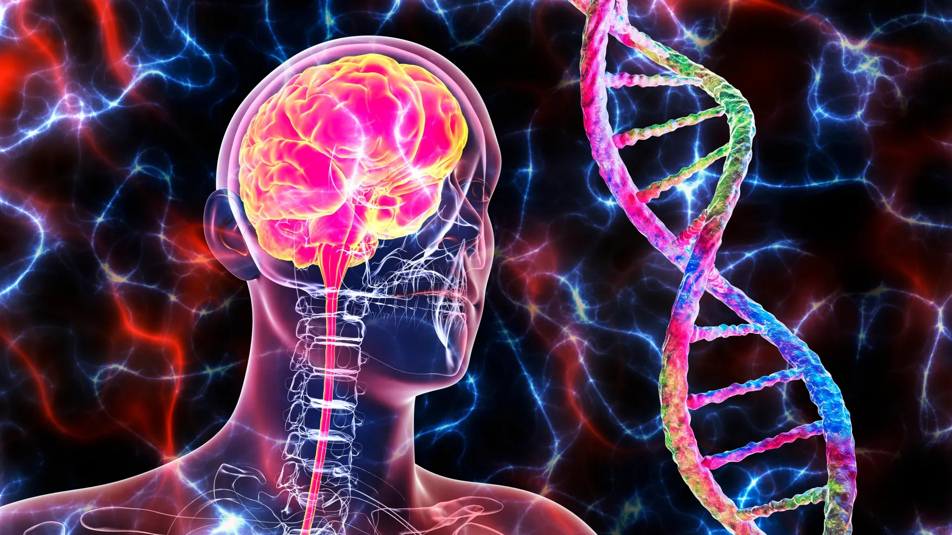
The human brain has long been regarded as a marvel of biological engineering, capable of storing a lifetime of experiences while maintaining a stable architecture of core memories. For decades, the prevailing consensus in neuroscience held that the intense plasticity required for rapid learning was a hallmark of the developing brain, eventually tapering off as an organism reached maturity. However, a groundbreaking study from neuroscientists at the Massachusetts Institute of Technology (MIT) has fundamentally challenged this narrative. Researchers have discovered that the adult brain contains millions of "silent synapses"—immature, inactive connections between neurons that remain dormant until they are recruited to form new memories.
This discovery, published in the journal Nature, reveals that approximately 30 percent of the synapses in the adult mouse cortex are silent. These connections provide a massive, hidden reserve of potential neural pathways, suggesting that the adult brain is far more flexible and capable of modification than previously understood. By maintaining this pool of uncommitted connections, the brain can acquire new information without disrupting or overwriting the stable, mature synapses that house long-term memories.
A Paradigm Shift in Neuroplasticity
The concept of silent synapses is not entirely new, but their presence in the adult brain was unexpected. First identified in the 1990s, silent synapses were traditionally associated with the "critical period" of early development. During this phase, the brain is highly sensitive to environmental stimuli, rapidly forming and pruning connections to build a foundational model of the world. In mice, these connections were thought to vanish by the twelfth day of life—roughly equivalent to the first few months of human infancy.
The traditional view posited that as the brain matures, these flexible connections are either converted into "functional" synapses—equipped with the necessary molecular machinery to transmit signals—or eliminated entirely. Once the brain reached adulthood, it was believed that learning occurred primarily through the modification of existing, active synapses. This process, known as synaptic plasticity, involves strengthening or weakening connections that are already operational.
However, the MIT team’s findings suggest a dual-track system for memory. While mature synapses provide the stability needed to retain essential information, the silent synapses provide the flexibility required for continuous learning. This mechanism solves a long-standing theoretical problem in neuroscience: how the brain remains plastic enough to learn new things throughout a lifespan without suffering from "catastrophic interference," where new data erases old, vital memories.
The Role of eMAP in Visualizing the Invisible
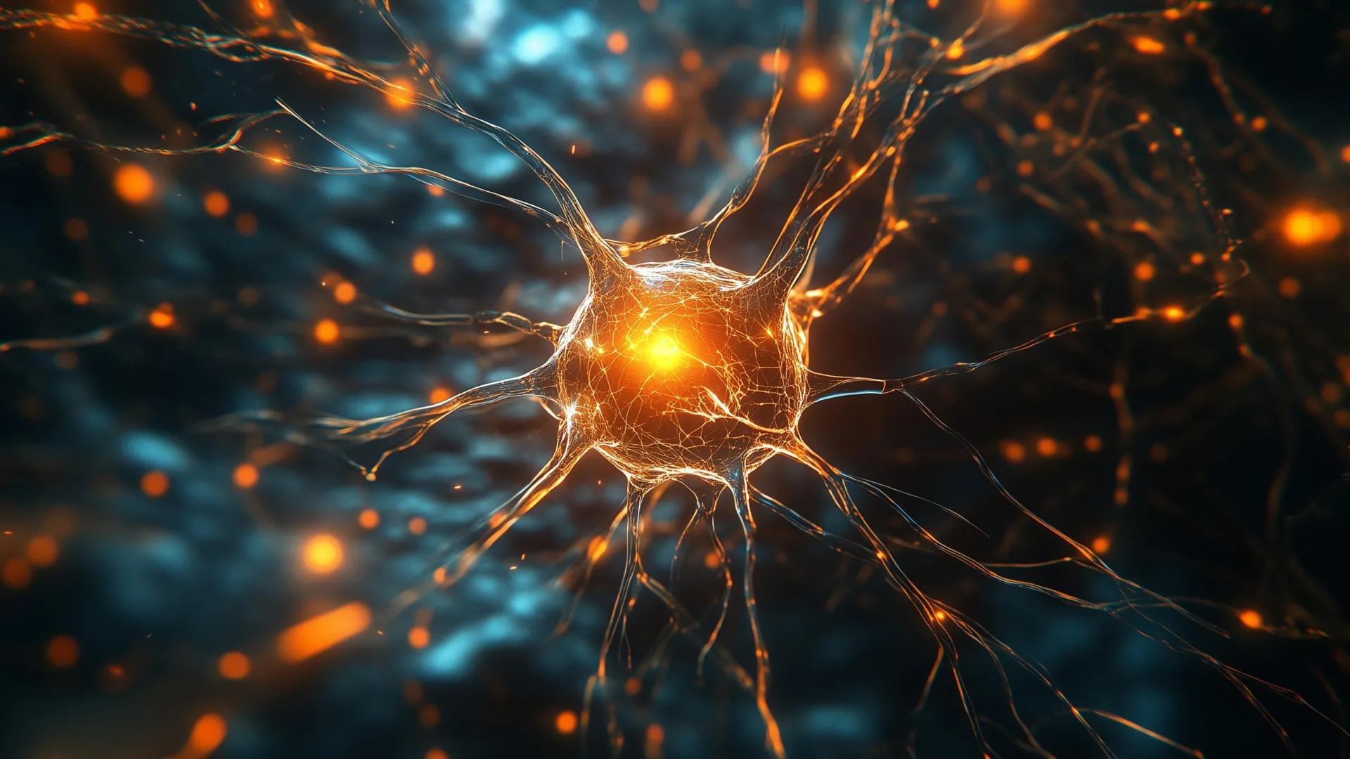
The discovery was made possible by a technological breakthrough in imaging. The MIT researchers, led by Dimitra Vardalaki, a graduate student in the Department of Brain and Cognitive Sciences, were initially investigating how dendrites—the branch-like extensions of neurons that receive signals—process information across different regions of the brain. To do this, they utilized a technique called eMAP (epitope-preserving Magnified Analysis of the Proteome), developed by co-author Kwanghun Chung, an associate professor of chemical engineering at MIT.
The eMAP technique is a form of expansion microscopy. It involves infusing brain tissue with a hydrogel that can be physically expanded, effectively enlarging the tissue samples to allow for high-resolution imaging of individual proteins. This allowed the researchers to see structures that were previously too small or too crowded to be distinguished using traditional light microscopy.
During their observations of the adult mouse visual cortex, the researchers noticed an abundance of filopodia—tiny, hair-like protrusions extending from the dendrites. While filopodia had been seen before, they were usually dismissed as artifacts of development or minor structural features of the adult brain. The high-resolution eMAP imaging revealed that these filopodia were ubiquitous, appearing at levels far higher than any previous study had documented.
The Molecular Anatomy of Silence
To determine if these filopodia were indeed silent synapses, the researchers analyzed their molecular composition. A functional synapse typically requires two types of neurotransmitter receptors to work in tandem: NMDA (N-methyl-D-aspartate) receptors and AMPA (α-amino-3-hydroxy-5-methyl-4-isoxazolepropionic acid) receptors.
When a neuron releases the neurotransmitter glutamate, it binds to both receptors. However, NMDA receptors are normally blocked by magnesium ions and cannot transmit an electrical signal on their own. They require the AMPA receptors to first depolarize the neuron, which displaces the magnesium and allows the NMDA receptors to open. A synapse that lacks AMPA receptors is effectively "silent"; it can receive glutamate, but it cannot pass an electrical signal forward.
Using eMAP, the MIT team found that the filopodia in the adult cortex were rich in NMDA receptors but completely lacked AMPA receptors. This molecular signature confirmed that the filopodia were acting as silent synapses. The researchers then expanded their search to other areas of the brain, finding similar concentrations of silent synapses in the prefrontal cortex and other regions associated with high-level cognitive functions.
Chronology of the Experiment: From Observation to Activation
Once the silent synapses were identified, the researchers sought to prove they could be "unsilenced" or converted into functional connections. The team employed a sophisticated version of patch-clamping, a technique used to measure the electrical activity of individual cells.
- Baseline Measurement: The researchers targeted individual filopodia and simulated the release of glutamate. As expected, no electrical signal was generated, confirming the connection was inactive.
- Experimental Unblocking: When the researchers experimentally removed the magnesium block from the NMDA receptors, the filopodia were able to transmit signals. This proved the NMDA receptors were functional and ready to be used.
- The Plasticity Protocol: To simulate learning, the researchers paired the release of glutamate with a high-frequency electrical pulse from the neuron’s body. This combination is a classic trigger for synaptic plasticity.
- Recruitment of AMPA Receptors: Following the stimulation, the researchers observed a rapid accumulation of AMPA receptors at the site of the filopodia. Within minutes, the previously silent connection had transformed into a functional synapse capable of transmitting signals independently.
Crucially, the researchers found that it was much easier to activate these silent synapses than it was to modify existing, mature synapses. Mature synapses required much stronger and more specific signals to change their strength, reflecting the brain’s need to keep established memories stable. In contrast, the filopodia were "primed" for recruitment, making them the primary vehicles for new memory formation.
Theoretical Alignment and Expert Reactions
The MIT study provides empirical weight to long-standing theoretical models in neuroscience. Stefano Fusi and Larry Abbott, prominent neuroscientists who have worked on mathematical models of memory, previously proposed that the brain must utilize a hierarchy of synapses with different levels of plasticity. Their work suggested that a pool of highly flexible connections was necessary to prevent the "overwriting" of stable memories.
Mark Harnett, an associate professor of brain and cognitive sciences at MIT and the senior author of the study, noted that this research is the first real evidence that such a system exists in the mammalian brain. "Filopodia allow a memory system to be both flexible and robust," Harnett stated. "You need flexibility to acquire new information, but you also need stability to retain the important information."
Other experts in the field have reacted with cautious optimism, noting that while the study was conducted on mice, the fundamental architecture of the mammalian cortex is highly conserved across species. The existence of a "reserve" of synapses could explain why humans are capable of learning complex new skills, such as a language or a musical instrument, well into their senior years.
Implications for Aging and Neurological Health
The discovery of a hidden reserve of synapses has profound implications for our understanding of the aging brain and neurodegenerative diseases. As humans age, cognitive flexibility often declines, making it harder to learn new tasks or break established habits. The MIT researchers are now investigating whether the number or "recruitability" of silent synapses decreases with age.
If the decline in cognitive plasticity is linked to a depletion of these silent synapses, it opens new avenues for therapeutic intervention. Scientists could potentially develop pharmacological or electrical stimulation therapies designed to preserve the pool of filopodia or make them easier to activate in older adults.
Furthermore, the research may shed light on conditions like Alzheimer’s disease. In the early stages of neurodegeneration, the brain’s ability to form new memories is often the first thing to fail. Understanding how silent synapses are maintained or lost could lead to early diagnostic tools or treatments that target the very beginning of memory impairment.
Future Research Directions
The MIT team is already moving forward with several follow-up studies. One priority is determining if these silent synapses are present in the human brain in similar proportions. Given the complexity of human cognition, some researchers speculate that the human reserve of silent synapses might be even larger than that of mice.
Additionally, the researchers want to explore the environmental factors that influence the density of silent synapses. It is possible that "enriched environments"—those filled with social interaction, physical activity, and cognitive challenges—may stimulate the production or maintenance of these connections, providing a biological basis for the "use it or lose it" theory of brain health.
The study concludes that the adult brain is not a static organ with a fixed number of connections. Instead, it is a dynamic system that keeps a massive "savings account" of potential neural pathways. This hidden reserve ensures that no matter how much we have already learned, the brain remains ready to adapt to the next new experience, striking a delicate balance between the memories of the past and the lessons of the future.


Leave a Reply