

The decline of the human sense of smell has long been observed as a precursor to the more debilitating cognitive symptoms of Alzheimer’s disease, often manifesting years before memory loss or disorientation take hold. Recent breakthroughs from a collaborative team of researchers at the German Center for Neurodegenerative Diseases (DZNE) and Ludwig-Maximilians-Universität München (LMU) have provided a biological explanation for this phenomenon, identifying the brain’s own immune system as the primary driver behind the destruction of olfactory-related nerve fibers. Published in the journal Nature Communications, the study details how the brain’s resident immune cells, known as microglia, mistakenly target and dismantle essential neural connections in the early stages of the disease. This discovery not only clarifies a long-standing medical mystery but also provides a potential window for early intervention using newly developed therapeutic agents.
The Biological Mechanism: Microglia and the Breakdown of Olfactory Pathways
At the heart of the research is the interaction between two specific regions of the brain: the olfactory bulb and the locus coeruleus. The olfactory bulb, situated in the forebrain, serves as the primary processing center for sensory information gathered by receptors in the nasal cavity. The locus coeruleus, located deep within the brainstem, acts as a regulatory hub, sending out long nerve fibers that distribute norepinephrine to various parts of the brain, including the olfactory bulb. These fibers are crucial for modulating sensory perception, sleep-wake cycles, and cerebral blood flow.
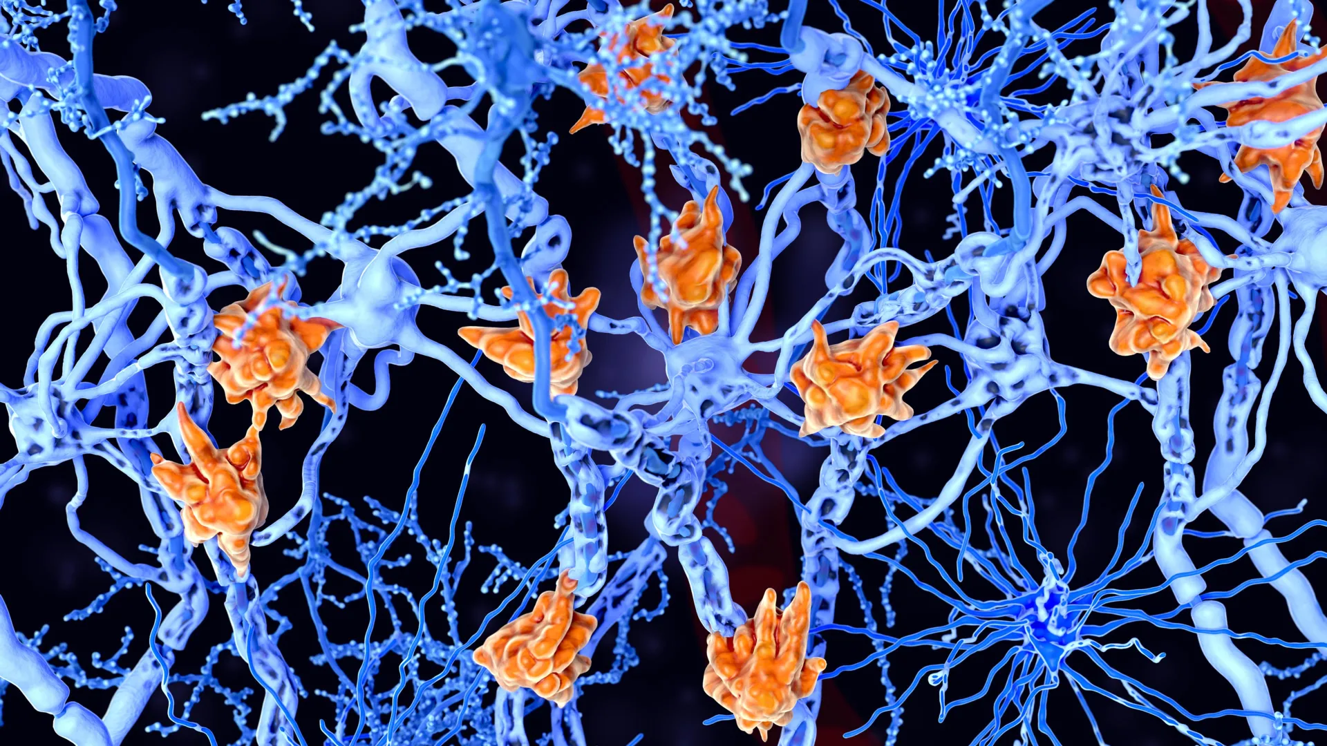
The study indicates that in the earliest phases of Alzheimer’s disease, the nerve fibers extending from the locus coeruleus to the olfactory bulb undergo pathological changes. These alterations act as a catalyst for the brain’s immune system. Microglia, which normally function as the brain’s maintenance crew by clearing away cellular debris and pathogens, begin to perceive these vital nerve fibers as defective or redundant.
Dr. Lars Paeger, a lead scientist in the study representing both DZNE and LMU, notes that the locus coeruleus is responsible for a wide array of physiological mechanisms. When the connectivity between the brainstem and the olfactory bulb is severed by microglial activity, the brain’s ability to process scents is compromised. This "synaptic pruning" or breakdown of connections is a normal part of brain development in childhood, but its occurrence in the adult brain under the influence of Alzheimer’s pathology represents a destructive departure from healthy neurological function.
The "Eat-Me" Signal: Molecular Shifts in Nerve Membranes
The researchers sought to understand exactly what triggers the microglia to attack these specific nerve fibers. The investigation led to the identification of a molecular shift on the surface of the neurons. Under normal conditions, a fatty molecule called phosphatidylserine is kept on the inner side of the neuron’s membrane. However, the study found that in the presence of Alzheimer’s pathology, this molecule migrates to the outer surface of the membrane.
In the language of neurobiology, the presence of phosphatidylserine on the exterior of a cell is a potent "eat-me" signal. This signal tells the microglia that the cell or connection is no longer functional and should be consumed and removed. The research suggests that this shift is likely caused by the hyperactivity of the affected neurons. In the early stages of Alzheimer’s, neurons often exhibit abnormal firing patterns, which stresses the cellular membrane and causes the internal molecules to flip to the outside. This biochemical error effectively marks healthy, albeit hyperactive, nerve fibers for destruction by the immune system.
Multi-Modal Research: From Animal Models to Human Brain Scans
To validate their findings, the research team employed a comprehensive, three-tiered approach that combined animal models, the analysis of human tissue, and advanced neuroimaging. This multi-modal strategy ensured that the observations made in a laboratory setting were reflective of the actual disease progression in human patients.
- Animal Models: The team utilized mice genetically engineered to exhibit Alzheimer’s-like features. These models allowed the researchers to observe the real-time interaction between microglia and nerve fibers, confirming the migration of phosphatidylserine and the subsequent microglial response.
- Human Tissue Analysis: Post-mortem examinations of brain tissue from deceased Alzheimer’s patients provided a direct link to human pathology. The researchers found evidence of the same microglial patterns and nerve fiber degradation in the olfactory regions that were observed in the mouse models.
- PET Scanning: The study incorporated Positron Emission Tomography (PET) scans from living individuals, including those diagnosed with Alzheimer’s and those with mild cognitive impairment (MCI). These scans allowed the team to visualize the activity of microglia and the density of nerve connections in the brain, correlating olfactory decline with the physical presence of immune-mediated damage.
By synthesizing data from these three sources, the researchers were able to confirm that the immunological mechanism they identified is a consistent feature of early-stage Alzheimer’s disease across species.
Chronology of Alzheimer’s Research and the Olfactory Connection
The understanding of Alzheimer’s disease has evolved significantly since Alois Alzheimer first described the condition in 1906. For decades, the focus remained primarily on the accumulation of amyloid-beta plaques and tau tangles. However, the timeline of discovery regarding sensory symptoms has been more gradual.
- 1980s-1990s: Clinical observations first began to consistently link the loss of smell (anosmia or hyposmia) to neurodegenerative disorders like Parkinson’s and Alzheimer’s.
- 2000s: Researchers identified that the olfactory bulb is one of the first areas to accumulate pathological protein aggregates, but the exact mechanism of nerve loss remained elusive.
- 2010s: The role of neuroinflammation and microglia gained prominence in the scientific community, shifting the focus from "protein clumps" to the brain’s immune response.
- 2023-2024: The current study by Paeger, Herms, and their colleagues provides the definitive link between microglial "eat-me" signals and the specific degradation of the locus coeruleus-olfactory bulb pathway.
This timeline illustrates a shift in the medical field toward "prodromal" diagnosis—identifying the disease in its silent, pre-symptomatic phase. The loss of smell is now recognized as a critical clinical marker in this early window.
Implications for Early Diagnosis and New Therapeutics
The findings have significant implications for the future of Alzheimer’s treatment, particularly regarding the use of amyloid-beta antibodies. In recent years, drugs such as lecanemab and donanemab have received regulatory attention and approval in various markets. These therapies are designed to clear amyloid-beta from the brain, but their efficacy is heavily dependent on early administration. Once significant cognitive decline has occurred, the damage to the brain’s architecture is often too extensive for these drugs to provide a meaningful reversal.
Prof. Dr. Jochen Herms, a research group leader at DZNE and LMU and a member of the "SyNergy" Cluster of Excellence, emphasizes that identifying patients at the earliest possible stage is the "holy grail" of current Alzheimer’s research. If a declining sense of smell can be used as a reliable biological "red flag," patients can undergo more intensive screening—such as PET scans or cerebrospinal fluid analysis—to confirm the presence of the disease before they begin to lose their memories or independence.
This would allow for a proactive treatment model. Instead of waiting for a patient to fail a cognitive test, clinicians could potentially intervene with immunological or amyloid-targeting therapies the moment the "eat-me" signals begin to trigger microglial destruction.
Analysis of Broader Impacts and Future Outlook
The socio-economic impact of this research is substantial. Alzheimer’s disease currently affects over 55 million people worldwide, a number expected to rise as the global population ages. The cost of care is astronomical, and the emotional toll on families is profound. By shifting the diagnostic window earlier, the medical community can potentially extend the "quality of life" years for millions of patients.
Furthermore, this study opens up a new avenue for drug development. If the "eat-me" signal (phosphatidylserine inversion) is the primary trigger for early nerve loss, researchers may look for ways to stabilize the neuronal membranes or inhibit the specific microglial receptors that recognize these signals. This would represent a shift from clearing plaques to preventing the immune system from overreacting in the first place.
The Munich-based "SyNergy" Cluster of Excellence, of which Prof. Herms is a part, continues to investigate how different systems in the brain—such as the immune system, the vascular system, and the nervous system—interact to drive neurodegeneration. This holistic view is increasingly seen as the future of neurology.
In conclusion, the work of Dr. Lars Paeger and his colleagues provides a clear biological roadmap for one of the most common early symptoms of Alzheimer’s. By identifying the immunological mechanism behind olfactory decline, the study offers hope for a future where the disease is detected through simple sensory screenings and treated before it can fundamentally alter a patient’s life. The transition of phosphatidylserine to the outer membrane and the subsequent microglial response is no longer just a laboratory observation; it is a critical target for the next generation of Alzheimer’s diagnostics and care.


Leave a Reply