

Investigators at the Johns Hopkins Kimmel Cancer Center and the Johns Hopkins Bloomberg School of Public Health have identified a critical mechanism by which certain combinations of rearranged genes drive the progression of a rare and aggressive form of kidney cancer. The study, published in the journal Cell Reports on April 22, details how fusion genes create "liquid droplets" within cell nuclei to hijack genetic machinery, a discovery that could pave the way for novel therapeutic interventions in a disease that currently lacks a standardized treatment protocol.
Supported by the National Institutes of Health (NIH), the research team demonstrated that proteins produced by these rearranged genes—known as fusion genes—form tiny, concentrated liquid condensates. These droplets function as localized command centers, turning specific genes on or off to promote the rapid growth and metastasis of cancer cells. By disrupting the formation of these droplets, the researchers were able to prevent the activation of oncogenic (cancer-promoting) genes, suggesting a new strategy for drug development that could extend beyond kidney cancer to other malignancies driven by similar genetic fusions.
Understanding Translocation Renal Cell Carcinoma
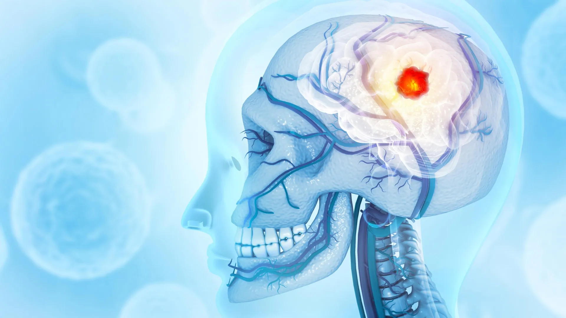
The specific focus of the study was translocation renal cell carcinoma (tRCC), a rare subtype of kidney cancer that accounts for a small percentage of adult kidney cancer cases but represents a significant portion of pediatric renal malignancies. Unlike the more common clear cell renal cell carcinoma, tRCC is characterized by chromosomal translocations—events where pieces of chromosomes break off and reattach to different chromosomes.
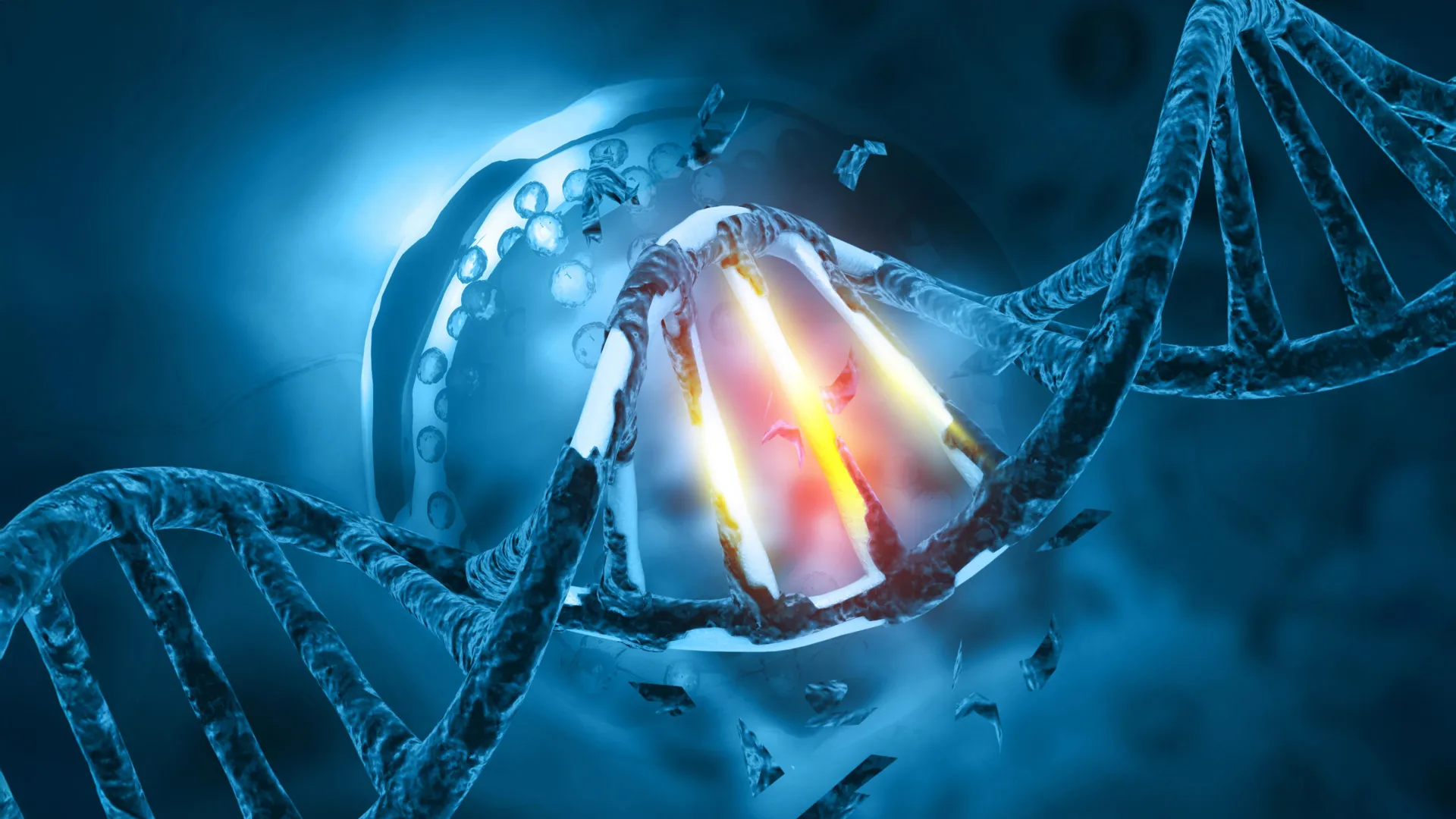
In tRCC, these translocations typically involve the TFE3 gene. When a chromosome rearranges, it swaps a DNA segment, combining the "tail" end of the TFE3 gene with the beginning of one of several "partner" genes, such as PRCC, NONO, or SFPQ. This genetic "shuffling" results in the creation of TFE3 fusion genes. These new genes produce fusion proteins that are not found in healthy cells. While the medical community has long recognized that these fusion proteins are the primary drivers of tRCC, the exact biochemical processes they use to trigger cancer remained elusive until now.
According to the study’s senior author, Danfeng "Dani" Cai, Ph.D., an assistant professor of biochemistry and molecular biology at the Johns Hopkins Bloomberg School of Public Health, the research focused on the most prevalent iterations of the disease. "There are about 20 fusion partners of TFE3 in translocation renal cell carcinoma, but we mainly focused on the two most common ones, NONO and SFPQ, which together make up approximately 40% of all TFE3 fusions," Cai stated.
The Role of Liquid Condensates in Cellular Malfunction
To visualize the activity of these fusion proteins, the research team employed advanced microscopic techniques. They attached fluorescent "glowing" tags to TFE3 fusion proteins within cells derived from kidney cancer patients. Observation through high-resolution microscopy revealed that these proteins did not distribute evenly throughout the nucleus; instead, they clustered into distinct "dots" or liquid condensates.
Liquid condensates are a relatively recent discovery in cell biology, often described as a form of "biomolecular phase separation"—similar to droplets of vinegar forming in oil. These condensates concentrate specific molecules within a small space to facilitate complex cellular processes. In the context of tRCC, the TFE3 fusion proteins use this phase separation to create a specialized environment where they can interact more effectively with DNA.
The team observed that these droplets also contained marker proteins typically associated with active gene transcription, as well as other proteins responsible for "switching on" genes. This indicated that the liquid droplets were not merely passive clusters but active regulatory hubs designed to facilitate the expression of genes that drive cancer cell proliferation.
Chromatin Remodeling and the Genomic Landscape
The research further explored how these fusion proteins interact with the cell’s genomic structure. DNA in the human cell is not a loose string; it is tightly packaged into a structure called chromatin, often described as "beads on a string." When the DNA is tightly wound around these "beads" (histones), the genes are inaccessible and remain "off." Conversely, when the chromatin is open or relaxed, the genes can be accessed and "turned on."
Cai collaborated with Eneda Toska, Ph.D., an assistant professor of oncology at the Johns Hopkins Kimmel Cancer Center, to map the interaction between TFE3 fusion proteins and the chromatin landscape. Using sophisticated genomic sequencing and mapping tools, they discovered that the fusion proteins act as master architects of the cell’s DNA structure.
"We found that these fusion proteins open and close different sites on the chromatin by making chemical modifications," Toska explained. "They bind, regulate, and redesign the chromosome landscape, interacting with target genes that promote cell proliferation and movement—functions that cancer needs to grow and spread."
By essentially "rewriting" the epigenetic code of the cell, these fusion proteins ensure that the machinery required for cancer growth is permanently activated, while genes that might normally suppress tumor growth are silenced.
Identifying the "Coiled-Coil" Achilles’ Heel
A pivotal moment in the study occurred when the researchers began dissecting the TFE3 fusion proteins to identify which specific segments were responsible for the formation of liquid condensates. Using CRISPR-based gene editing and protein engineering, the team systematically removed different parts of the protein.
They identified a small segment known as a "coiled-coil" domain—a structural motif where two or more alpha-helices wrap around each other. This domain is located in the region that connects the TFE3 tail to its fusion partner (such as NONO or SFPQ). When this specific coiled-coil segment was deleted, the fusion proteins lost their ability to form liquid droplets. More importantly, without the formation of these condensates, the proteins were no longer able to activate the genes responsible for cancer progression.
"Individually, all the protein components found in the TFE3 fusions, including full-length TFE3, NONO, and SFPQ, are typically involved in the cell machinery that turns on genes to make proteins," Cai noted. "However, we found when in the form of these fusion proteins, they acquire an even stronger ability to control what genes get turned on."
This finding suggests that the physical state of the protein—its ability to form a liquid droplet—is just as important as its genetic sequence. This "phase-dependent" activity provides a new target for drug developers: if a small molecule could be designed to disrupt the coiled-coil interaction or dissolve the condensate, it could potentially "turn off" the cancer without needing to alter the patient’s DNA.
Broader Implications for Oncology and Rare Disease Research
The implications of the Johns Hopkins study extend far beyond the niche of translocation renal cell carcinoma. Many other forms of cancer, including Ewing sarcoma (a bone cancer primarily affecting children) and various types of leukemia, are also driven by fusion genes.
"Other cancers are caused by fusion genes as well," Cai said. "It’s possible that these fusion genes form similar droplets, or condensates, that regulate genes in these cancers and could react to similar treatment strategies."
The study contributes to a growing body of evidence suggesting that "condensate pathology" may be a unifying theme in oncology. If researchers can identify common structural motifs—like the coiled-coil domain—that facilitate droplet formation across different cancer types, it could lead to the development of a new class of "condensate-disrupting" drugs. This would represent a significant shift toward precision medicine, where treatments are tailored to the physical behavior of proteins within a cell.
Statistical Context and Future Directions
Kidney cancer is among the ten most common cancers in both men and women. According to the American Cancer Society, there are approximately 81,800 new cases of kidney cancer diagnosed in the United States annually. While tRCC represents only a fraction of these cases (estimated at 1% to 5% of adult renal cell carcinomas), its resistance to conventional therapies like chemotherapy and radiation makes it a high-priority area for research. In pediatric populations, tRCC is much more prevalent, accounting for up to 50% of renal cell carcinomas in children and adolescents.
The current lack of a "standard of care" for tRCC means that patients are often treated with therapies designed for other types of kidney cancer, which frequently yield sub-optimal results. The discovery of the TFE3 condensate mechanism offers the first clear roadmap for developing tRCC-specific therapies.
The research team at Johns Hopkins plans to continue their work by identifying other molecular components that reside within these liquid droplets. By understanding the full "inventory" of proteins and RNA molecules inside the condensates, they hope to conduct high-throughput screenings for existing drugs or new small molecules that can safely disrupt these structures in humans.
Research Support and Acknowledgments
The study was a multi-institutional effort involving contributors from the Johns Hopkins Bloomberg School of Public Health, the Johns Hopkins University School of Medicine, and the National Cancer Institute. Co-authors included Choon Leng So, Ye Jin Lee, Wanlu Chen, Binglin Huang, Emily De Sousa, Yangzhengyu Gao, Marie Elena Portuallo, Sumaiya Begum, Kasturee Jagirdar, Vito Rebecca, Hongkai Ji, Bujamin Vokshi, and W. Marston Linehan.
Funding for the research was provided by several branches of the National Institutes of Health, including the National Institute of General Medical Sciences, the National Cancer Institute, and the National Human Genome Research Institute. Additional support was provided by the Department of Defense Kidney Cancer Idea Development Award, a Jayne Koskinas Ted Giovanis grant, and a Johns Hopkins Provost Catalyst Award.
Dr. Eneda Toska disclosed that she has received grants and consulting fees from AstraZeneca and Menarini, reflecting the ongoing interest of the pharmaceutical industry in translating these laboratory findings into clinical applications. As the medical community moves forward, the focus remains on transforming this fundamental biological insight into a life-saving reality for patients facing rare and difficult-to-treat cancers.


Leave a Reply