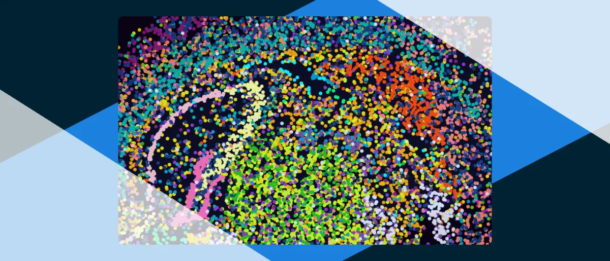
The newly released eBook, supported by Takara Bio, offers an in-depth exploration into the revolutionary advancements shaping the field of biomedical research, with a particular focus on innovative tools designed to capture and integrate true single-cell spatial omics data. This comprehensive resource delves into critical areas, including the intricate cellular origins of cancer and neurodevelopmental diseases, the profound impact of spatial proteomics and artificial intelligence (AI) on cancer research, and provides a detailed, streamlined protocol for conducting single-cell spatial transcriptomics on human kidney tissue. The publication arrives at a pivotal moment in science, as researchers increasingly recognize the necessity of understanding biological processes not just at a cellular level, but also within their native tissue context, thereby unlocking unprecedented insights into health and disease.
The Dawn of Spatial Omics: A Paradigm Shift in Biological Understanding
For decades, traditional molecular biology techniques, such as bulk sequencing, provided a foundational understanding of genomic, transcriptomic, and proteomic profiles by averaging signals across millions of cells. While immensely valuable, this approach inherently obscured cellular heterogeneity and the critical spatial relationships between cells within tissues. The advent of single-cell omics technologies began to address the former, allowing scientists to characterize individual cells. However, it was the subsequent development of single-cell spatial omics that truly marked a paradigm shift, enabling the study of molecular profiles—DNA, RNA, and proteins—at single-cell resolution while preserving their precise anatomical location within a tissue section. This capability is paramount because cellular function and fate are profoundly influenced by a cell’s immediate microenvironment and its interactions with neighboring cells.
The concept of spatial biology has roots extending back to classical histology and immunohistochemistry, which provided visual cues about cellular organization and protein distribution. However, these methods were often limited in their multiplexing capabilities and quantitative precision. The modern era of spatial omics, which began gaining significant traction in the mid-2010s, leverages a combination of advanced microscopy, molecular barcoding, and sophisticated computational analysis to generate high-resolution maps of molecular expression across entire tissues. Technologies like Visium spatial gene expression (10x Genomics), GeoMx Digital Spatial Profiling (NanoString Technologies), and various in situ sequencing and imaging-based methods (e.g., MERFISH, seqFISH+, CODEX) have emerged as powerful tools, each offering distinct advantages in terms of resolution, throughput, and the type of molecular information captured. The ongoing innovation in this space is focused on achieving "true" single-cell spatial omics, meaning methods that can simultaneously provide comprehensive molecular profiling (e.g., hundreds or thousands of RNA transcripts or proteins) with subcellular resolution and high sensitivity, integrated across large tissue areas.
Unraveling the Complexities of Cancer at Spatial Resolution
One of the most immediate and impactful applications of single-cell spatial omics is in cancer research. Cancer is not merely a disease of malignant cells but a complex ecosystem involving tumor cells, immune cells, stromal cells, vasculature, and the extracellular matrix—collectively known as the tumor microenvironment (TME). Understanding the spatial organization and interactions within the TME is crucial for deciphering tumor initiation, progression, metastasis, and response to therapy. Traditional bulk analysis often misses rare but critical cell populations, such as cancer stem cells or specific immune subsets, or averages out their signals. Spatial omics, conversely, allows researchers to precisely localize these cells and characterize their molecular states in relation to their neighbors.
For instance, spatial transcriptomics can reveal distinct transcriptional programs within different regions of a tumor, identifying areas of proliferation, invasion, or immune exclusion. This has led to the discovery of novel biomarkers for disease stratification and prediction of treatment response. Spatial proteomics complements this by directly visualizing the distribution and activity of key proteins, such as immune checkpoints (e.g., PD-1, PD-L1) or signaling molecules, providing insights into resistance mechanisms to immunotherapies or targeted drugs. The eBook’s focus on this area underscores its importance in moving beyond a simplistic view of cancer to a holistic understanding of the cellular choreography that drives malignancy. For example, recent studies using spatial omics have shown how the spatial arrangement of T-cells and tumor cells within melanoma lesions can predict patient response to anti-PD-1 therapy with greater accuracy than bulk immune profiling. Similarly, in pancreatic cancer, spatial analysis has helped delineate distinct stromal subtypes with varying prognostic implications. The ability to map these interactions in situ provides an unparalleled view into the dynamic processes that dictate tumor behavior, offering unprecedented opportunities for targeted therapeutic development.
Illuminating Neurodevelopmental Disease Mechanisms
Beyond oncology, single-cell spatial omics is proving instrumental in unraveling the intricate origins of neurodevelopmental diseases. The brain is an extraordinarily complex organ, characterized by a vast diversity of neuronal and glial cell types, organized into highly specific circuits. Disruptions in the development, migration, or function of these cells, or in the formation of their connections, can lead to severe conditions such as autism spectrum disorders, schizophrenia, intellectual disabilities, and epilepsy. Investigating these diseases has historically been challenging due to the brain’s structural complexity and the difficulty in obtaining and analyzing human brain tissue.
Spatial omics offers a powerful lens to examine these pathologies. By mapping gene expression or protein distribution at single-cell resolution within affected brain regions, researchers can identify subtle alterations in cell type composition, neuronal connectivity, or gene regulatory networks that might underpin disease symptoms. For example, in autism spectrum disorder, studies have begun to use spatial transcriptomics to identify specific cortical layers or subregions where neuronal gene expression is perturbed, potentially pinpointing the cellular origins of synaptic dysfunction. In schizophrenia, researchers are applying these technologies to post-mortem brain tissue to characterize the spatial patterns of altered gene expression in specific neuronal populations, shedding light on the molecular mechanisms of neuroinflammation or impaired neurotransmission. The eBook’s inclusion of this topic highlights the potential for spatial omics to transform our understanding of these debilitating conditions, paving the way for more targeted diagnostic tools and therapeutic interventions. The ability to precisely localize molecular changes within specific brain structures, such as the hippocampus or prefrontal cortex, offers an unprecedented opportunity to dissect the cellular pathology of these disorders. This level of detail is critical for developing therapies that address the root causes of these complex conditions.
The Pivotal Role of Spatial Proteomics in Cancer Research
While genomics and transcriptomics provide insights into the potential for cellular function, proteomics directly measures the proteins—the workhorses of the cell—that execute biological processes. Spatial proteomics, therefore, adds another crucial layer of information by mapping the location and abundance of thousands of proteins within a tissue. This is particularly vital in cancer research, where protein modifications (e.g., phosphorylation, glycosylation) and protein-protein interactions drive signaling pathways, cellular responses, and drug efficacy.
Technologies such as imaging mass cytometry (IMC) and multiplexed immunofluorescence (MIF) allow for the simultaneous detection of dozens to over a hundred protein markers in a single tissue section, preserving spatial context. This enables a detailed characterization of cellular phenotypes, immune cell states, and signaling pathway activation within the TME. For instance, spatial proteomics can identify specific immune cell populations that are infiltrating a tumor, their activation status, and their proximity to cancer cells or other stromal components. This information is critical for understanding why some patients respond to immunotherapies while others do not, and for identifying novel therapeutic targets that modulate protein activity or interactions. The synergy between spatial transcriptomics (revealing gene expression patterns) and spatial proteomics (revealing protein function) provides a comprehensive molecular snapshot of a tissue, offering a more complete understanding of disease mechanisms. For example, spatial proteomic profiling of tumor biopsies has identified distinct immune cell niches within tumors that correlate with patient prognosis and response to checkpoint inhibitors, providing actionable insights for precision oncology. The direct measurement of functional molecules in their native context provides a level of mechanistic insight that is otherwise unattainable.
Artificial Intelligence: The Catalyst for Unlocking Omics Data
The sheer volume and complexity of data generated by single-cell spatial omics technologies necessitate sophisticated analytical approaches. This is where Artificial Intelligence (AI) and machine learning (ML) become indispensable. AI algorithms are revolutionizing how researchers extract meaningful insights from vast datasets, moving beyond manual interpretation to automated, unbiased analysis.

In spatial omics, AI is employed across multiple stages:
- Image Analysis and Segmentation: AI-powered computer vision algorithms can accurately segment individual cells from complex tissue images, even in densely packed or irregular regions, and quantify their molecular features. This is a significant improvement over traditional methods, which can be time-consuming and prone to human error.
- Cell Type Identification and Annotation: Machine learning models can be trained on known cell markers to automatically classify and annotate different cell types and states based on their spatial molecular profiles, accelerating the identification of novel or rare cell populations.
- Spatial Pattern Recognition: AI can identify subtle, non-obvious spatial patterns and relationships between cells and their molecular features within a tissue. For example, it can detect clusters of specific cell types, predict cell-cell interaction networks, or identify spatially defined disease signatures that might be missed by human observers.
- Data Integration and Multi-modal Analysis: Integrating data from different spatial omics modalities (e.g., spatial transcriptomics, proteomics, epigenomics) is a major challenge. AI algorithms, particularly deep learning models, are adept at finding hidden correlations and patterns across these diverse datasets, building a more holistic picture of biological systems.
- Predictive Modeling: In cancer research, AI can leverage spatial omics data to build predictive models for patient prognosis, treatment response, and drug resistance, facilitating personalized medicine approaches.
The eBook’s emphasis on AI underscores its transformative potential, not just as a tool for data processing, but as a genuine accelerator for scientific discovery, enabling researchers to uncover previously hidden biological insights from complex spatial information. The ongoing development of open-source AI tools and platforms is also democratizing access to these powerful analytical capabilities, fostering broader engagement across the scientific community. The synergy between advanced data generation and intelligent analysis is pivotal for realizing the full promise of spatial omics.
A Streamlined Protocol for Single-Cell Spatial Transcriptomics on Human Kidney Tissue
A specific highlight of the eBook is a detailed, streamlined protocol for conducting single-cell spatial transcriptomics on human kidney tissue. The inclusion of such a practical guide is invaluable for researchers aiming to apply these cutting-edge technologies in their own laboratories. Human kidney tissue presents unique challenges for spatial omics due to its delicate structure, complex cellular architecture, and the prevalence of fibrotic changes in many kidney diseases.
The kidney is a vital organ responsible for filtration, waste removal, and maintaining electrolyte balance. A wide array of conditions, including chronic kidney disease, acute kidney injury, diabetic nephropathy, and transplant rejection, are characterized by complex cellular interactions and pathological changes that are best understood in their spatial context. For instance, understanding how immune cells infiltrate specific regions of the kidney during inflammation or how different renal cell types contribute to fibrosis requires high-resolution spatial mapping of gene expression.
A streamlined protocol offers several advantages:
- Reproducibility: Standardized steps ensure consistent and reliable results across experiments and laboratories, critical for validating findings.
- Efficiency: Optimized workflows reduce hands-on time and material costs, making the technology more accessible to a wider range of research institutions.
- Data Quality: Carefully validated steps minimize technical artifacts and improve the quality and interpretability of the spatial transcriptomic data.
- Accessibility: Simplifying complex procedures allows researchers with varying levels of expertise to adopt these advanced techniques, accelerating discovery.
By providing a protocol specifically tailored for human kidney tissue, the eBook facilitates research into the cellular mechanisms of renal diseases, the identification of novel biomarkers for early detection and progression, and the development of more effective therapies. For example, applying this protocol could enable researchers to precisely map the transcriptional changes in different segments of the nephron during injury, or to characterize the spatial distribution of immune cell populations in kidney allografts experiencing rejection. This practical guide serves as a crucial bridge between theoretical advancements and their real-world application in clinical and translational research, directly addressing a significant need within the nephrology community.
Takara Bio: Empowering Scientific Discovery
This insightful eBook is supported by Takara Bio, a global leader in providing innovative tools and services for life science research. Takara Bio has a long-standing reputation for its expertise in molecular biology, genomics, and cell biology, developing reagents, kits, and instruments that enable groundbreaking discoveries. Their support for this eBook underscores their commitment to advancing scientific understanding and empowering researchers with the knowledge and tools necessary to tackle complex biological questions.
A spokesperson for Takara Bio, while not directly quoted in the provided material, would likely emphasize the company’s dedication to facilitating cutting-edge research in spatial omics and its applications. Such a statement might highlight how Takara Bio’s product portfolio, which includes solutions for single-cell analysis, gene expression, and advanced genomics, complements the themes explored in the eBook. The company’s vision aligns with the growing need for high-resolution, spatially resolved biological insights, and this collaboration serves as a testament to their role in fostering innovation and knowledge dissemination within the scientific community. By supporting resources like this eBook, Takara Bio contributes to the education and training of the next generation of scientists, ensuring the continued progress of biomedical research and the translation of fundamental discoveries into clinical impact.
Broader Implications and the Future Landscape of Biomedical Research
The themes addressed in this eBook—single-cell spatial omics, its application in cancer and neurodevelopmental diseases, the power of spatial proteomics, and the enabling force of AI—collectively point towards a transformative era in biomedical research. The implications are far-reaching, promising to revolutionize our understanding of fundamental biological processes and accelerate the development of personalized medicine.
- Personalized Medicine: The ability to characterize disease at an unprecedented resolution within individual patients’ tissues will enable more precise diagnostics, prognostics, and tailored therapeutic strategies, moving away from "one-size-fits-all" treatments towards truly individualized care.
- Drug Discovery and Development: Spatial omics can identify novel drug targets with higher confidence, evaluate drug efficacy in a spatially resolved manner, and uncover mechanisms of drug resistance, leading to more effective and safer pharmaceuticals with fewer side effects.
- Fundamental Biology: Beyond disease, these technologies are shedding new light on normal tissue development, regeneration, and physiological function, providing a deeper understanding of life itself and how complex systems maintain homeostasis.
Despite the rapid advancements, challenges remain. The high cost of some spatial omics platforms, the computational intensity of data analysis, and the need for standardized protocols across different technologies are ongoing hurdles that the scientific community and industry partners are actively working to overcome. Ethical considerations related to data privacy and the responsible use of human tissue samples also require careful navigation and robust regulatory frameworks.
However, the trajectory is clear. The field is rapidly moving towards even higher resolution, multi-modal integration (simultaneously measuring DNA, RNA, proteins, and metabolites in a spatially resolved manner), and potentially even in vivo spatial omics, allowing for real-time monitoring of biological processes within living organisms. The insights offered by this eBook serve as a valuable guide for researchers navigating this exciting and complex landscape, underscoring the collaborative effort required from scientists, technologists, and industry partners like Takara Bio to unlock the full potential of these revolutionary approaches. The future of medicine is increasingly spatial, and resources like this eBook are crucial for guiding that journey, fostering innovation, and ultimately improving human health outcomes.
The full eBook is available for those interested in delving deeper into these cutting-edge topics. Readers can access the form to download the eBook directly or through the provided alternative link if the embedded form is unavailable.