
An international research consortium, spearheaded by scientists from the Max Planck Institute of Molecular Cell Biology and Genetics (MPI-CBG) in Dresden, Germany, and the Weizmann Institute of Science in Rehovot, Israel, has unveiled a groundbreaking imaging technique named Lipid-CLEM. This innovative methodology offers an unprecedented view into the intricate world of lipids within cellular membranes, enabling researchers to visualize their organization at the nanoscale and providing critical insights into their previously enigmatic behavior. This breakthrough addresses long-standing challenges in cellular biology, where the dynamic and elusive nature of lipids has historically hampered detailed investigation.
The Enigmatic World of Cellular Membranes and Nanodomains
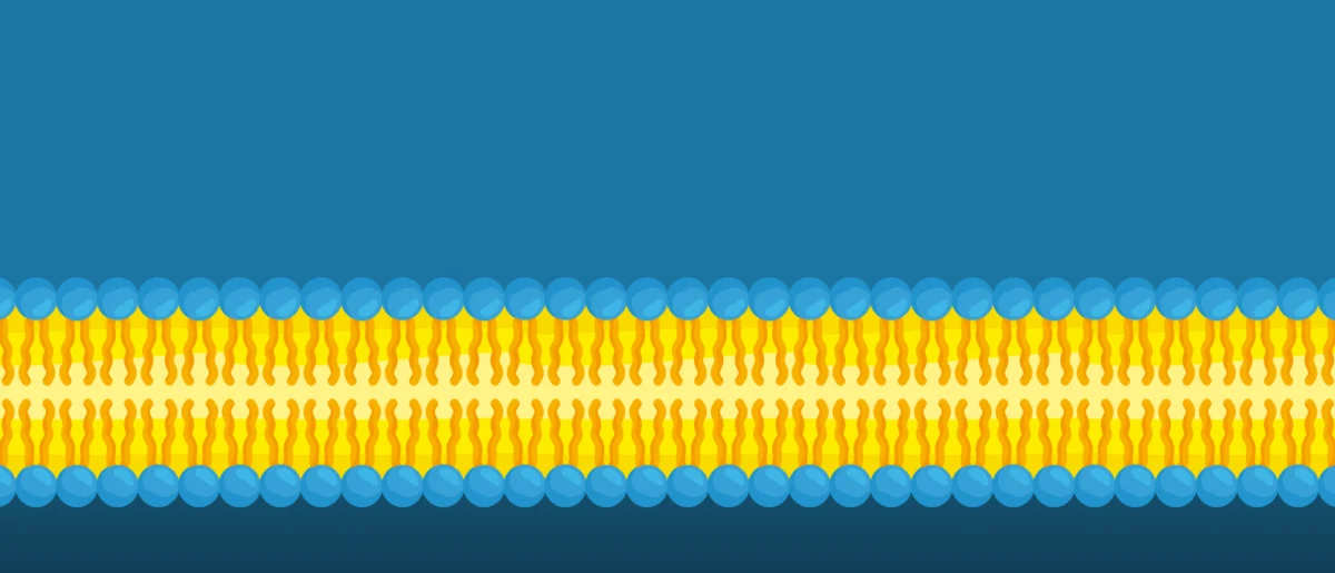
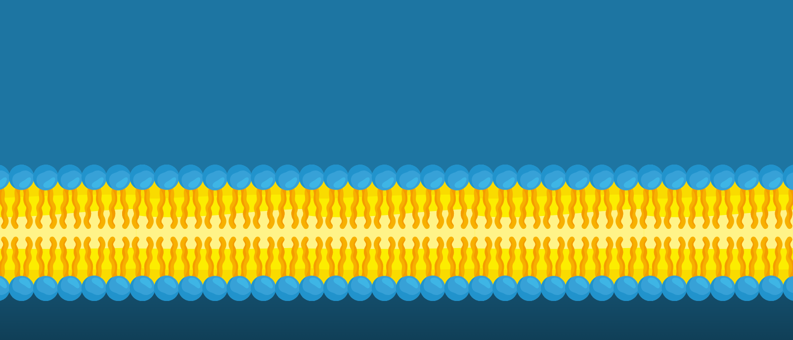
Cellular membranes, the dynamic boundaries that define cells and their internal organelles, are far from simple barriers. Often described by the "fluid mosaic model," they are complex, highly organized structures composed primarily of a diverse array of lipids and proteins. These components are not randomly distributed but rather coalesce into specialized, tiny regions known as nanodomains. These nanodomains, sometimes referred to as "lipid rafts," are believed to serve as critical hubs for a multitude of cellular functions, including signal transduction, material sorting, receptor clustering, and the transport of molecules across the membrane.
While the protein components within these specialized regions have been extensively studied and are relatively well-understood, the organization and behavior of lipids themselves have remained largely shrouded in mystery. This knowledge gap stems from several inherent difficulties. Lipids are incredibly dynamic, undergoing rapid lateral diffusion, rotation, and even "flip-flop" movements between membrane layers. Furthermore, their minute size makes individual lipid species exceptionally challenging to visualize with the resolution required to discern their precise distribution within nanoscale domains. Existing methods have often fallen short, either lacking the necessary spatial resolution, perturbing the delicate membrane structure, or being unable to distinguish between the vast array of different lipid types. The ability to precisely map where specific lipids reside and how they behave within these nanodomains is fundamental to understanding their roles in maintaining cellular health and contributing to disease pathogenesis.
Overcoming Methodological Hurdles: The Genesis of Lipid-CLEM
For decades, scientists have grappled with the limitations of available microscopy techniques when attempting to peer into the nanoscale world of lipids. Traditional light microscopy, while excellent for observing living cells and larger structures, simply lacks the resolving power to visualize individual lipid molecules or the fine architecture of nanodomains. Electron microscopy (EM), on the other hand, provides exquisite structural detail at the nanoscale but traditionally struggles with the specific identification of molecular species without the use of heavy metal labels that can sometimes distort the very structures they aim to reveal.
The concept of Correlative Light and Electron Microscopy (CLEM) emerged as a powerful solution to bridge this gap, combining the molecular specificity of fluorescence microscopy (a form of light microscopy) with the ultra-high resolution of electron microscopy. In CLEM, a specific molecule or structure is first identified and localized using fluorescent tags under a light microscope. Subsequently, the same region of the sample is imaged with an electron microscope to reveal its detailed ultrastructure. The two sets of images are then precisely correlated, offering a comprehensive view that combines both molecular identity and structural context.
However, even advanced CLEM methods presented their own set of challenges when applied to the notoriously difficult task of lipid visualization. Previous iterations often involved sample preparation techniques that could inadvertently damage the fragile membrane structures, making accurate observation problematic. Some methods were limited to studying the outer surface of the cell, failing to provide access to the intricate internal membrane systems of organelles. Crucially, distinguishing between individual lipid species remained a significant hurdle, as generic lipid labels often failed to provide the necessary specificity.
It was against this backdrop of persistent methodological limitations that Mathilda Lennartz, a co-corresponding and lead author of the study, alongside a dedicated team of researchers in the group of André Nadler at MPI-CBG and the group of Ori Avinoam at the Weizmann Institute of Science, embarked on developing a more refined and effective approach. Their collective efforts culminated in the development of Lipid-CLEM, a technique meticulously designed to overcome these historical barriers.
The Mechanics of Lipid-CLEM: A Multi-Stage Molecular GPS System
The ingenuity of Lipid-CLEM lies in its sophisticated, multi-stage approach, which effectively acts as a molecular GPS system for lipids within living cells. The core components of this technique are:
-
Bifunctional Lipid Probes: These are not just any labels; they are carefully engineered, slightly modified lipid molecules that can seamlessly integrate into the cell’s natural membranes. The "bifunctional" aspect refers to their dual purpose: one part of the probe contains a photo-activatable group, and another part carries a chemical handle for subsequent labeling. By mimicking natural lipids, these probes minimize perturbation to the cell’s physiology, ensuring that the observed lipid behavior is as close to natural as possible.
-
Photo-Crosslinking: Once the bifunctional lipid probes are incorporated into the membranes of living cells, the next critical step is to "freeze them in place." This is achieved through photo-crosslinking, where specific wavelengths of light activate the photo-activatable group on the probes. This causes the probes to form covalent bonds with adjacent molecules, essentially locking them into their positions at a specific moment in time. This rapid immobilization is crucial for capturing a snapshot of the dynamic lipid distribution, preventing their fast movements from blurring the image.
-
Click Chemistry: After the lipids are frozen in place and the cells are prepared for imaging, a highly specific and efficient chemical reaction known as "click chemistry" is employed. This bio-orthogonal reaction allows researchers to attach a fluorescent tag to the chemical handle on the now-immobilized lipid probes. The beauty of click chemistry is its high specificity and efficiency, occurring without interfering with other biological processes in the sample. This step enables the precise fluorescent labeling of the specific lipid species of interest.
-
Correlative Light and Electron Microscopy (CLEM): With the specific lipids fluorescently labeled and fixed, the samples are then subjected to the correlative imaging process. Light microscopy is used to quickly identify the regions where the labeled lipids are concentrated. Subsequently, the exact same regions are imaged using electron microscopy to reveal the ultrastructural details of the membrane nanodomains. The precise alignment of these two image sets provides an unparalleled view, showing where specific lipids are located at the molecular level within the high-resolution structural context of the membrane.
Mathilda Lennartz elaborated on the methodological advancements, particularly regarding sample preparation for internal cellular structures: "To study lipid sorting in early endosomes — a key sorting station inside the cell — cells must be rapidly frozen to stop lipids in their tracks and to preserve the membrane of the cells. Later, these lipids can be labeled on very thin slices of the sample, termed ‘sections’, of cells using click chemistry. These sections are what we then image using the Lipid-CLEM approach." This rapid cryo-fixation technique is vital for preserving the delicate and dynamic architecture of cellular membranes, ensuring that the captured images faithfully represent the cell’s native state.
First Insights: Sphingomyelin and the Divergence of Sorting Pathways
The immediate application of Lipid-CLEM has already yielded fascinating new insights into cellular processes. The research team focused their initial investigations on early endosomes, which are critical intracellular sorting stations responsible for processing internalized cargo and membrane components. By applying Lipid-CLEM, they were able to precisely map the distribution of a specific lipid called sphingomyelin within these endosomes.
Mathilda Lennartz shared a key finding: "With Lipid-CLEM, we observed that a specific lipid called sphingomyelin is more common in small vesicles inside the endosome and less common in tubular membrane domains. This separation has already been observed for some proteins." The significance of this observation is profound. It demonstrates unequivocally that lipids, much like proteins, are actively sorted and organized within different subdomains of cellular organelles.
Even more strikingly, Lennartz highlighted a critical divergence: "What we concluded from this is that at least some lipids, just like proteins, must also be sorted in the endosome. Interestingly, in our study, sphingomyelin and a protein cargo arrive at the same time in the early endosome but separate into different domains, indicating that lipid and protein trafficking routes can diverge during this sorting." This finding challenges a purely protein-centric view of membrane trafficking and organization, suggesting that lipids play a more active and independent role in directing cellular processes than previously understood. It implies a complex interplay, where lipids can act as independent sorting signals, influencing the fate of membrane components and potentially influencing downstream signaling events.
The Power of International and Interdisciplinary Collaboration
The successful development and application of Lipid-CLEM stand as a testament to the indispensable role of interdisciplinary and international collaboration in modern scientific discovery. The expertise required to develop such a sophisticated technique spans multiple fields, from synthetic chemistry for probe design to advanced microscopy and cell biology.
Ori Avinoam, whose team at the Weizmann Institute brought their deep expertise in correlative light and electron microscopy to the project, emphasized this synergy: "This study highlights how essential collaborations are for driving research forward. Bringing together complementary expertise allowed us to establish a method that made it possible to uncover fundamental principles of lipid sorting that were previously inaccessible." The MPI-CBG group, led by André Nadler, likely contributed significant expertise in lipid biology, membrane dynamics, and the development of the bifunctional probes, complementing the microscopy prowess of the Israeli team. This seamless integration of diverse scientific backgrounds was crucial in overcoming the complex technical hurdles associated with visualizing lipids at such high resolution.
Broader Impact and Future Horizons
The advent of Lipid-CLEM is poised to have a transformative impact across numerous fields of biological and biomedical research. As André Nadler, corresponding author, concluded: "Our Lipid-CLEM workflow enables 3D visualization of lipid densities in membrane nanodomains, offering a new way to study lipid organization in complex cellular structures. We finally can look at lipid sorting in membranes with the resolution we need. We believe that our new method Lipid-CLEM will help us to better understand how lipids work in cells, as it allows us to study both lipids and proteins together during membrane organization and function. This may also contribute to a better understanding of membrane dysfunction-related diseases."
The implications are far-reaching:
- Fundamental Cell Biology: This technique will undoubtedly deepen our understanding of basic cellular processes. Researchers can now explore the precise roles of different lipid species in membrane biogenesis, organelle identity, cell-cell communication, and the formation and function of various membrane domains beyond endosomes, such as the Golgi apparatus, endoplasmic reticulum, and the plasma membrane.
- Disease Mechanisms: Membrane dysfunction is implicated in a wide array of human diseases. Lipid-CLEM can provide crucial insights into the pathogenesis of:
- Neurodegenerative Disorders: Many conditions like Alzheimer’s, Parkinson’s, and Huntington’s diseases involve altered lipid metabolism and membrane trafficking, impacting neuronal health and function.
- Metabolic Diseases: Disorders such as diabetes, obesity, and cardiovascular diseases are intrinsically linked to lipid metabolism and membrane integrity.
- Infectious Diseases: Viruses and bacteria often exploit specific lipid nanodomains on host cell membranes for entry, replication, and egress. Understanding lipid organization could pave the way for novel antiviral and antibacterial strategies.
- Cancer: Cancer cells often exhibit altered lipid metabolism and membrane composition, which contributes to their uncontrolled growth, metastasis, and drug resistance.
- Drug Discovery and Development: A more precise understanding of lipid organization in cellular membranes could revolutionize drug design. Many drugs target membrane proteins, and their efficacy can be influenced by the surrounding lipid environment. Furthermore, lipid-based nanoparticles are increasingly used as drug delivery vehicles, including the highly successful mRNA vaccines. Lipid-CLEM could help in optimizing the design and targeting of these delivery systems by allowing researchers to visualize their interaction with cellular membranes at an unprecedented resolution.
- Advanced Imaging Capabilities: The potential for 3D visualization of lipid densities within membrane nanodomains opens up entirely new avenues for quantitative analysis, enabling researchers to build comprehensive spatial maps of lipid organization in complex cellular structures.
In essence, Lipid-CLEM represents a monumental leap forward in our ability to probe one of the cell’s most fundamental yet elusive components. By providing a high-resolution window into the dynamic and complex world of membrane lipids, this technique is set to catalyze a wave of new discoveries, fundamentally reshaping our understanding of cellular function and providing new targets and strategies for addressing a myriad of human diseases. The collaborative spirit that forged this innovation promises continued advancements as scientists globally begin to apply Lipid-CLEM to answer long-standing questions in cell biology.


Leave a Reply