
A landmark study led by the Wellcome Sanger Institute has redefined the scientific understanding of chronic myeloid leukemia (CML), uncovering that the cancer cells responsible for the disease proliferate at explosive rates years before a clinical diagnosis is ever made. Published in the journal Nature, the research utilizes advanced whole-genome sequencing to map the evolutionary trajectory of CML, revealing that the genetic spark for the disease typically occurs between three and 14 years prior to detection. Perhaps most striking is the discovery that these cancerous clones can grow by more than 100,000 percent annually, a rate of expansion that far outstrips most other forms of cancer, which generally accumulate mutations over several decades. By tracing the "family trees" of individual blood cells, the international research team has provided a new window into the life cycle of leukemia, offering potential explanations for why some patients fail to respond to standard therapies.
The Genetic Catalyst: Understanding the Philadelphia Chromosome
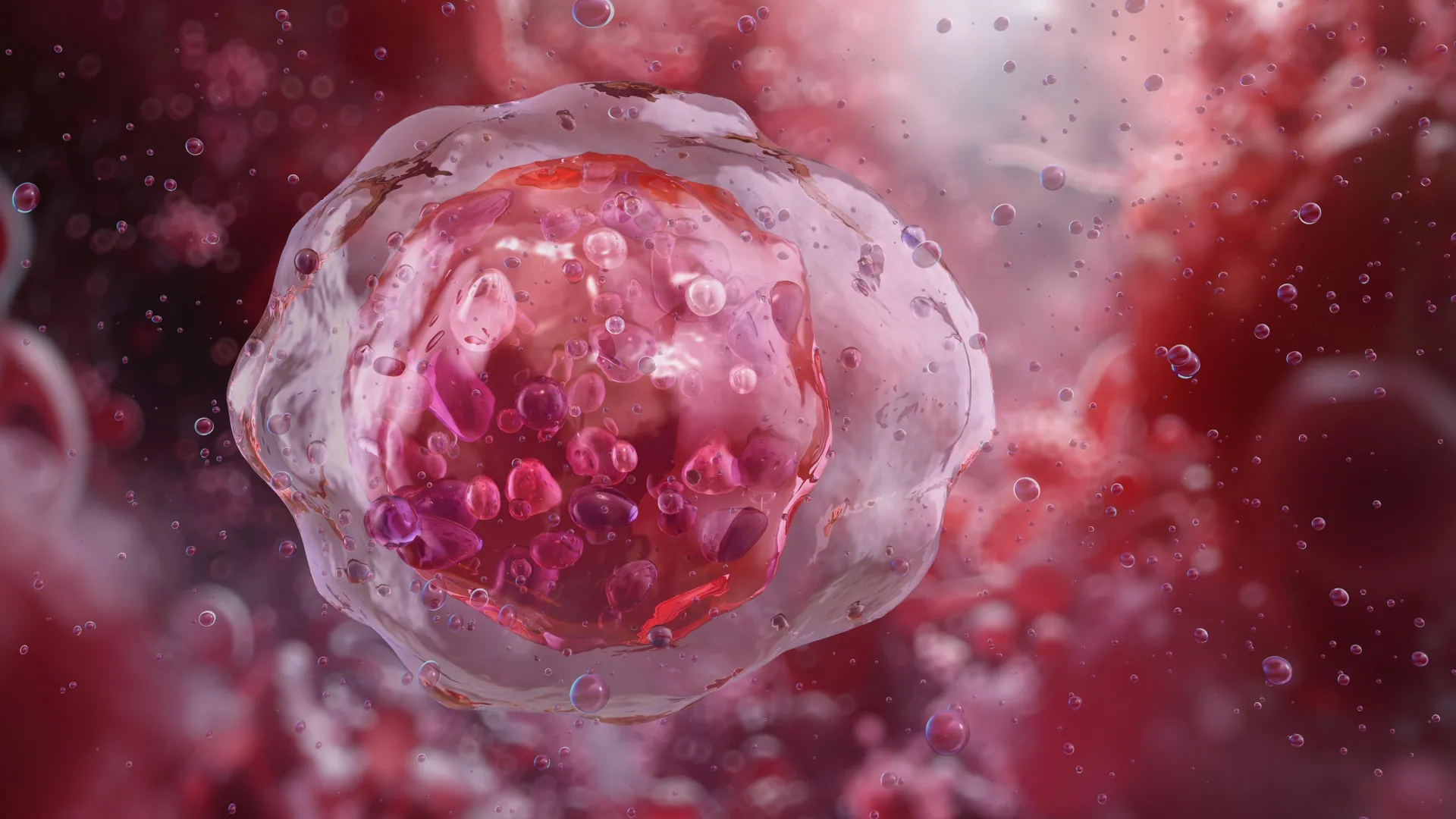
Chronic myeloid leukemia is a primary cancer of the bone marrow and blood, characterized by the overproduction of white blood cells. At the heart of nearly every CML case is a specific genetic abnormality known as the Philadelphia chromosome. This occurs when a piece of chromosome 9, containing the ABL1 gene, breaks off and attaches to the BCR gene on chromosome 22. The resulting fusion, the BCR::ABL1 gene, acts as a "permanently on" switch for cell division.
While the existence of this fusion gene has been known for decades—marking a turning point in oncology that led to the development of targeted therapies—the actual "natural history" of the gene’s evolution remained elusive until now. Scientists previously struggled to determine exactly when the fusion event occurred in a patient’s life and how the population of cells behaved in the intervening years before the patient felt ill. The Sanger Institute study addressed this by analyzing over 1,000 whole genomes of single blood cells from nine patients, ranging in age from 22 to 81. This high-resolution approach allowed researchers to look past the point of diagnosis and reconstruct the ancestral history of the cancer.
A Timeline of Malignancy: From First Mutation to Diagnosis
The research team employed phylogenetic analysis, a method typically used by evolutionary biologists to study the relationships between species, to create "family trees" of blood cells. By identifying shared somatic mutations—tiny, harmless genetic changes that accumulate naturally over a lifetime—the researchers could determine which cells were related and when the BCR::ABL1 fusion first appeared in a common ancestor cell.
The findings indicate a remarkably consistent timeline: the initial genetic fusion event occurred between three and 14 years before the patients were diagnosed with CML. This "latency period" is significantly shorter than that observed in many solid tumors or other blood cancers, such as certain myeloproliferative neoplasms, which can simmer in the body for 30 to 40 years before manifesting as disease.
Furthermore, the study highlighted that CML is unique because it is often driven by this single, powerful genetic event. While most cancers require a "multi-hit" process—the accumulation of five to ten distinct mutations that gradually erode cellular safeguards—the BCR::ABL1 fusion is so potent that it can drive a healthy cell toward malignancy almost entirely on its own.
Explosive Growth Rates and Age-Related Variation
Once the BCR::ABL1 fusion gene is formed, the resulting "clone" of cancer cells begins to multiply with unprecedented speed. The study recorded growth rates exceeding 100,000 percent per year. This means that a single mutated cell can expand into billions of leukemia cells in a relatively short timeframe. This rapid expansion explains why CML can appear to emerge suddenly in patients who had normal blood counts just a year or two prior.
However, the data also revealed significant variation in growth rates among different patients, with age playing a critical role. Younger patients in the study exhibited substantially higher rates of cell multiplication compared to older patients. Specifically, the cancerous clones in younger individuals tended to expand more aggressively, while the growth in older patients, though still rapid by oncological standards, was comparatively slower.
Scientists speculate that this could be due to the different environments of the bone marrow in younger versus older individuals. In younger people, the bone marrow is generally more "active," potentially providing a more fertile ground for the BCR::ABL1 gene to exert its influence. Conversely, the "crowded" or more exhausted marrow environment in older age might naturally constrain the speed at which even a potent cancer clone can expand.
Clinical Implications and Treatment Resistance
The discovery of varying growth rates has immediate implications for the clinical management of CML. Since the early 2000s, the standard of care for CML has been Tyrosine Kinase Inhibitors (TKIs), such as Imatinib (Gleevec). These drugs specifically target the protein produced by the BCR::ABL1 gene, effectively turning off the signal for cell growth. While TKIs have transformed CML from a terminal illness into a manageable chronic condition for many, approximately 20 percent of patients do not respond well to the treatment or eventually develop resistance.
The Sanger Institute study found a direct correlation between the speed of cancer growth and treatment efficacy. Patients whose leukemia cells showed the fastest pre-diagnosis growth rates were significantly less likely to achieve a deep molecular response to TKI therapy. This suggests that the inherent "fitness" or aggressiveness of the cancer clone, established years before the first dose of medicine is taken, may dictate the ultimate success of the treatment.
Dr. Aleksandra Kamizela, a co-first author of the study and resident doctor at Lister Hospital, emphasized the gap between current diagnostic tools and these new findings. "In a clinical setting, healthcare professionals will perform a reverse transcription polymerase chain reaction (RT-PCR) test to measure a patient’s response to CML treatment," Kamizela noted. "However, they are not able to routinely see differences in the genetic cause of CML in patients at the DNA level, which we have been able to highlight in our study."
Analysis of the "All of Us" Cohort
To determine if the BCR::ABL1 fusion could exist in the general population without causing disease, the researchers turned to the "All of Us" Research Program, a massive health database in the United States. They screened the genomic data and health records of over 200,000 participants for the presence of the BCR::ABL1 fusion.
The results were telling: the fusion gene was extremely rare in the general population. Furthermore, in the few instances where it was detected in individuals not yet diagnosed with CML, those individuals almost invariably went on to develop a blood disorder shortly thereafter. This confirms that the BCR::ABL1 fusion is not a benign "passenger" mutation; rather, its presence in a blood cell is a near-certain harbinger of impending leukemia. This distinguishes CML from other conditions like "Clonal Hematopoiesis of Indeterminate Potential" (CHIP), where people can carry mutated blood cells for decades without ever developing cancer.
Expert Reactions and Future Directions
The study has been hailed by the hematology community as a breakthrough in understanding the kinetics of cancer. Dr. Jyoti Nangalia, senior author of the study and a hematologist at the University of Cambridge, noted that CML stands out as a biological "outlier."
"What our study suggests is that chronic myeloid leukemia is an outlier compared to other cancers—both solid tumors and other blood cancers," Dr. Nangalia said. "We have shown that chronic myeloid leukemia cells undergo incredibly rapid growth within a few years to a decade before diagnosis, whereas for most cancers, the timeline from start to clinical presentation is several decades."
The ability to look back in time using a patient’s own cells as a "molecular clock" opens new avenues for precision medicine. If clinicians could eventually estimate the growth rate of a patient’s cancer at the time of diagnosis, they might be able to tailor the intensity of the treatment or choose specific drug combinations more effectively.
Conclusion: A New Paradigm for Leukemia Research
The Wellcome Sanger Institute’s research provides a comprehensive narrative of Chronic Myeloid Leukemia that was previously invisible to science. By proving that the disease is the result of an explosive, short-term expansion driven by a single genetic event, the study shifts the focus from the moment of diagnosis to the years of silent growth that precede it.
While further research with larger patient cohorts is required to validate the use of growth-rate metrics in daily clinical practice, the foundation has been laid for a more nuanced approach to blood cancer. The work not only clarifies the unique potency of the BCR::ABL1 fusion gene but also provides a roadmap for optimizing treatment for the one-fifth of patients who currently face poor outcomes. As genomic technology becomes more accessible, the transition from observing these cellular family trees in a lab to using them in a hospital may be the next great leap in the fight against leukemia.


Leave a Reply