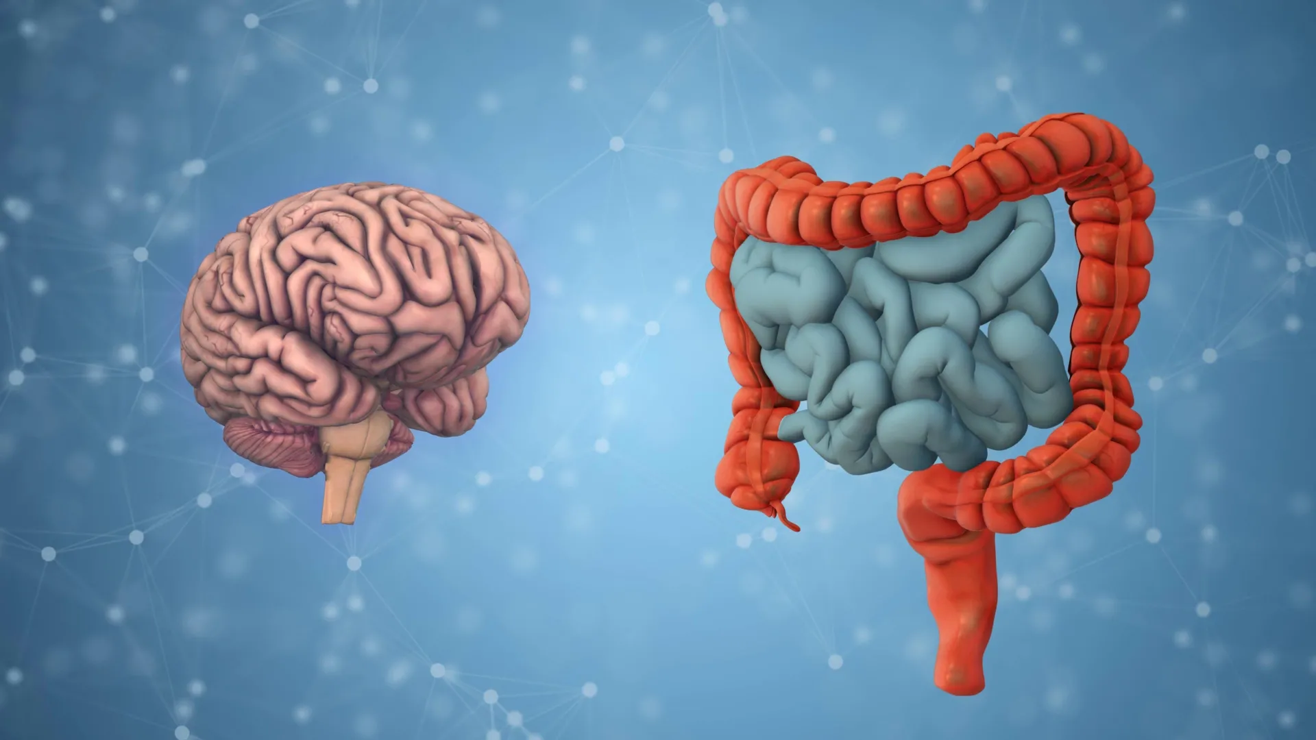
Recent neurobiological research from the Monell Chemical Senses Center has unveiled a critical distinction in how the human brain processes two of the most common sugars in the modern diet: fructose and glucose. While these two simple sugars are chemically similar and provide an identical caloric load of four calories per gram, the study suggests that the body’s internal signaling systems do not treat them as equals. The findings, published in the peer-reviewed journal Neuron, demonstrate that fructose and glucose communicate with the brain through entirely separate gut-brain pathways, a discovery that carries profound implications for understanding dietary preferences, the obesity epidemic, and the neurological basis of appetite regulation.
For decades, the prevailing wisdom in nutrition science was built upon the "isocaloric" principle—the idea that the source of a calorie matters less than the total energy provided. However, the Monell team, led by senior author Amber Alhadeff, PhD, has provided evidence that challenges this assumption. By identifying a specific signaling route that allows fructose to talk to the brain, the researchers have shown that the brain’s "hunger center" responds with significantly less intensity to fructose than it does to glucose. This disparity in neural suppression may explain why diets high in certain sweeteners fail to provide the same level of satiety as more traditional sugar sources, leading to overconsumption and metabolic imbalance.
The Biological Mechanisms of Sugar Sensing
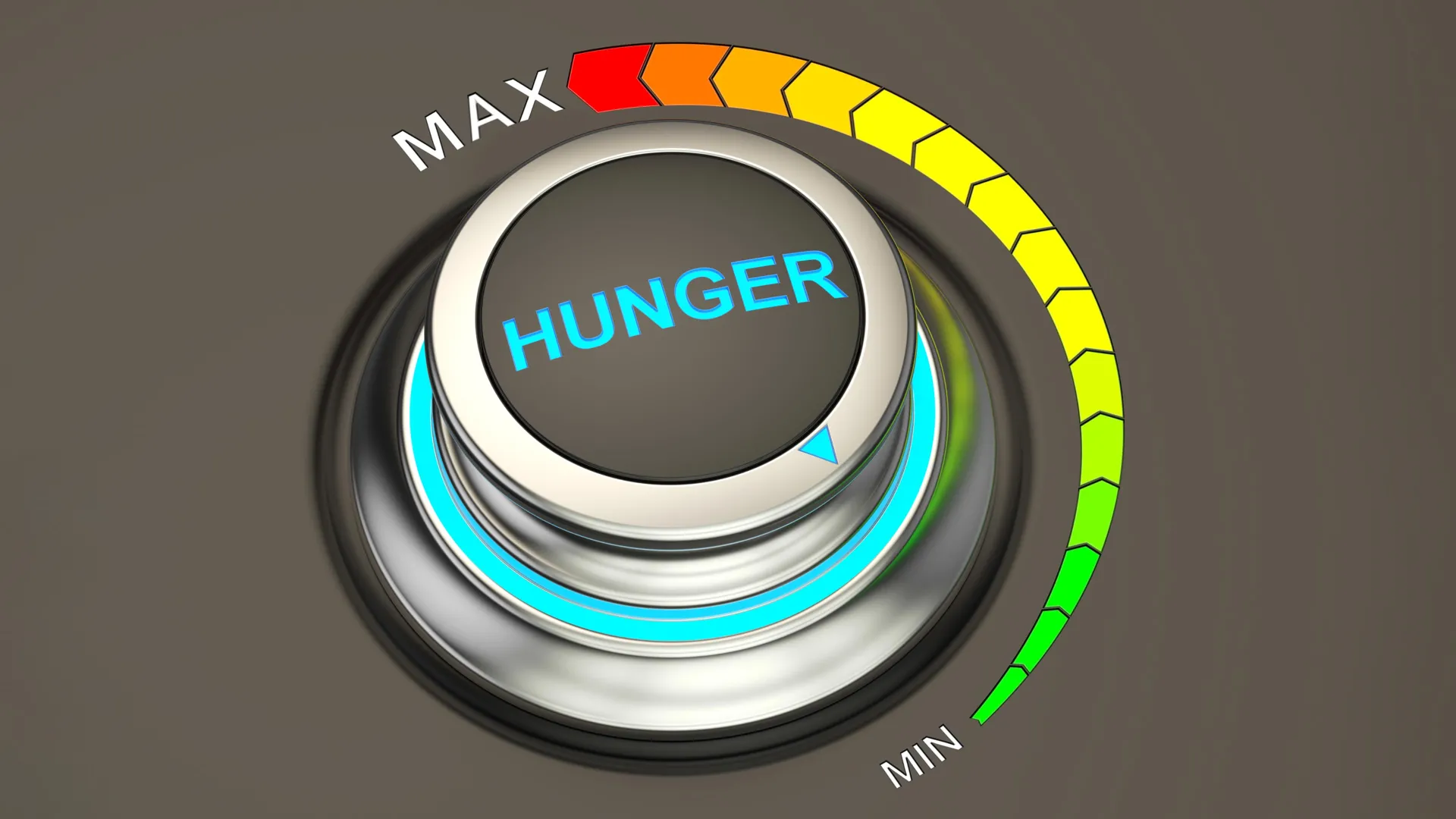
To understand why the brain distinguishes between these sugars, it is necessary to examine the specific cellular pathways involved in nutrient sensing. The Monell study focused on the agouti-related protein (AgRP) neurons, located in the hypothalamus. These neurons are often referred to as the "hunger neurons" because their activity drives the search for food; when AgRP neurons are active, an organism feels hungry, and when their activity is suppressed, the sensation of hunger diminishes.
In controlled laboratory experiments involving murine models, the researchers observed that the ingestion of glucose led to a rapid and robust suppression of AgRP neuron activity. This response was not dependent on the specific hormone pathway used by fructose. Instead, glucose appears to utilize a more direct or high-potency signaling mechanism that effectively tells the brain that energy has been consumed, thereby shutting off the hunger signal.
Conversely, the pathway for fructose was found to be far more circuitous and less effective. The researchers discovered that when fructose enters the gut, it triggers the release of a hormone called peptide YY (PYY). This hormone then sends a signal through the vagus nerve—the primary "information highway" between the gut and the brain. While this PYY-vagus nerve pathway does eventually reach the AgRP neurons, the resulting reduction in hunger-related activity is modest at best. In essence, while glucose acts as a loud "stop eating" signal, fructose provides only a faint whisper, leaving the hunger neurons relatively active despite the intake of calories.
The Role of High-Fructose Corn Syrup in Modern Diets
One of the most significant aspects of the study involved the analysis of High-Fructose Corn Syrup (HFCS), a sweetener that has become a staple of the global food supply since its introduction in the 1970s. HFCS is typically a blend of both fructose and glucose, designed to mimic the sweetness of sucrose (table sugar) but at a lower production cost.
The researchers found that mice showed a marked preference for HFCS over pure fructose. More importantly, the combination of the two sugars in HFCS suppressed AgRP neuron activity more strongly than fructose alone. This suggests a synergistic effect where the presence of glucose "primes" the brain to respond, while the fructose adds to the overall palatability and caloric density. Dr. Alhadeff noted that this enhanced effect on hunger-related neurons might explain the unique appeal of HFCS-sweetened products. Because these products provide a complex signal to the brain—combining a partial satiety signal with a high reward value—they may be specifically engineered by biological evolution (and industrial food science) to be more difficult to stop consuming.
Chronology of Gut-Brain Research and the Monell Study
The discovery of these separate pathways is the culmination of years of research into the "gut-brain axis," a field that has expanded rapidly over the last decade.
- Early 2000s: Research focused primarily on the hormonal signals like leptin and ghrelin, which regulate long-term energy balance.
- 2010-2015: Advances in optogenetics and in vivo imaging allowed scientists to observe individual neurons in the hypothalamus in real-time as animals consumed different nutrients.
- 2018-2022: Studies began to suggest that the gut possesses "neuropod cells" that can distinguish between nutritive and non-nutritive sweeteners almost instantly.
- June 10, 2024: The Monell Chemical Senses Center publishes the Neuron study, providing the first definitive map of how the brain differentiates between the two primary simple sugars, fructose and glucose, through distinct vagal and hormonal routes.
This timeline illustrates a shift from viewing the gut as a simple organ of digestion to recognizing it as a sophisticated sensory organ capable of biochemical analysis.
Supporting Data and Experimental Evidence
The data supporting the Monell findings were derived from sophisticated neural recording techniques. In the study, researchers utilized fiber photometry to monitor the calcium dynamics of AgRP neurons, which serves as a proxy for neural activity.
When mice were given a 10% glucose solution, AgRP activity dropped by approximately 60-70% within minutes of ingestion. In contrast, a 10% fructose solution resulted in a drop of only 25-30%. When the researchers genetically or chemically disrupted the PYY receptors or the vagus nerve, the fructose-induced suppression was nearly eliminated, while the glucose-induced suppression remained largely intact. This provided the "smoking gun" evidence that glucose does not rely on the PYY-vagus pathway to the same extent that fructose does.
Furthermore, the behavioral data indicated that over time, the mice learned to associate the "feeling" of glucose with a greater reduction in hunger. Even when the tastes were masked or matched, the mice eventually gravitated toward the sugar that provided the most significant neural "reward" in terms of hunger suppression.
Broader Implications for Public Health and Nutrition
The implications of this research extend far beyond the laboratory. As global rates of obesity and Type 2 diabetes continue to rise, understanding the neurological drivers of sugar consumption is a public health priority.
- The "Empty Calorie" Debate: While fructose is not "empty" of energy, it may be "empty" of satiety. If the brain does not register the consumption of fructose as effectively as glucose, individuals consuming high-fructose beverages may consume significantly more calories before feeling full.
- Refining Dietary Guidelines: Current dietary guidelines often group all "added sugars" into a single category. This research suggests that the specific ratio of fructose to glucose in those sugars could be a major factor in their metabolic impact.
- Targeting the Vagus Nerve: The identification of the PYY-vagus nerve pathway opens new doors for pharmacological interventions. If scientists can find ways to enhance the PYY signal or the brain’s sensitivity to it, they might be able to create treatments that increase satiety for individuals on high-fructose diets.
- Food Industry Transparency: There may be increased pressure on food manufacturers to disclose the specific fructose-to-glucose ratios in their products, as the biological response to a 50/50 blend may differ significantly from a 90/10 blend.
Analysis of Nutrient Sensing Complexity
The study highlights the extreme complexity of the human body’s nutrient-sensing apparatus. It suggests that the body has evolved multiple, redundant systems to ensure that it can identify and capitalize on various energy sources. Glucose, being the primary fuel for the brain and muscles, has a high-priority signaling route. Fructose, which is primarily metabolized in the liver and was historically less common in the human diet (found mostly in fruit), appears to have a secondary, less urgent signaling route.
In the context of the modern food environment, where fructose is ubiquitous in the form of refined sweeteners, this evolutionary mismatch becomes a liability. The "modest" reduction in hunger neurons caused by fructose is simply not enough to counteract the high-calorie environment of the 21st century.
Official Responses and Collaborative Support
The research was a multi-institutional effort, reflecting the interdisciplinary nature of modern neuroscience. Dr. Amber Alhadeff emphasized that this work is a critical step in understanding the "neural systems involved in appetite." The study received support from several prestigious organizations, including the National Institutes of Health (NIH), the American Heart Association, and the New York Stem Cell Foundation.
Representatives from the nutrition science community have noted that the study provides a biological framework for what many clinicians have observed in practice: that patients often find it much harder to regulate their intake of sugary sodas and processed snacks compared to whole foods. The Monell Chemical Senses Center, an independent non-profit research institute, continues to lead the way in investigating how the senses of taste and smell influence human health.
As researchers look toward the future, the next step will be to determine if these same pathways are present and dominant in humans. While murine models are highly predictive of human metabolic pathways, clinical trials will be necessary to confirm that the PYY-vagus nerve route is the primary regulator of fructose satiety in the human brain. If confirmed, this will represent a landmark shift in our understanding of the relationship between what we eat and how our brains decide when we have had enough.