

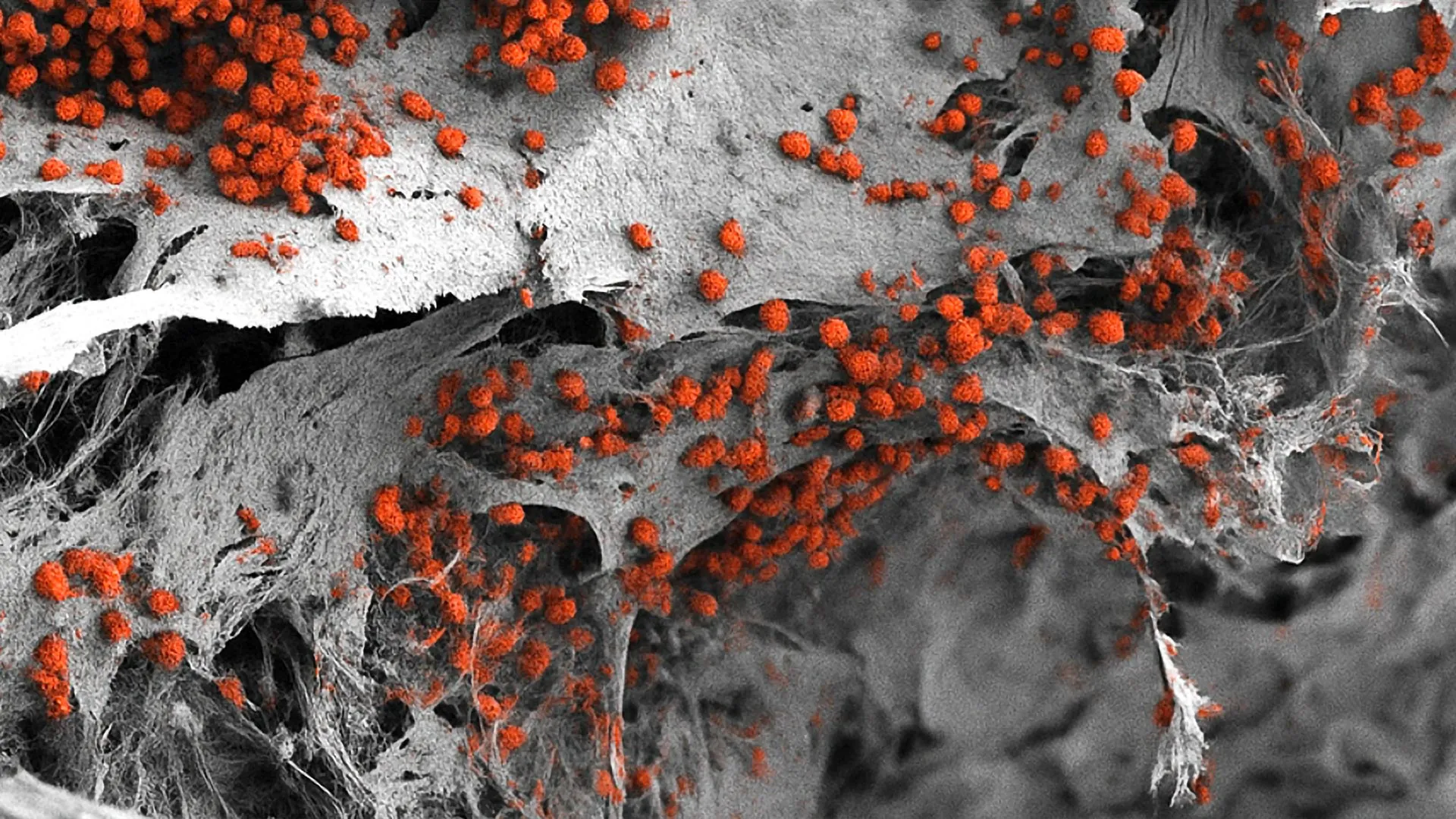
In a landmark achievement for regenerative medicine and hematology, researchers from the University of Basel and University Hospital Basel have successfully engineered a fully functional, three-dimensional model of human bone marrow using exclusively human cells. This breakthrough, recently detailed in the prestigious journal Cell Stem Cell, represents the first time the intricate architecture of the human "blood factory" has been replicated with such biological fidelity in a laboratory setting. By recreating the complex network of bone cells, nerves, and blood vessels that characterize the bone marrow environment, the research team has provided the scientific community with a powerful new tool to study blood-related diseases, accelerate drug development, and significantly reduce the reliance on animal testing.
The Biological Complexity of the Bone Marrow Environment
Bone marrow is one of the most complex and vital tissues in the human body. Located within the cavities of bones, it serves as the primary site for hematopoiesis—the continuous process of blood cell production. On average, a healthy adult produces hundreds of billions of new blood cells every day, including oxygen-carrying red blood cells, infection-fighting white blood cells, and clot-forming platelets. This massive logistical undertaking is managed by a specialized microenvironment known as the hematopoietic niche.
Within this niche, various cell types interact in a highly coordinated fashion. Bone-forming cells (osteoblasts), blood vessel cells (endothelial cells), and nerve fibers create a protective and regulatory "home" for hematopoietic stem cells. These stem cells are the precursors to all blood types, and their behavior—whether they remain dormant, self-renew, or differentiate into specific blood lineages—is governed by the physical and chemical signals they receive from their surroundings.
Historically, studying these interactions has proven exceptionally difficult. Because bone marrow is encased in hard mineralized bone, it is inaccessible for real-time observation in living humans. For decades, scientists have relied on two main alternatives: two-dimensional (2D) cell cultures and animal models, primarily mice. While 2D cultures are easy to manage, they fail to capture the 3D structural complexity and mechanical stresses of real tissue. Conversely, while animal models provide a living system, they are not perfectly analogous to human biology. Significant differences in immune signaling and genetic expression between mice and humans often lead to "translational failure," where drugs that appear successful in mice fail to show efficacy or safety in human clinical trials.
Methodology: Engineering a Human Endosteal Niche
The research team, led by Professor Ivan Martin and Dr. Andrés García García at the Department of Biomedicine, focused their efforts on replicating a specific area of the marrow known as the endosteal niche. This region, located near the inner surface of the bone, is of particular interest to oncologists. It is widely believed that certain blood cancers, such as leukemia, utilize the endosteal niche as a sanctuary, where cancer cells can remain dormant and escape the effects of chemotherapy, leading to eventual relapse.
To build this model, the researchers adopted a multi-step bioengineering approach. The process began with the creation of a three-dimensional scaffold made of hydroxyapatite. Hydroxyapatite is a naturally occurring mineral form of calcium apatite that constitutes the bulk of human bone and teeth. By using this material, the team provided a chemically and physically accurate "skeleton" for the cells to inhabit.
The most innovative aspect of the study involved the use of human induced pluripotent stem cells (iPSCs). These are adult cells that have been genetically "reprogrammed" back into an embryonic-like state, giving them the ability to transform into any cell type in the body. By applying specific growth factors and environmental cues, the Basel team guided these pluripotent cells to differentiate into the various components of the bone marrow, including the stromal cells, bone-forming cells, and vascular precursors.
Chronology of the Research Development
The development of this model was the culmination of several years of interdisciplinary collaboration. The timeline of the project highlights the rigorous validation required to move from theoretical design to a functional biological system:
- Phase I: Scaffold Optimization: The team initially experimented with various synthetic polymers but ultimately selected hydroxyapatite for its superior biocompatibility and its ability to mimic the mineral density of human trabecular bone.
- Phase II: Stem Cell Programming: Researchers refined the protocols for differentiating iPSCs. This was a critical hurdle, as the model required multiple cell lineages to develop simultaneously within the same scaffold to ensure they could form a cohesive tissue network.
- Phase III: System Integration: Once the individual cell types were successfully integrated into the 3D scaffold, the team monitored the development of the "niche." They observed the formation of rudimentary blood vessel structures and the establishment of neural signaling pathways within the engineered tissue.
- Phase IV: Functional Testing: In the final stage of the study, the researchers introduced human hematopoietic stem cells into the model. They were able to demonstrate that the engineered environment could sustain the production and maturation of blood cells for several weeks, proving that the system was not just a structural replica, but a functional biological unit.
Technical Specifications and Comparative Data
The Basel model is notable not only for its composition but also for its scale. Measuring approximately eight millimeters in diameter and four millimeters in thickness, it is significantly larger than previous "organ-on-a-chip" or microfluidic bone marrow models. This increased volume allows for a more realistic distribution of cells and a more accurate representation of the gradients of oxygen and nutrients found in natural bone marrow.
Data from the study indicates that the engineered niche successfully maintained the "stemness" of hematopoietic cells—meaning it kept a pool of stem cells in their undifferentiated state, which is a hallmark of healthy marrow function. In traditional 2D cultures, stem cells often differentiate too quickly, exhausting the supply and making long-term studies impossible. The Basel model’s ability to sustain this population for weeks provides a much longer window for observing disease progression and drug response.
Reducing Reliance on Animal Experimentation
One of the most significant implications of this research is its potential to advance the "3Rs" principle in science: the replacement, reduction, and refinement of animal testing. The University of Basel has been a vocal proponent of these ethical standards, and this project represents a tangible step toward those goals.
Professor Ivan Martin emphasized that while mouse models have provided foundational knowledge, they are limited by species-specific biological traits. "Our model brings us closer to the biology of the human organism," Martin stated. By using human cells to study human diseases, researchers can bypass many of the ethical and scientific complications associated with animal research. Furthermore, because the model is derived from human stem cells, it can be standardized in a way that animal populations—which can vary significantly between strains—cannot.
Broader Impact on Medicine and Drug Development
The potential applications for this technology are vast, ranging from basic science to clinical diagnostics:
1. Blood Cancer Research:
The model provides an unprecedented look at how leukemia and multiple myeloma cells interact with their environment. By simulating the endosteal niche, scientists can investigate how cancer cells "hide" in the bone marrow and test new drugs designed to "flush" these cells out into the bloodstream, where they can be targeted by conventional therapies.
2. High-Throughput Drug Testing:
Pharmaceutical companies can use these models to screen new drug candidates for toxicity and efficacy before they ever reach human trials. While Dr. Andrés García García noted that the current 8mm model might be too large for massive, automated screening, the team is already looking at ways to miniaturize the system. Smaller versions of the model could be used in "plates" containing hundreds of miniature bone marrow niches, allowing for the simultaneous testing of thousands of different chemical compounds.
3. Personalized Medicine:
Perhaps the most exciting future prospect is the creation of patient-specific models. In this scenario, a patient with a rare or treatment-resistant form of blood cancer would provide a small sample of their own cells. These cells would be used to create a "personalized bone marrow" in the lab. Doctors could then test various combinations of chemotherapy or immunotherapy on the patient’s own engineered tissue to see which treatment is most effective before administering it to the patient. This would eliminate the "trial and error" approach that often characterizes cancer treatment today.
Analysis of Challenges and Future Outlook
Despite the success of the study, several challenges remain before the model can be widely adopted in clinical settings. The current system, while complex, does not yet include every single component of the human body, such as a full immune system or a connection to other organs like the liver or kidneys, which play a role in drug metabolism.
Furthermore, the cost of producing iPSC-derived models remains high. Scaling the production of these niches so they are affordable for routine use in hospitals and research labs will require advancements in automated bio-manufacturing.
However, the scientific community has reacted with optimism to the Basel study. Industry experts suggest that this model could become the gold standard for "pre-clinical validation." By providing a more accurate bridge between laboratory research and human clinical trials, this technology has the potential to reduce the failure rate of new drugs, which currently stands at over 90% for oncology treatments entering Phase I trials.
In conclusion, the creation of a fully human, 3D bone marrow model is a transformative milestone. It represents a shift away from traditional, simplified research methods toward a more integrated, bio-accurate approach. As the team at the University of Basel continues to refine the system—improving its scalability and adding further layers of biological complexity—the medical community moves one step closer to a future where blood cancers are more effectively treated, animal testing is minimized, and personalized medicine becomes a reality for patients worldwide.

















