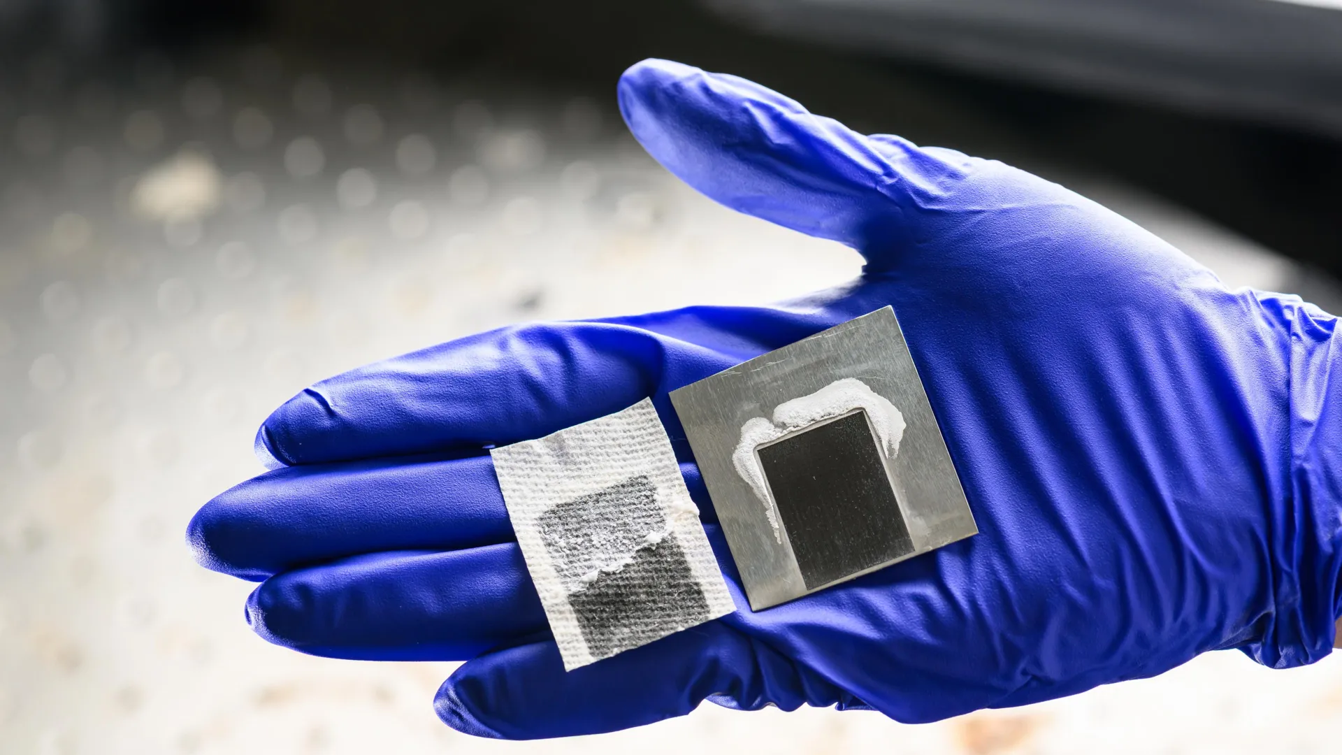
The landscape of regenerative medicine and pediatric reconstructive surgery may be on the verge of a profound paradigm shift. Researchers at the University of Sydney, working in close collaboration with the University of Queensland, have successfully engineered a novel biodegradable "nanobone" material designed to prompt the human body to heal its own skeletal defects. Published in the peer-reviewed journal ACS Nano, this breakthrough offers a prospective alternative to traditional bone grafting techniques—procedures that, despite their widespread application, have remained largely unchanged for more than half a century.
Traditional bone grafting typically requires surgeons to harvest healthy bone tissue from another site on a patient’s body or rely on animal-derived structural matrices. These conventional approaches are frequently invasive, painful, and prone to significant recovery complications. The newly developed calcium-aluminosilicate nanomaterial bypasses these hurdles by acting as a biological catalyst rather than a mere structural filler. By stimulating the body’s intrinsic healing mechanisms, the technology has demonstrated an unprecedented ability to accelerate bone regeneration in preclinical models, potentially paving the way for safer, faster, and much less invasive treatments for millions of patients worldwide.
Understanding the Scale of the Clinical Challenge
To fully appreciate the significance of this technological leap, one must examine the profound clinical challenges associated with severe bone defects, most notably cleft lip and palate. Affecting approximately one in every 700 children globally, cleft lip and palate occurs when the tissue of the upper lip or the roof of the mouth fails to fuse completely during fetal development. While soft-tissue surgeries can be performed in early infancy, repairing the associated gaps in the jawbone is notoriously complex.
Under current medical standards, children born with jawbone gaps must endure years of functional impairment before they can even be considered for corrective intervention. Because a growing child’s facial skeleton is still developing, surgeons are generally forced to wait until the patient reaches 10 to 12 years of age. During this protracted waiting period, children frequently grapple with severe difficulties regarding eating, breathing, and speech development. Furthermore, the visible and functional impacts of untreated jaw defects can exert a devastating toll on a child’s psychological well-being, social integration, and self-confidence.
The introduction of a biocompatible material capable of prompting early-stage bone regeneration could theoretically eliminate the need for these prolonged delays. By allowing clinicians to intervene much earlier in a child’s development, regenerative nanobone technology could significantly mitigate both the physical and emotional burdens historically associated with congenital facial deformities.
Inside the Mechanism: How the Nanobone Activates Healing
The ingenuity of the University of Sydney team’s approach lies in its biomimetic strategy. Rather than attempting to manufacture and deliver external growth factors—a costly and complex process often hindered by rapid protein degradation within the body—the research team engineered a nanomaterial that awakens dormant healing signals already present within human tissues.
The core of this mechanism involves a naturally occurring growth factor known as latent Transforming Growth Factor Beta 1 (TGF-β1). In a healthy human body, TGF-β1 often remains inactive until it is triggered by specific physiological events. The newly formulated calcium-aluminosilicate nanomaterial acts as an intelligent switch. When introduced to an injury site, the material specifically targets and activates latent TGF-β1, setting off a biochemical cascade that fundamentally alters the local cellular environment.
Once activated, this growth factor acts as a powerful beacon, recruiting bone-forming stem cells from surrounding tissues and guiding them directly to the site of the defect. Furthermore, the biochemical environment stimulated by the nanomaterial encourages these recruited stem cells to differentiate into osteoblasts—the specialized cells responsible for synthesizing bone matrix and mineralization. Over time, as the body continues its natural remodeling cycle, the biodegradable nanomaterial is gradually resorbed and replaced entirely by healthy, native host bone tissue.
In addition to directing cellular differentiation, the material exhibits another critical property: rapid hemostasis. Preclinical evaluations revealed that the nanomaterial promotes blood clotting at the injury site within approximately 30 seconds of application. This rapid coagulation is vital, as it stabilizes the wound environment during the crucial initial phases of trauma recovery, preventing excessive bleeding and establishing a stable scaffold for subsequent cellular infiltration.
Preclinical Findings and Quantitative Efficacy
The research team, drawing upon expertise from the University of Sydney School of Dentistry, the Charles Perkins Centre, and Sydney Nano, alongside institutional colleagues in Brisbane, subjected the material to rigorous preclinical validation. The results of these controlled trials have provided robust quantitative support for the material’s clinical efficacy.

According to data published in ACS Nano, testing within a standardized preclinical bone model demonstrated that the calcium-aluminosilicate nanomaterial generated approximately 80% more new bone tissue than standard material controls after an eight-week observation period. Even more striking was the material’s ability to trigger key repair metrics: it successfully activated the target bone-repair growth factor at levels roughly ten times higher than those achieved through conventional treatment methodologies.
Chun Xu, a Sydney Horizon Fellow within the Faculty of Medicine and Health and lead researcher on the project, emphasized the elegance of this biological harnessing. The human body, Xu noted, already possesses all the requisite genetic and cellular instructions needed to repair complex tissue damage; the historical limitation has been the medical community’s inability to deliver precise activation signals precisely where and when they are required. By bypassing the need for exogenous growth factor delivery, the Sydney team has designed a system that leverages the patient’s own biological machinery with exceptional precision.
A Chronology of Innovation and Collaborative Development
The path toward this discovery represents a convergence of materials science, nanotechnology, and advanced clinical dentistry. The collaborative effort began years prior, as researchers sought to address the systemic limitations of traditional autografts and xenografts.
- Initial Conceptualization: Researchers recognized that static structural fillers used in standard bone grafts failed to actively participate in the biological healing process, often resulting in prolonged integration times or outright graft failure.
- Nanomaterial Formulation: Chemists and materials scientists at Sydney Nano and the Charles Perkins Centre synthesized various silicate and calcium-based formulations, seeking a composition that exhibited both high biocompatibility and strong protein-interaction capabilities.
- In Vitro and Preclinical Testing: Laboratory assays confirmed the material’s rapid blood-clotting properties, followed by advanced preclinical bone model evaluations that quantified the unprecedented 80% increase in bone generation.
- Publication and Peer Review: The findings were formally documented and submitted to ACS Nano, culminating in the public release of the breakthrough data and opening the door for future translational research phases.
Broader Applications Across Regenerative Medicine
While the initial impetus for the research centered on alleviating the suffering of children with cleft lip and palate, the clinical implications of the nanobone extend far beyond pediatric dentistry. Globally, more than 4 million bone repair procedures are performed annually, addressing everything from high-impact orthopedic trauma and tumor resection recovery to advanced periodontal disease and tooth loss.
Because the calcium-aluminosilicate nanomaterial can be manufactured and deployed uniformly, it holds immense promise as a standardized, off-the-shelf therapeutic option for emergency trauma care, military battlefield medicine, and routine orthopedic surgeries. Furthermore, the material’s unique biochemical profile makes it an ideal candidate for integration into next-generation surgical interventions, including custom-engineered medical devices.
The Future of Personalized 3D-Printed Scaffolds
Looking toward the horizon, Xu and his research collaborators are already investigating ways to marry their nanobone technology with advanced additive manufacturing techniques. Human skeletal defects are rarely uniform; every patient presents a unique anatomical geometry dictated by the nature of their injury, genetics, and overall health status.
To address this variability, the research team is exploring the incorporation of the calcium-aluminosilicate nanomaterial into personalized, patient-specific 3D-printed scaffolds. By utilizing high-resolution medical imaging data—such as CT and MRI scans—engineers can design porous scaffolds digitally that precisely match the exact dimensions of an individual patient’s bone void. When embedded with the nanobone formulation, these custom-printed matrices could be surgically implanted to provide immediate structural support while simultaneously orchestrating rapid, localized tissue regeneration.
Next Steps Toward Human Clinical Trials
Despite the overwhelmingly positive preclinical outcomes, researchers stress that the technology remains firmly within the preclinical development phase. Before human clinical trials can commence, the team must complete comprehensive long-term biocompatibility assessments, toxicological profiling, and large-animal translational studies to satisfy regulatory bodies such as the Therapeutic Goods Administration (TGA) and the Food and Drug Administration (FDA).
These upcoming phases will evaluate the material’s performance under complex physiological loads and ensure its safety profile remains uncompromised over extended healing timelines. Nonetheless, the scientific community views the initial data as a monumental milestone. If subsequent trials successfully replicate the preclinical findings in human patients, medical science may soon witness the obsolescence of invasive donor-site bone grafting, ushering in an era where the human body is effectively taught to heal itself with microscopic precision.