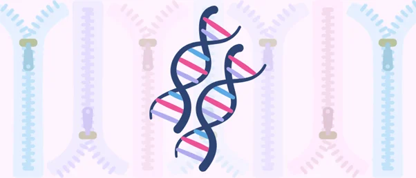
For the first time in scientific history, researchers have successfully captured the exact moment two DNA molecules zip together on camera, putting to rest a fundamental biochemical debate that has persisted for more than twenty years. A collaborative team of biophysicists and molecular scientists from the University of Sheffield and the University of York in the United Kingdom has finally resolved the structural paradox of DNA-DNA pairing. By combining cutting-edge atomic force microscopy with advanced molecular dynamics simulations, the research group has provided the first direct, empirical visualization of how genetic material overcomes its inherent electrical repulsion to lock tightly into place. This landmark discovery sheds light on critical intracellular mechanics such as chromosomal condensation and genome organization, while simultaneously opening unprecedented pathways for synthetic biology, nanotechnology, and genetic engineering.
The core paradox that has baffled generations of molecular biologists centers on basic physics. Every single molecule of deoxyribonucleic acid carries a uniform, strongly negative electrical charge along its sugar-phosphate backbone. According to the foundational laws of physics, like charges repel one another. For decades, scientists faced a profound contradiction: if two identical or homologous DNA strands naturally repel each other due to these overlapping negative fields, how do they manage to recognize one another, align with pinpoint precision, and zip together during vital biological processes such as genetic recombination and DNA repair?
The Chronology of a Decades-Long Hypothesis
To understand the weight of this recent breakthrough, it is necessary to examine the historical trajectory of the field. The journey toward solving this biophysical enigma began in earnest at the turn of the century. In 2001, a leading theoretical framework known as the electrostatic DNA zipper model—frequently referred to in literature as the helical coherence model—was first formally proposed.
At the time, theorists hypothesized that positively charged ions present in the cellular environment, known as counterions, could bind tightly to the helical grooves of the DNA strands. This binding action was theorized to create intricate, alternating charge patterns along the exterior of the duplexes. In theory, these patterns would generate localized zones of electrostatic attraction that could successfully override the macroscopic repulsion of the backbones, allowing the positively charged grooves of one double helix to interlock seamlessly with the negatively charged backbone of another.
Despite its elegance and widespread acceptance as a theoretical baseline, the electrostatic zipper model remained stubbornly unproven for over twenty years. Traditional biochemical techniques lacked the spatial resolution required to witness individual molecular duplexes interacting in real time. Because the interactions occur on a scale of nanometers and transpire within fractions of a second in fluid environments, researchers could only infer the mechanics of the process through indirect biochemical assays and thermodynamic measurements. The precise molecular choreography remained an elusive shadow, documented extensively in theory but entirely absent from direct experimental observation.
Bridging Microscopy and Computer Simulations
The stalemate was broken when the British research consortium deployed a dual-pronged investigative strategy that merged ultra-high-resolution experimental imaging with powerful computational modeling.
On the experimental front, the team utilized high-resolution atomic force microscopy (AFM). Unlike traditional light microscopy, which is constrained by the wavelength of visible light, AFM physically scans surfaces with a microscopic probe tip, allowing scientists to generate topographical maps of biological macromolecules down to the nanometer scale. By placing DNA samples into a carefully controlled solution containing divalent ions—specifically, positively charged nickel cations (Ni2+)—the researchers were able to capture the physical alignment of double-stranded DNA molecules in real time.
The resulting AFM imaging revealed that the major and minor grooves of paired double-stranded DNA matched up almost perfectly across adjacent duplexes. This physical alignment served as the first tangible proof that divalent ions function precisely as the electrostatic zipper model predicted, acting as structural mediators that align the grooves of neighboring DNA molecules.
To validate and expand upon these physical observations, the team turned to atomistic molecular dynamics simulations. These high-powered computer models simulated the physical movements and interactions of every individual atom within the DNA fragments when solvated by nickel cations. The computational results corroborated the microscopy data with astonishing precision. Across nearly all simulations containing Ni2+ ions, the DNA pairs spontaneously self-assembled and interacted with one another, even when the underlying nucleotide sequences were non-homologous.

Furthermore, the molecular dynamics simulations exposed the microscopic mechanics of the stabilization process. The computer models demonstrated that specific positively charged ions interact with both DNA duplexes simultaneously. These ions lodge themselves securely within designated coordination sites inside the minor groove, acting effectively as a molecular bridge that rivets the two separate strands together. Crucially, both the imaging data and the computational models confirmed that these coordination sites do not form at random locations; instead, their precise positions vary depending on the underlying genetic sequence and the specific chemical identity of the divalent ion involved.
Perspectives from the Research Front
The significance of capturing this microscopic event has resonated deeply throughout the scientific community, eliciting strong reactions from the primary investigators who spearheaded the project.
"To be able to directly visualize this long-hypothesized mechanism for the first time was incredible," remarked Thomas Catley, a researcher at the University of Sheffield and co-lead author of the groundbreaking study. Catley emphasized that the methodological breakthrough was largely driven by recent leaps in analytical hardware and software. "The advanced imaging techniques at our disposal have allowed us to uncover these key DNA interactions which have implications in many key cellular processes. It opens the door to studying other DNA interactions that, until now, have only existed as theory."
Beyond satisfying decades of academic curiosity, Catley and his colleagues are already looking toward the practical applications of their findings. By understanding the precise parameters that govern how DNA molecules recognize and bind to one another via ionic bridges, scientists can begin to harness these forces for technological innovation.
"Hopefully, these programmable interactions could eventually help engineers design custom DNA structures for future biotechnology, such as DNA origami, and shed light on how DNA is actually packaged inside cells," Catley added.
Broader Implications for Biology and Nanotechnology
The resolution of this twenty-year-old mystery carries far-reaching implications that extend well beyond theoretical biophysics, touching upon several distinct scientific disciplines.
First and foremost, the discovery fundamentally advances our comprehension of internal cellular organization. Eukaryotic genomes are remarkably vast; inside a human cell, nearly two meters of DNA must be tightly packaged into a microscopic nucleus roughly six micrometers in diameter. Understanding how DNA strands interact, condense, and organize themselves on a structural level without tangling or malfunctioning is essential for mapping out healthy cellular maintenance. Errors in chromosomal condensation and DNA recombination are directly linked to a host of human diseases, including various forms of cancer, congenital developmental disorders, and genetic abnormalities. By illuminating the baseline physics of how DNA duplexes find and lock onto one another, medical researchers gain a clearer window into the mechanical origins of these pathologies.
Second, the findings represent a major leap forward for the burgeoning field of structural DNA nanotechnology. For years, bioengineers have utilized DNA as a programmable building material—a technique famously known as DNA origami—to construct intricate two-dimensional and three-dimensional nanoscale shapes for drug delivery systems, molecular sensors, and nano-electronic devices. Until now, the design of these structures has relied heavily on empirical trial and error regarding inter-duplex forces. Armed with precise data concerning how divalent ions mediate electrostatic zipping and groove alignment, bioengineers can now refine their predictive models. This will allow for the fabrication of vastly more complex, stable, and functionally precise nanoscale architectures with tailored mechanical properties.
Finally, the success of this interdisciplinary study validates the powerful synergy between modern experimental biophysics and computational atomistic modeling. By demonstrating that high-resolution atomic force microscopy and molecular dynamics simulations can be synchronized to solve longstanding structural enigmas, the Sheffield and York teams have established a new methodological standard. Researchers across the life sciences are likely to adopt similar combinatorial approaches to investigate other elusive biological phenomena—such as complex protein-DNA binding dynamics and transient enzymatic states—that have historically resisted traditional experimental capture.
As the scientific community digests these findings, the peer-reviewed study stands as a testament to the power of patience, technological refinement, and cross-disciplinary collaboration. What began as a theoretical postulate in the early days of twenty-first-century biophysics has now been firmly anchored in observational reality, closing a two-decade chapter of scientific inquiry while simultaneously opening an expansive new frontier for genetic research and nanotechnological design.