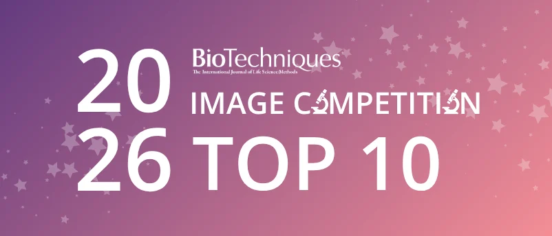
The intersection of advanced scientific methodology and visual artistry has once again captured the attention of the global research community as the BioTechniques Image Competition officially announces its shortlisted finalists. Following weeks of rigorous deliberation by an international panel of microscopy experts, cellular biologists, and imaging specialists, the contest organizers have unveiled the definitive selection of top entries. Originally conceived as a strict top-ten showcase, the extraordinary caliber and technical precision of this year’s submissions forced an unexpected revision: due to a formidable three-way statistical tie for the final position, the showcase has officially been expanded to feature twelve exceptional pieces of visual science.
This year’s competition drew an exceptionally high volume of entries from academic institutions, private research laboratories, and biotechnology firms spanning multiple continents. The sheer breadth of the portfolio reflects rapid advancements in microscopy hardware, fluorophore engineering, and computational image processing. As scientific inquiry increasingly relies on high-resolution visualization to decode complex biological pathways, competitions of this nature serve a vital dual purpose. They not only celebrate the aesthetic beauty hidden within the microscopic universe but also highlight the cutting-edge technical innovations driving modern life sciences research forward.
The Evaluation Process and Rigorous Judging Criteria
Selecting finalists from a vast pool of global submissions required a standardized, multifaceted evaluation framework. The judging panel—comprising distinguished leaders in imaging technology, academic research, and scientific publishing—assessed each candidate file against three core metrics: scientific impact, image quality, and the subjective yet critical “wow factor.”
Scientific impact measured how effectively each image communicated a significant biological phenomenon, pathway, or structural relationship. Reviewers looked for visuals that went beyond mere aesthetic appeal to provide genuine insight into cellular mechanics, tissue architecture, or molecular interactions. Image quality evaluated the technical execution of the capture, scrutinizing parameters such as signal-to-noise ratio, optical clarity, resolution limits, depth of field, and the appropriate application of staining or fluorescent labeling techniques. Finally, the wow factor captured the immediate visual resonance of the piece, recognizing images that challenge conventional perspectives and evoke a profound appreciation for the complexity of natural systems.
The scoring process was characterized by intense debate, particularly during the final ranking stages. The density of high-scoring entries near the bottom of the initial cutoff threshold resulted in the unprecedented three-way tie. Rather than deploying arbitrary tie-breaking metrics that could disadvantage any of the deserving candidates, the editorial and judging teams made the executive decision to expand the showcase to a top-twelve format, ensuring that all exceptional contributions received the recognition they warranted.
Competition Timeline and Key Milestones
The trajectory of the BioTechniques Image Competition 2026 followed a structured timeline designed to ensure equitable participation and thorough evaluation:
- Launch and Submission Phase: Opening early in the year, the submission portal invited researchers worldwide to upload their highest-quality microscopy and imaging data, accompanied by detailed technical descriptions of the methodologies utilized.
- Initial Screening and Technical Review: Editorial staff conducted preliminary audits of all incoming files to verify technical specifications, ownership rights, and adherence to competition guidelines.
- Expert Panel Deliberation: Qualified entries were forwarded to the judging panel for independent scoring across the three core criteria, culminating in several rounds of comparative review.
- Finalist Announcement: The shortlisted entries were officially compiled into a comprehensive digital showcase and PDF portfolio, marking the commencement of the public voting phase.
- Public Voting Window: Open immediately following the announcement, the public voting period runs through Tuesday, October 6, closing strictly at 5:00 PM BST (12:00 PM EDT / 9:00 PM PDT).
- Winner Declaration: Following the tabulation of verified public votes combined with the judges’ initial scores, the ultimate competition winners will be announced in a forthcoming publication.
Technical Innovations Reflected in the 2026 Shortlist
A retrospective analysis of the shortlisted images reveals compelling trends in contemporary scientific imaging. Confocal laser scanning microscopy, super-resolution techniques such as stimulated emission depletion (STED) microscopy, and advanced light-sheet fluorescence microscopy dominated the technical metadata submitted by entrants. Furthermore, the integration of machine learning algorithms for image denoising and deconvolution was prominently featured, enabling researchers to extract unprecedented structural detail from challenging, low-light biological samples.
The subject matter represented in the top-twelve portfolio spans diverse branches of biology, including neurobiology, developmental biology, immunology, and oncology. Several entries captured dynamic cellular processes in real-time, utilizing time-lapse live-cell imaging to illustrate phenomena such as mitosis, cellular migration, and immune response mechanisms. Others focused on macro-to-micro structural architectures, employing advanced tissue-clearing techniques alongside multi-channel labeling to render intact organs transparent while preserving their internal cellular geography.
These technical milestones underscore a broader paradigm shift in life sciences research. Visualization is no longer merely a descriptive tool employed at the conclusion of an experiment; it is an active driver of hypothesis generation. High-resolution imaging allows scientists to interrogate biological questions with spatial and temporal precision that was unimaginable decades ago, bridging the gap between molecular biochemistry and macroscopic physiology.
Perspectives from the Organizing Committee and Scientific Community
While formal statements from individual submitters remain tied to their respective institutional affiliations, organizers and representatives from BioTechniques have emphasized the cultural and educational value of the competition.
"The standard of submissions for the 2026 competition has completely redefined our expectations," noted a representative involved in the administration of the event. "We are witnessing a convergence of art and rigorous science. The images selected by our judges do not just illustrate data; they tell compelling stories about the fundamental nature of life at the cellular and molecular levels. The fact that our judges experienced such difficulty narrowing the field—resulting in a three-way tie for the tenth spot—is a testament to the extraordinary talent and dedication operating within laboratories across the globe."
The research community has similarly embraced the initiative. Academic laboratories frequently utilize such platforms to enhance public engagement with science. By translating complex, jargon-heavy datasets into visually striking, universally accessible imagery, competitions help demystify scientific research for the general public, fostering broader scientific literacy and inspiring the next generation of researchers.
Public Voting Mechanics and Integrity Protocols
With the finalists now public, attention shifts to the community at large to determine the ultimate favorites. Organizers have established a secure online voting portal, accessible via dedicated registration forms, allowing peers, enthusiasts, and the wider scientific community to review the portfolio and cast their ballots.
To maintain the absolute integrity of the voting process, strict protocols have been instituted. Most notably, participation is limited to a single vote per individual email address recorded in the system. Automated systems monitor incoming ballots to detect and invalidate duplicate entries, scripting attempts, or anomalous voting patterns. This safeguards the legitimacy of the competition, ensuring that the final standings authentically reflect community preference.
Voters are encouraged to carefully examine the comprehensive PDF portfolio detailing the top-twelve finalists before making their selections. The deadline for casting votes is firmly set for Tuesday, October 6, at 5:00 PM BST (12:00 PM EDT / 9:00 PM PDT). Any submissions recorded after this threshold will not be factored into the final tally.
Broader Implications for Science Communication and Imaging Technology
The sustained success and growth of the BioTechniques Image Competition highlight a critical evolution in how scientific discoveries are communicated. In an era dominated by digital media and rapid information consumption, visual literacy has become an essential competency for researchers. High-impact journals increasingly prioritize clear, aesthetically compelling figures that can communicate complex findings at a glance.
Furthermore, the economic and technological implications of advancements in bio-imaging are profound. The market for advanced microscopy hardware, fluorescent probes, and computational analysis software continues to expand rapidly, driven by the demands of academic and pharmaceutical research. Competitions like this one provide an invaluable showcase for emerging technologies, validating the utility of novel hardware and software platforms in real-world research environments.
As the voting window progresses toward its October deadline, anticipation builds within the global scientific community. The eventual winners will not only secure formal recognition for their technical and artistic achievements but will also contribute to a growing archive of visual milestones that document the ongoing advancement of the life sciences. Through the lens of the microscope, these researchers continue to illuminate the unseen mechanisms of life, providing clarity where there was once only shadow.