A groundbreaking study led by researchers at Stanford Medicine has demonstrated that targeting a specific protein linked to cellular aging can successfully restore lost knee cartilage in older mice and prevent the onset of osteoarthritis following severe joint injuries. Published in the scientific journal Science, the study opens new therapeutic avenues for millions of patients suffering from degenerative joint diseases. By administering an inhibitor against a protein known as 15-PGDH—categorized by researchers as a "gerozyme"—the scientific team observed not only structural recovery in aged animal models but also encouraging regenerative activity in human tissue samples retrieved during knee replacement surgeries.
The implications of these findings extend far beyond veterinary medicine, pointing toward a future where debilitating joint degeneration caused by aging or trauma could be treated effectively with non-invasive local injections or oral medications. If these preclinical successes translate safely and effectively to human clinical trials, the approach could drastically reduce the annual volume of total knee and hip replacement surgeries performed worldwide. Notably, an oral iteration of this therapeutic candidate has already advanced to clinical evaluation for age-related muscle weakness, establishing a precedent for human safety and pharmacokinetics.
Understanding the Burden of Osteoarthritis
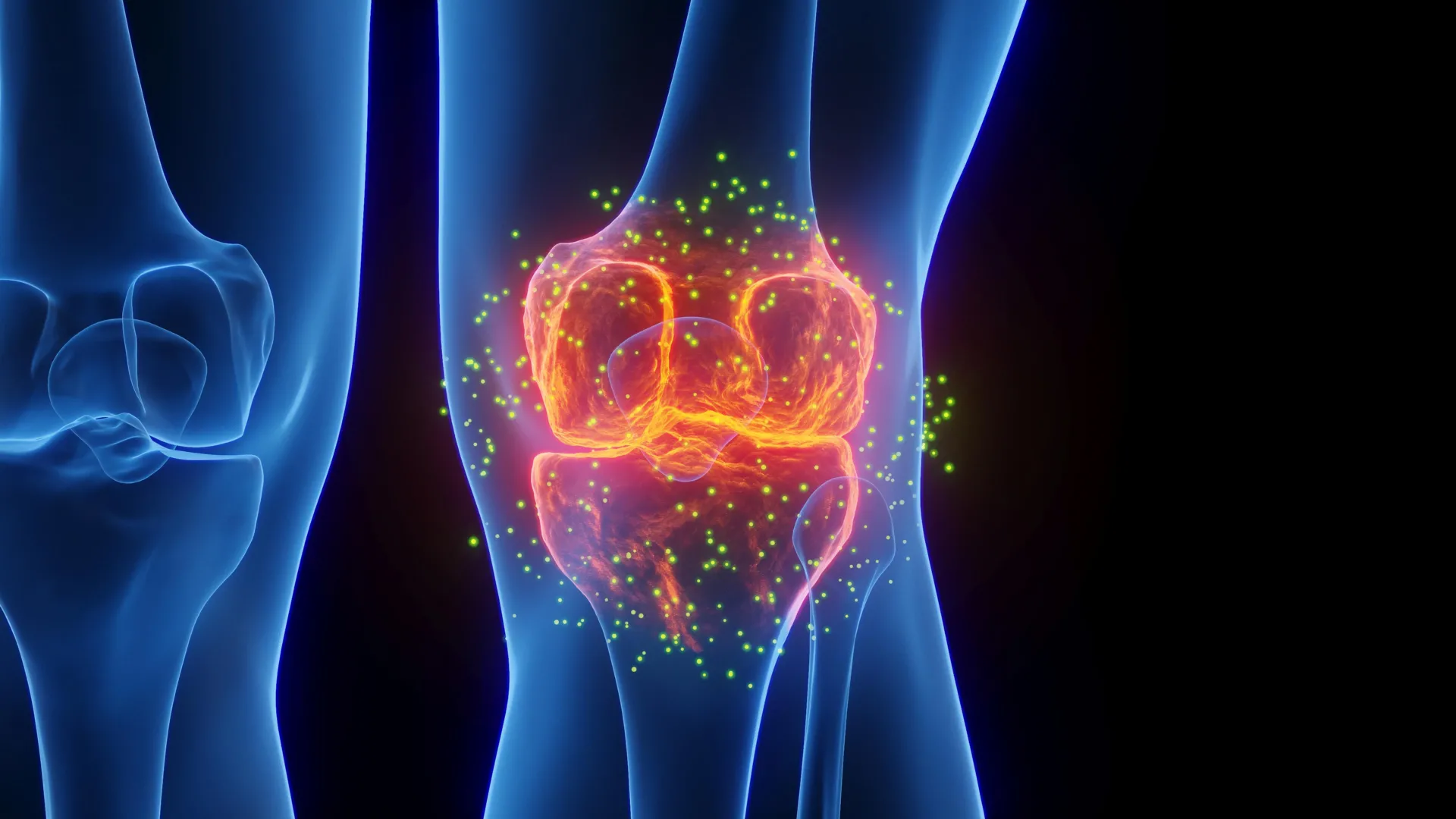
Osteoarthritis stands as the most prevalent form of joint disease, impacting approximately one in five adults across the United States. Characterized by the gradual, relentless degradation of articular cartilage—the smooth, slippery tissue that cushions the ends of bones and enables frictionless joint movement—the condition manifests clinically through persistent pain, joint stiffness, and chronic swelling. Beyond the significant toll it takes on patient mobility and overall quality of life, osteoarthritis imposes a massive economic burden on the healthcare system, generating an estimated $65 billion in direct annual medical expenditures in the U.S. alone.
For decades, the standard of care for osteoarthritis has remained largely palliative, focusing heavily on symptom management through physical therapy, nonsteroidal anti-inflammatory drugs (NSAIDs), corticosteroid injections, and, ultimately, invasive joint replacement surgery for advanced cases. Historically, modern medicine has lacked any approved pharmacological intervention capable of altering the fundamental disease trajectory. No existing pharmaceutical agent can reliably slow, halt, or reverse the progressive destruction of articular cartilage, leaving clinicians with limited options as joint integrity deteriorates over time.
The Mechanism of Action: Unmasking the Gerozyme 15-PGDH
The core of the Stanford discovery centers on the protein 15-hydroxyprostaglandin dehydrogenase (15-PGDH), which researchers classify as a gerozyme. Gerozymes are a specific class of proteins that naturally accumulate within tissues as an organism ages, actively driving down cellular function and contributing to systemic physiological decline.
The trajectory toward this discovery began years prior. In 2023, the same Stanford research collective first identified the physiological significance of gerozymes, specifically noting that 15-PGDH serves as a principal driver of age-related muscle deterioration in mice. When scientists pharmacologically blocked this protein, aged animals demonstrated significant recoveries in muscle mass, strength, and physical endurance. Conversely, artificially upregulating 15-PGDH in young, healthy mice induced rapid muscle atrophy and weakness. Subsequent investigations have progressively linked 15-PGDH activity to the regulation of regenerative pathways in diverse tissues, including bone, nerve, and blood cells.
To understand its role in joint health, the research team examined the biochemical pathway involving prostaglandin E2, a molecule critical for maintaining tissue and stem cell vitality. Because 15-PGDH acts as the primary degrader of prostaglandin E2, researchers hypothesized that suppressing the enzyme might similarly protect aging joints. Upon analyzing cartilage samples from mice of varying ages, the team discovered that concentrations of 15-PGDH approximately doubled as the animals aged.
Experimental Findings in Animal Models
To test their hypothesis, investigators administered a small-molecule drug designed to inhibit 15-PGDH activity to a cohort of aged mice. The treatment was delivered via systemic abdominal injections, which exposed the entire body to the therapeutic agent, and localized injections directly into the knee joints.
Both administration routes yielded striking physiological improvements. Cartilage that had thinned and degraded as a consequence of age regenerated significantly across the entire joint surface. Histological analyses confirmed that the newly grown tissue was hyaline cartilage—the specialized, resilient articular tissue required for proper joint mechanics—rather than fibrocartilage, which is tougher but less effective at shock absorption.
Building upon these findings, the research team evaluated whether the therapy could proactively shield joints from trauma-induced osteoarthritis. Using a murine model designed to replicate anterior cruciate ligament (ACL) tears—a common athletic injury frequently sustained in sports involving abrupt deceleration, pivoting, or jumping—the scientists administered the gerozyme inhibitor twice weekly for four weeks post-injury.
While human patients suffering ACL tears face roughly a 50% probability of developing osteoarthritis in the damaged joint within 15 years, the treated mice experienced a dramatically lower incidence of the disease. Untreated control animals exhibited post-injury 15-PGDH levels twice as high as uninjured counterparts and developed classic signs of osteoarthritis within four weeks. Furthermore, treated mice demonstrated immediate functional recoveries, exhibiting more symmetrical gait patterns and placing significantly more weight on the recovering limb.
A Novel Paradigm in Tissue Regeneration
The cellular mechanics driving this recovery challenged prevailing biological assumptions regarding how adult tissues heal. In many organ systems, structural regeneration is heavily dependent on the proliferation and differentiation of resident stem or progenitor cells. However, detailed single-cell analyses revealed a different mechanism at play within articular cartilage.
Rather than recruiting a dormant stem cell population, existing cartilage-producing cells known as chondrocytes appeared to undergo transcriptional reprogramming, shifting their gene expression profiles to revert to a functionally youthful state.
Older chondrocytes typically exhibit upregulated expression of genes tied to inflammation, matrix destruction, and the pathological conversion of cartilage into bone, alongside downregulated expression of structural maintenance genes. Following treatment with the 15-PGDH inhibitor, these age-related patterns reversed. Cell populations driving cartilage breakdown dropped from 8% to 3%, while cells associated with fibrocartilage production decreased from 16% to 8%. Concurrently, the proportion of cells dedicated to synthesizing hyaline cartilage and maintaining the extracellular matrix expanded from 22% to 42%.
This cellular reprogramming effectively rejuvenates the existing cellular architecture of the joint without requiring the intervention of stem cells, offering a highly efficient target for clinical intervention.
Translating Findings to Human Tissue
To validate the translational relevance of their preclinical data, the Stanford team acquired human cartilage samples harvested from patients undergoing total knee replacement surgeries due to end-stage osteoarthritis.
When exposed to the 15-PGDH inhibitor ex vivo for a period of one week, the human tissue samples exhibited marked biological improvements. The treated samples showed a notable reduction in cartilage-degrading cells and decreased activity among genes responsible for matrix breakdown and fibrocartilage formation. Most importantly, the human tissue began generating new articular cartilage in response to the treatment.
Senior researchers emphasized the profound shift this discovery represents for orthopedic medicine. By demonstrating that a large, pre-existing pool of resident joint cells can be pharmacologically coaxed into a regenerative state, the study presents a viable therapeutic target for human clinical application.
Broader Implications and Future Clinical Outlook
The study was co-led by Helen Blau, PhD, professor of microbiology and immunology and director of the Baxter Laboratory for Stem Cell Biology, and Nidhi Bhutani, PhD, associate professor of orthopedic surgery. Mamta Singla, PhD, instructor of orthopedic surgery, and Yu Xin (Will) Wang, PhD, former postdoctoral scholar and current assistant professor at the Sanford Burnham Prebys Medical Discovery Institute, served as lead authors of the research paper.
Financial support for the investigation was provided by prominent institutional and philanthropic sources, including multiple grants from the National Institutes of Health, the Baxter Foundation for Stem Cell Biology, the Li Ka Shing Foundation, the Stanford Cardiovascular Institute, the Milky Way Research Foundation, the Canadian Institutes of Health Research, and various fellowship programs. In accordance with institutional policies, Stanford University has filed patent applications concerning 15-PGDH inhibition for tissue rejuvenation and cartilage repair, which have been licensed to Epirium Bio, with several study authors holding equity or co-founding affiliations with the enterprise.
As researchers look toward the horizon, the primary objective remains the clinical translation of these findings into human trials. Because a Phase 1 clinical trial evaluating a 15-PGDH inhibitor for age-related muscle weakness has already established an initial safety profile and biological activity in healthy human volunteers, investigators are optimistic that cartilage-focused trials can be expedited. If successful, this pharmacological strategy could fundamentally alter the landscape of musculoskeletal medicine, offering a non-invasive pathway to regenerate damaged joints, restore mobility, and circumvent the necessity for major surgical interventions.