Bone health has long been understood as a dynamic biological process heavily influenced by physical activity. For decades, medical science has prescribed weight-bearing exercise as the primary defense against bone degradation. However, the precise molecular mechanisms by which physical force is translated into cellular instructions for bone growth remained a persistent mystery. Now, a team of researchers from the Department of Medicine at the School of Clinical Medicine, LKS Faculty of Medicine, University of Hong Kong (HKUMed), has successfully decoded this biological pathway. Their landmark discovery identifies a specific protein that functions as the body’s internal exercise sensor, paving the way for revolutionary pharmacological treatments that could replicate the benefits of physical movement for individuals who are unable to exercise.
The findings, published in the prestigious international journal Signal Transduction and Targeted Therapy, arrive at a critical juncture for global healthcare systems grappling with rapidly aging populations. By pinpointing how mechanical forces stimulate bone formation and suppress fat accumulation within the bone marrow, the research consortium has unlocked a viable therapeutic target. This breakthrough offers unprecedented hope for elderly populations, bedridden patients, and those suffering from debilitating chronic illnesses who face a high risk of debilitating fractures and a subsequent loss of independence.
The Anatomy of an Aging Skeleton and the Global Burden of Osteoporosis
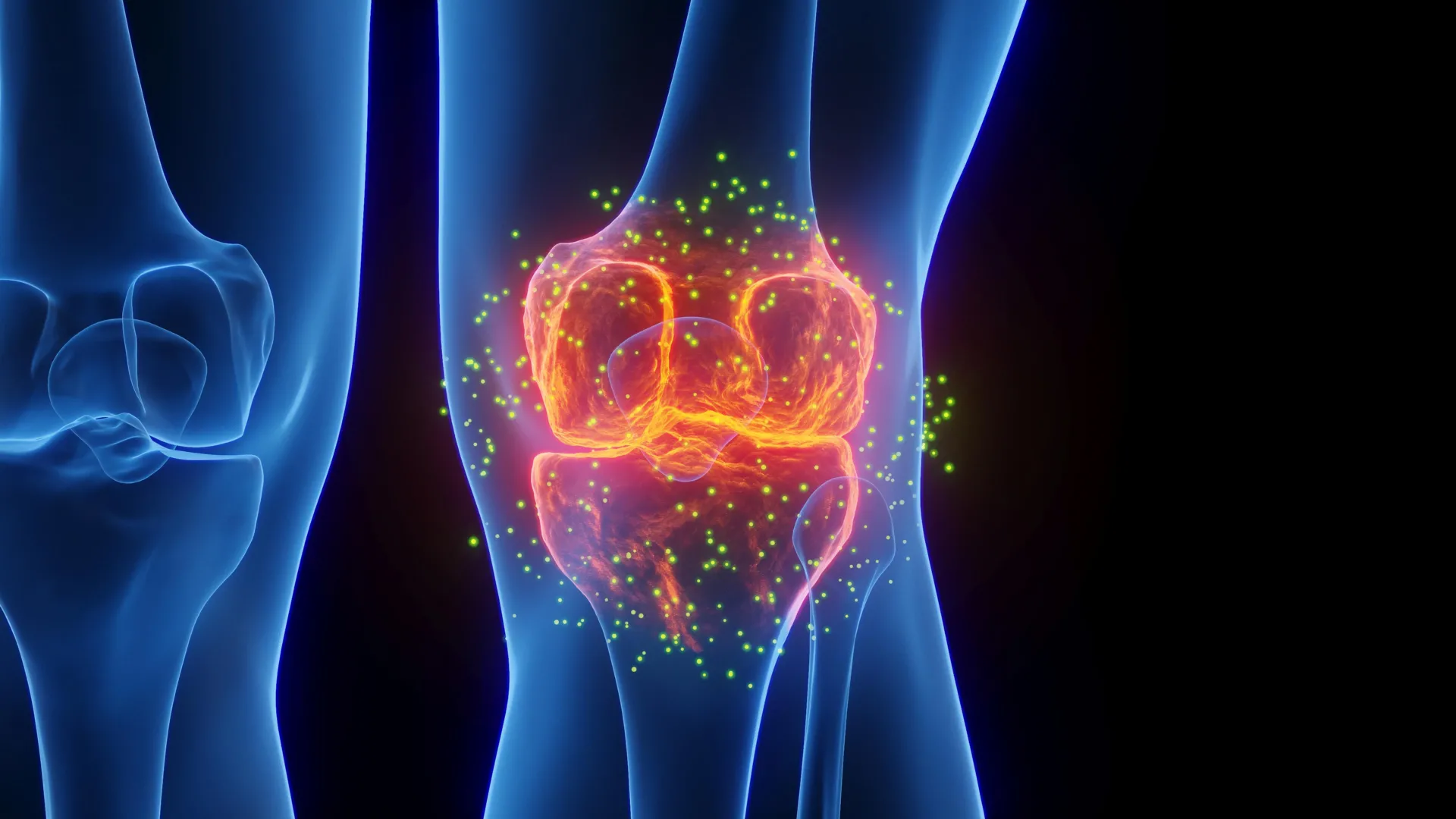
To understand the magnitude of the HKUMed discovery, one must examine the profound public health crisis posed by osteoporosis and age-related bone degradation. According to epidemiological data compiled by the World Health Organization (WHO), approximately one in three women and one in five men over the age of 50 will experience an osteoporotic fracture in their lifetime. These injuries are far from benign; they frequently trigger a downward spiral of chronic pain, immobility, long-term hospitalization, and premature mortality, while placing an immense financial strain on global healthcare infrastructure.
The impact is acutely felt in densely populated urban centers with rapidly aging demographics, such as Hong Kong. Local health statistics indicate that osteoporosis affects an alarming 45% of women and 13% of men aged 65 and older. As life expectancies rise globally, the societal and economic costs associated with bone fragility are projected to escalate dramatically unless innovative prophylactic and therapeutic measures are developed.
At the microscopic level, bones are far from static structures; they undergo a continuous process of remodeling, wherein old bone tissue is resorbed and replaced by new matrix. This delicate balance is managed within the bone marrow, a spongy tissue housed inside the bones that contains mesenchymal stem cells (MSCs). These versatile progenitor cells possess the capacity to differentiate into various cell types, primarily osteoblasts (bone-forming cells) or adipocytes (fat cells).
In youth, physical forces—such as gravity, muscle contraction, and mechanical impact—naturally stimulate MSCs to develop into bone tissue, maintaining skeletal density and strength. However, as the human body ages, a fundamental shift occurs in this cellular microenvironment. Aging, combined with a lack of physical stimuli, skews the differentiation pathway of mesenchymal stem cells, causing them to increasingly transform into fat cells rather than bone cells.
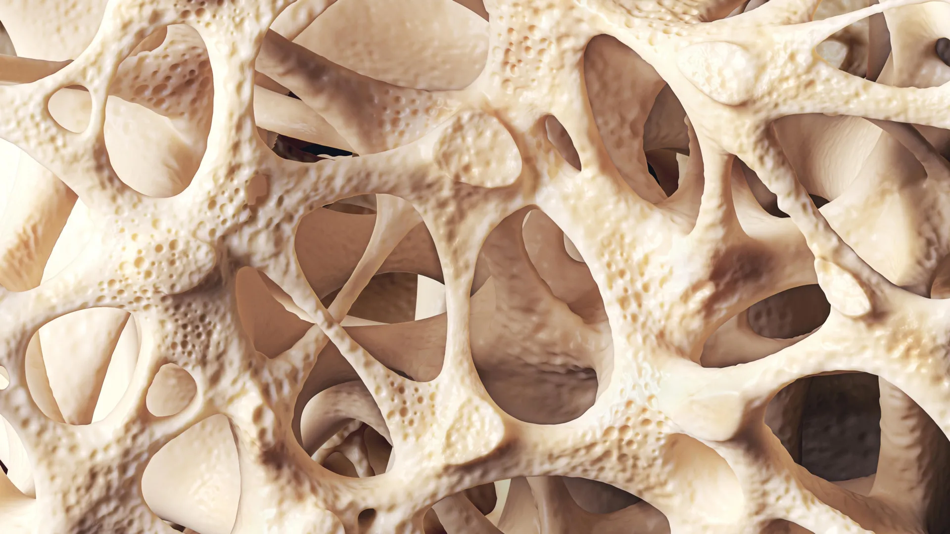
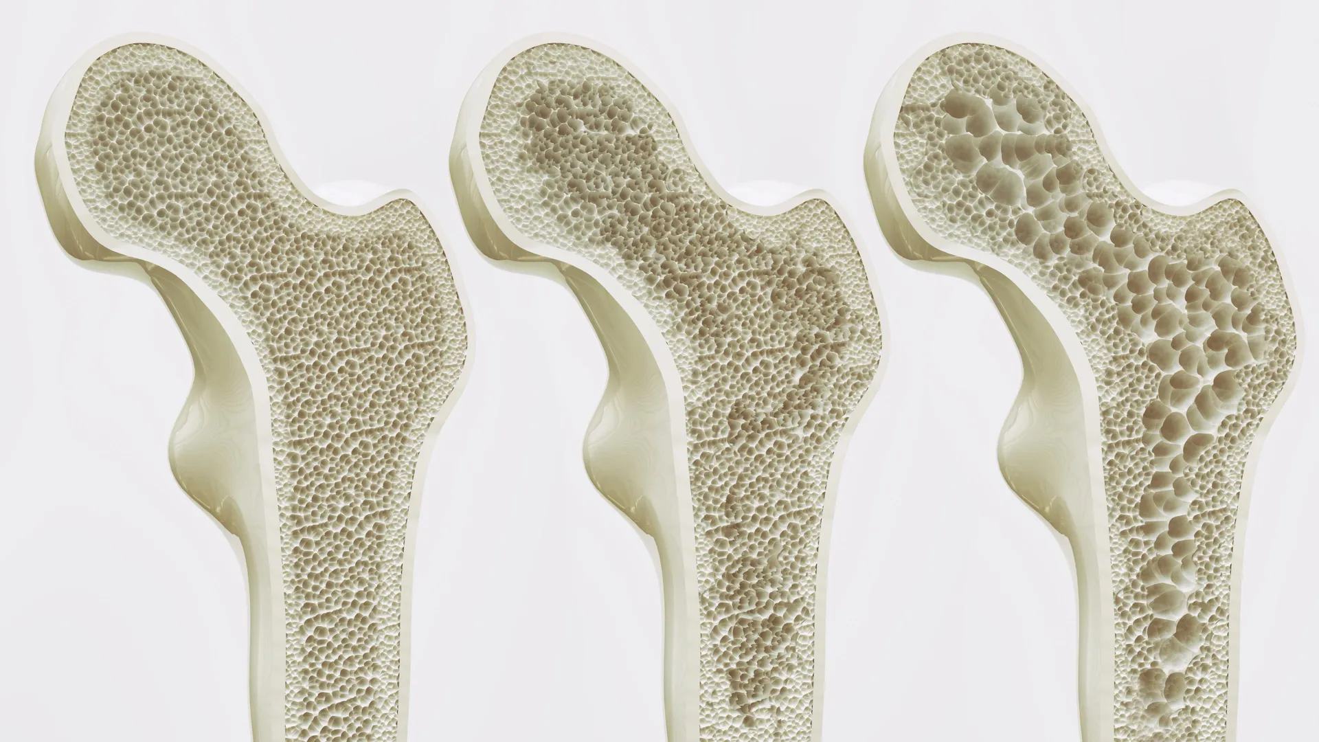
This accumulation of fat within the bone marrow is not a neutral byproduct of aging; it actively exacerbates skeletal deterioration. As adipose tissue builds up inside the bone marrow cavity, it physically crowds out healthy bone-forming cells and disrupts the structural integrity of the trabecular bone network. This creates a vicious biological cycle of progressive bone loss, increased porosity, and heightened fracture susceptibility that conventional therapies struggle to reverse, particularly when patients cannot engage in weight-bearing exercise.
Piezo1: The Molecular Switch That Translates Movement into Bone Strength
The core of the HKUMed breakthrough centers on the identification of a mechanoreceptor protein known as Piezo1. Through a series of rigorous experiments utilizing advanced mouse models and human stem cells, the research team discovered that Piezo1 is expressed on the surface of mesenchymal stem cells residing within the bone marrow.
Piezo1 acts as a highly sensitive mechanical transducer. When an individual engages in physical activity, the resulting mechanical forces physically deform the cell membrane, activating the Piezo1 protein. Once triggered, Piezo1 initiates a cascade of intracellular signaling events that actively suppress the pathways responsible for adipogenesis (fat cell formation) while simultaneously promoting osteogenesis (bone tissue creation).
Conversely, when physical activity is absent—such as in bedridden patients, astronauts in microgravity, or individuals with severe mobility impairments—Piezo1 remains inactive. Without this mechanical stimulation, mesenchymal stem cells default to fat production. Furthermore, the absence of Piezo1 activity triggers the upregulation and release of specific inflammatory signaling molecules, namely Ccl2 and lipocalin-2. These inflammatory cues further accelerate fat accumulation within the bone marrow microenvironment and actively inhibit bone growth, locking the skeletal system into a state of accelerated degradation.
Crucially, the research team demonstrated that pharmacologically blocking these inflammatory signals or artificially stimulating the Piezo1 pathway can effectively halt this destructive cycle, restoring a healthier balance to bone marrow stem cell differentiation even in the absence of mechanical movement.
Translational Medicine and the Promise of Exercise Mimetics
The identification of Piezo1 and its associated signaling networks bridges a longstanding gap in musculoskeletal research by establishing a direct causal link between physical biomechanics and molecular biology.
"We have essentially decoded how the body converts movement into stronger bones," stated Professor Xu Aimin, Director of the State Key Laboratory of Pharmaceutical Biotechnology, Chair Professor in the Department of Medicine at HKUMed, and leader of the study. "We have identified the molecular exercise sensor, Piezo1, and the signalling pathways it controls. This gives us a clear target for intervention. By activating the Piezo1 pathway, we can mimic the benefits of exercise, effectively tricking the body into thinking it is exercising, even in the absence of movement."
This capability to chemically replicate the physiological benefits of physical exertion introduces an entirely new pharmaceutical category to medical science: exercise mimetics. While exercise mimetics have been hypothesized in metabolic research regarding muscle and liver function, applying this concept directly to skeletal preservation represents a pioneering frontier in bone biology.
Dr. Wang Baile, Research Assistant Professor in the Department of Medicine at HKUMed and co-leader of the study, underscored the profound implications of these findings for vulnerable patient cohorts. "This discovery is especially meaningful for older individuals and patients who cannot exercise due to frailty, injury, or chronic illness," Dr. Wang noted. "Our findings open the door to developing exercise mimetics—drugs that chemically activate the Piezo1 pathway to help maintain bone mass and support independence."
International Collaboration and Multidisciplinary Methodology
The success of this research underscores the power of global scientific collaboration. The project was co-led by Professor Xu Aimin and Dr. Wang Baile of HKUMed, working in close partnership with Professor Eric Honoré, a globally recognized authority on mechanosensitive ion channels and Team Leader at the Institute of Molecular and Cellular Pharmacology within the French National Centre for Scientific Research (CNRS), Université Côte d’Azur (UniCA), and the French National Institute of Health and Medical Research (Inserm), who also serves as a Visiting Professor in the Department of Pharmacology and Pharmacy at HKUMed.
Professor Honoré emphasized the broader strategic implications of the team’s collaborative work for clinical practice. "This offers a promising strategy beyond traditional physical therapy," Professor Honoré remarked. "In the future, we could potentially provide the biological benefits of exercise through targeted treatments, thereby slowing bone loss in vulnerable groups such as bedridden patients or those with limited mobility, and substantially reducing their risk of fractures."
The research methodology combined cutting-edge genetic knock-out mouse models with human primary stem cell cultures, allowing the investigators to validate their findings across both preclinical models and human cellular biology. This rigorous dual approach ensures that the identified mechanisms are not merely laboratory phenomena but hold genuine translational relevance for human therapeutics.
Roadmap to Clinical Applications and Future Horizons
Following the publication of their findings in Signal Transduction and Targeted Therapy, the research consortium has shifted its focus toward translational development. The immediate objective is to design small-molecule compounds and targeted pharmacological agents capable of selectively activating the Piezo1 pathway in human bone marrow without triggering adverse systemic side effects.
Developing such therapeutics requires navigating complex pharmacological challenges, including ensuring targeted delivery to the bone marrow microenvironment and verifying long-term safety profiles. However, the clear molecular target provided by Piezo1 transforms what was once an elusive physiological mystery into a tractable drug discovery program.
The financial and institutional support underpinning this research reflects its high priority within the global scientific community. The project was generously funded by a coalition of prestigious agencies, including the Areas of Excellence Scheme and the General Research Fund of the Research Grants Council; the Health and Medical Research Fund under the Health Bureau of the Government of the Hong Kong Special Administrative Region; the National Key R&D Program of China; the National Natural Science Foundation of China; the Human Frontier Science Program; the French National Research Agency; Fondation de France; Fondation pour la Recherche Médicale; and the Macau Science and Technology Development Fund.
As aging populations continue to expand worldwide, the imperative to maintain functional independence among older adults grows ever more urgent. By translating the mechanical language of physical movement into the biochemical language of molecular pharmacology, the HKUMed-led research team has laid the groundwork for a new generation of therapeutics. These future treatments promise to protect the skeletal health of millions who cannot move, offering a biochemical shield against the fragility of age and preserving dignity and mobility for generations to come.