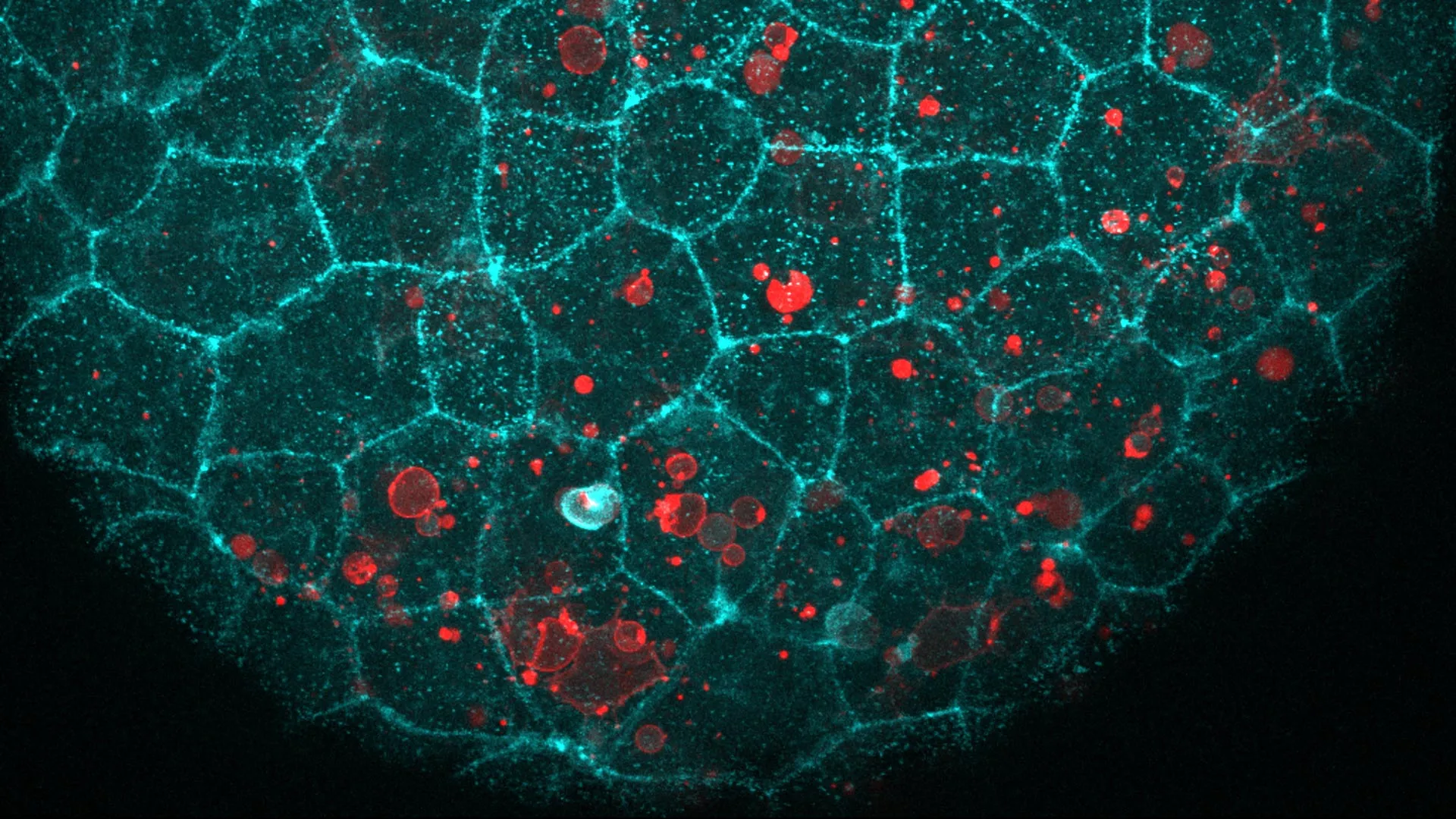
The integrity of the human body relies on epithelial cells—the tightly packed, specialized units that form the protective linings of our skin, gut, lungs, and various organ structures. For decades, the E-cadherin protein complex has been celebrated by the scientific community primarily for its role as the molecular “glue” that stitches these cells together, maintaining the structural resilience of our tissues. However, a groundbreaking study published in the journal Nature Communications has unveiled a startling dual functionality for this protein: it acts not only as a structural anchor but also as a critical orchestrator of cellular cleanup, enabling epithelial cells to engulf and dispose of neighboring dead cells without compromising the integrity of the protective barrier.
The Mechanism of Cellular Cleanup
Led by ICREA Research Professor Verena Ruprecht, the research team investigated the behavior of epithelial tissues in both zebrafish and mouse embryos. By utilizing advanced live-imaging techniques, the scientists observed that when a cell undergoes programmed death—a process known as apoptosis—the E-cadherin machinery reconfigures itself. Rather than merely reinforcing the connections between living neighbors, the protein complex migrates to the contact point between the dying cell and the surrounding tissue.
This discovery challenges the traditional understanding of how tissues manage cellular waste. Previously, it was assumed that specialized immune cells, such as macrophages, were the primary agents responsible for “eating” cellular debris. While macrophages do perform this function, the study demonstrates that epithelial cells are capable of an autonomous, localized cleanup process. By repurposing their adhesion machinery, these cells perform a delicate mechanical feat: they wrap their membranes around the dying cell to internalize it while maintaining a seal with their neighboring cells.
Chronology and Experimental Validation
The investigation into this phenomenon began with the hypothesis that E-cadherin might be involved in more than just static adhesion. Over several years of observation, the researchers established a clear timeline of events:
- Detection: Upon the detection of an apoptotic signal, the E-cadherin complex accumulates at the interface between the healthy epithelial cell and the dying cell.
- Deformation: The epithelial cell initiates a physical remodeling process. Imaging showed that while the “upper” surface—the side facing the lumen or external environment—remains rigid to preserve the barrier, the “lower” surface undergoes significant deformation, stretching and bending to engulf the debris.
- Integration: The internalized cell is then processed within the epithelial cell, preventing the release of intracellular components into the surrounding tissue space.
To confirm that the E-cadherin complex was the driving force, the team performed a series of rigorous experiments. In one instance, they introduced dying cells that had been stripped of E-cadherin; the epithelial tissues cleared them just as efficiently as normal cells, suggesting the machinery is primed to react to the presence of a dying neighbor regardless of the neighbor’s own surface proteins. In a further test, they presented the tissue with protein-free fat droplets coated with the chemical “eat-me” signals typical of dying cells. The epithelial cells successfully engulfed these droplets, confirming that the physical machinery of the cell is triggered by specific biochemical cues and executed through the E-cadherin framework.
The Role of Molecular Tethers and Brakes
The physical mechanics of this process are highly sophisticated. The research team identified that one protein within the E-cadherin complex functions as a "molecular rope," or tether. This protein links the adhesion assembly to the internal cytoskeleton of the cell, providing the necessary mechanical force to pull the dying cell into the epithelial layer. When the researchers genetically modified the tissues to lack this tether, or removed the specific region of the protein that connects to the skeleton, the epithelial cells lost their ability to swallow the debris.
Equally important is the component that acts as a “brake” on the cell’s contractile machinery. Interestingly, the study found that removing this brake did not accelerate the cleanup. Instead, it rendered the cell too rigid, preventing the fluid movement required to engulf the target. This suggests a finely tuned balance of force and flexibility is essential for successful debris removal, a finding that adds a layer of complexity to our understanding of cellular biomechanics.
Implications for Human Health and Chronic Inflammation
The broader implications of this study are significant, particularly concerning the pathogenesis of chronic inflammatory diseases. Chronic inflammation is often fueled by the accumulation of cellular debris that has not been properly cleared by the body’s natural processes. When dead cells remain in the tissue, they can undergo secondary necrosis, rupturing and releasing inflammatory signals into the surrounding environment. This can lead to tissue damage, persistent immune activation, and, in some cases, the development of autoimmune or degenerative conditions.
By revealing that epithelial cells take an active role in their own waste management, the research opens new avenues for therapeutic intervention. If the E-cadherin-dependent cleanup process is found to be impaired in adult tissues, it could explain why certain chronic inflammatory conditions persist. Experts in the field suggest that boosting this autonomous cleanup mechanism could potentially mitigate inflammation in the lungs, gut, and other epithelial-lined organs.
Broadening the Scope: From Embryos to Adults
While the study focused on zebrafish and mouse embryos due to their optical transparency and rapid development, the research team is optimistic about the universality of the mechanism. E-cadherin is highly conserved across vertebrates, meaning its structure and function have remained consistent throughout millions of years of evolution. Given that adult tissues in the human retina, colon, and mammary glands already exhibit active clearing of apoptotic cells, it is highly probable that the E-cadherin mechanism is a fundamental feature of adult tissue homeostasis as well.
The research was supported by a coalition of international funding bodies, including the Spanish Ministry of Science and Innovation, the Human Frontier Science Program, the European Union’s Horizon Europe program, and the “la Caixa” Foundation. The use of the CRG Core Facilities for Advanced Light Microscopy was instrumental in capturing the high-resolution footage of the cell-eating process, providing the visual evidence needed to validate the team’s mechanical theories.
Looking Toward Future Clinical Applications
The work led by Professor Ruprecht and her colleagues—including joint first authors Hanna-Maria Häkkinen, Marta Batet Palau, and Laura F. Bianchi—represents a major shift in cell biology. By viewing epithelial cells not as static walls, but as active participants in immune defense and tissue maintenance, the scientific community gains a more nuanced perspective on how the body sustains itself.
Future research will likely focus on whether this process can be stimulated in human adult tissues to treat inflammatory diseases. If scientists can identify the precise molecular triggers that activate this "cleanup choreography," they may eventually be able to develop drugs or therapies that enhance the natural ability of tissues to maintain a clean and healthy environment. As the study concludes, the mechanics of how dying cells are removed is of paramount relevance to human health, bridging the gap between basic developmental biology and the clinical management of chronic, systemic inflammation. The choreography of the cell, it seems, is far more complex and vital than previously imagined.