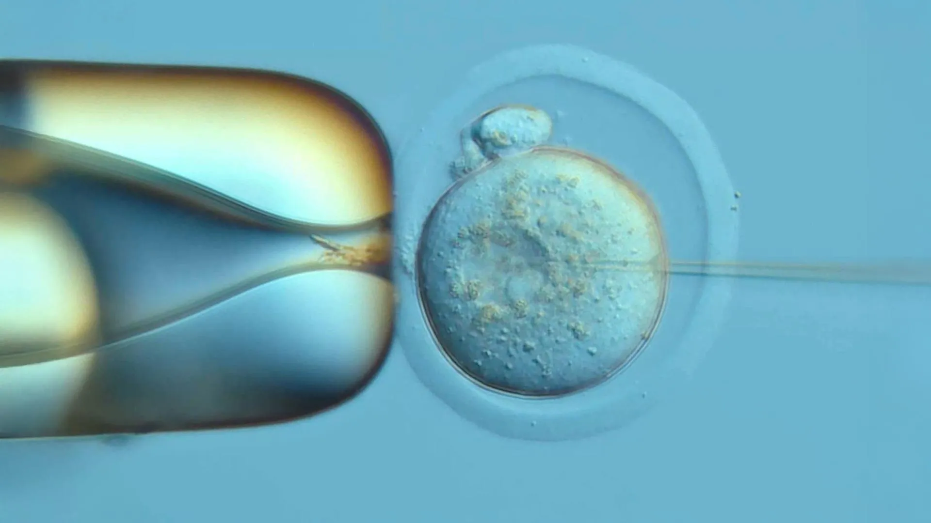
In a landmark achievement for developmental biology, an international research team has successfully reconstructed a comprehensive, high-resolution family tree of a mouse embryo, tracing the lineage of its cells back to the initial division of the fertilized egg. Published in the journal Science on October 8, 2026, the study introduces a sophisticated genetic recording technology dubbed "DNA Typewriter." This tool allows researchers to document the precise sequence of cell divisions and fate decisions as an embryo transforms from a single-celled zygote into a complex organism with distinct tissues and organ systems. By providing an unprecedented view of early mammalian development, the findings offer a new lens through which scientists can investigate the origins of congenital malformations, neurodevelopmental disorders, and the cellular mechanisms underpinning cancer.
The research was spearheaded by Dr. Jay Shendure, a professor of genome sciences at the University of Washington School of Medicine and a Howard Hughes Medical Institute Investigator, in collaboration with Dr. Chengxiang Qiu, a molecular and systems biologist at Dartmouth College. Together, the team has navigated a long-standing obstacle in biological research: the inability to track the life history of individual cells in a live, developing mammal without disrupting the very processes they aim to observe.
The Mechanism of the DNA Typewriter
At the heart of this breakthrough is a system designed to bypass the limitations of traditional lineage tracing. Conventional methods often rely on fluorescent labeling or DNA-cutting techniques that can inadvertently damage the genome or provide only snapshots of development. In contrast, the DNA Typewriter acts as a permanent, internal ledger. Developed by Shendure alongside Junhong Choi of the Sloan Kettering Cancer Center, the technology inserts a specialized "tape"—a sequence of synthetic DNA—into the genome of a fertilized mouse egg.
As the embryo develops, this tape records the history of cell division. Haedong Kim, a postdoctoral scientist and co-first author of the study, likens the process to an actual mechanical typewriter. Each time a cell divides, the system strikes a new character onto a blank segment of the DNA tape. Because these characters are added in strict, linear order and are inherited by all daughter cells, the resulting genetic sequence serves as a biological diary. Researchers can later extract this "tape" from individual cells and, by identifying shared markings, reconstruct the entire genealogical tree of the embryo.
This iteration of the DNA Typewriter featured a redesigned recording sequence that significantly improved the ease and accuracy of data recovery. By embedding the recorder directly into the germline of the mouse, the scientists ensured that the process remained non-invasive, allowing the embryo to develop normally while the genetic archive was compiled.
A Chronology of Development
The study focused on the first 13.5 days of mouse embryonic development. While a full mouse gestation period typically lasts between 19 and 21 days, the 13.5-day window provided enough developmental data to observe the transition from undifferentiated stem cells to the formation of specialized tissue foundations.
To conduct the study, the researchers initiated the recording process in 100 fertilized eggs. Out of this cohort, 10 embryos progressed successfully to the point of analysis. Among these, the embryo designated "No. 3" yielded the most robust data, becoming the focal point of the team’s detailed reconstruction.
The analysis of Embryo No. 3 revealed a remarkable insight into the earliest moments of life. By examining the DNA record, the researchers identified a definitive mark that differentiated the two founding cells resulting from the very first division of the zygote. The data showed that while one of these two founding cells contributed a larger number of total descendants, both cells gave rise to an almost identical distribution of specialized cell types. This suggests that the early divergence of cell lineages is not necessarily tied to a functional bias, but rather follows a pattern of balanced differentiation despite variations in population size.
Uncovering the Schedules of Cell Commitment
One of the primary objectives of the research was to determine exactly when cells "commit" to their future roles. In developmental biology, this transition from a pluripotent state—where a cell can become anything—to a specialized state is critical. Through their lineage mapping, the researchers observed that this process is not a uniform event across the embryo.
The team found that different tissues follow distinct biological timelines. For instance, cells destined to become blood and retinal tissues committed to their identities relatively early in the development cycle. Conversely, the progenitor cells of the outer skin layer remained unspecialized for a significantly longer period. This suggests that the "decision-making" process of a cell is decentralized, with different parts of the embryo following unique, independent schedules. This discovery challenges the notion of a synchronized, global commitment phase and highlights the complex, modular nature of mammalian organogenesis.
Historical Context and Technical Advancements
The reconstruction of cellular lineages has long been the "holy grail" of developmental biology. Decades ago, researchers achieved a similar feat in the nematode Caenorhabditis elegans, a tiny roundworm with a rigid, predictable cell lineage and a total of only about 1,000 cells. However, mammals are exponentially more complex, consisting of millions of cells that interact in dynamic and often unpredictable environments.
Previous attempts to map mammalian lineage—such as those using CRISPR-based DNA editing—often suffered from "information loss." These methods typically cut the DNA to create marks, which can lead to double-strand breaks that damage the cell or delete previous records. Furthermore, because these marks were often created in a non-sequential fashion, the temporal order of events was difficult to determine. The DNA Typewriter solves these issues by maintaining a steady, sequential writing process that does not require lethal cutting of the genetic material, allowing for a resolution that was previously unattainable.
Implications for Clinical Medicine
The ability to map the life of a cell with such granular detail has profound implications for clinical research and regenerative medicine. Many congenital malformations, such as those affecting the heart or brain, occur when cell commitment or migration goes awry during the embryonic stage. By having a clear map of normal development, scientists can now compare the "logbooks" of healthy embryos against those with developmental disruptions.
Furthermore, the study provides a new framework for cancer research. Cancer is often viewed as a disease of misregulated development, where cells revert to a more primitive, proliferative state or fail to properly differentiate. Understanding the precise pathways and "checkpoints" that govern cell fate could enable researchers to identify the exact moments where cellular regulation fails, potentially opening new avenues for therapeutic intervention.
In the realm of stem cell engineering, this technology could act as a quality control mechanism. As scientists work to grow organs or tissues from stem cells in a laboratory setting, the DNA Typewriter could provide a way to monitor whether the cells are following the correct developmental trajectory, ensuring they reach their intended specialized state without unexpected mutations or detours.
Future Research Directions
While the current findings are confined to mouse models, the success of the DNA Typewriter serves as a proof of concept for higher-order biological systems. The researchers acknowledge that translating this technology to larger organisms or humans presents significant ethical and technical challenges. However, the foundational knowledge gained from this study—specifically the observation that cell fate is a series of scheduled decisions rather than a single event—provides a vital roadmap for future studies.
The research team, which includes additional co-first authors Qi Yu and Sophie Seidel, has emphasized that this study is only the beginning. With the Seattle Hub for Synthetic Biology serving as a central node for these efforts, the group plans to expand the use of DNA Typewriters to investigate how external environmental factors, such as toxins or nutrient availability, influence the "writing" of these developmental logs.
By bridging the gap between molecular biology and systems engineering, the DNA Typewriter represents a significant shift in how we approach the study of life. It moves the field away from descriptive observation and toward a quantitative, data-driven understanding of the biological events that define the journey from a single cell to a complex, living organism. As the technology matures, it promises to decode the intricate instructions that guide the formation of the human body, potentially transforming our approach to treating disorders that begin long before birth.