

A groundbreaking discovery by researchers at the Perelman School of Medicine at the University of Pennsylvania has fundamentally shifted scientific understanding of cellular biology, revealing that a protein traditionally associated with the protection of chromosome ends also plays a critical, unexpected role in maintaining muscle stem cell functionality and tissue regeneration. Published in the peer-reviewed journal Science Advances, the study demonstrates that the protein TRF2 (telomere repeat-binding factor 2) acts far beyond its conventional domain at the tips of chromosomes. Within skeletal muscle stem cells, TRF2 preserves the vital genetic instructions necessary to define cell identity and orchestrate muscle repair following physical trauma.
This revelation not only opens prospective avenues for the development of novel therapeutic interventions for degenerative conditions such as Duchenne muscular dystrophy, but it also sheds light on broader oncological puzzles, particularly regarding why skeletal muscle maintains an exceptional capacity for self-repair while remaining remarkably resilient to primary tumor formation.
Main Facts and the Core Discovery
For decades, the consensus within molecular biology was that TRF2 served a singular, highly specialized purpose: guarding telomeres. These repetitive DNA sequences and associated proteins cap the ends of linear chromosomes, shielding them from degradation, illegitimate recombination, and erroneous recognition by cellular DNA damage response pathways as broken double-stranded DNA. Without functional telomeres, cells typically undergo senescence, apoptosis, or chromosomal fusion, accelerating aging and driving various pathologies.
However, the Penn Medicine research team, led by senior author Foteini Mourkioti, PhD, an associate professor of Orthopedic Surgery, discovered that TRF2’s portfolio of responsibilities is significantly more expansive than previously appreciated. In skeletal muscle stem cells—also known as satellite cells—TRF2 dynamically regulates the expression of genes that dictate cellular identity.
Skeletal muscle possess a remarkable capacity for regeneration, driven almost exclusively by a quiescent population of satellite cells. Under normal physiological conditions, these stem cells remain dormant, nestled between the basal lamina and the sarcolemma of mature muscle fibers. When physical trauma, exercise-induced micro-tears, or pathological injury occurs, these dormant cells are rapidly awakened. They proliferate extensively, differentiate into myoblasts, fuse to repair the damaged myofiber, and concurrently replenish the stem cell pool through asymmetric division, ultimately returning to a quiescent state to maintain homeostatic reserve.
The Science Advances study revealed that TRF2 levels fluctuate in a precisely timed, oscillatory pattern as satellite cells navigate this complex life cycle. The concentration of the protein rises and falls systematically during the transitions between quiescence, activation, tissue repair, and self-renewal. This temporal choreography strongly implies that TRF2 acts as a molecular conductor, coordinating the multi-step regenerative program required to restore structural integrity to injured skeletal muscle.
Chronology of the Investigation and Experimental Methodology
The path to this discovery involved a rigorous, multi-phase experimental timeline designed to dissect the consequences of TRF2 manipulation in both in vitro models and in vivo murine systems.
Initially, the research team mapped the expression kinetics of TRF2 during various stages of muscle regeneration in healthy murine models following controlled cardiotoxin-induced muscle injuries. This temporal mapping established the cyclical nature of TRF2 regulation, prompting the hypothesis that the protein played an active role in the regeneration cascade rather than merely serving a structural role at chromosomal termini.
To test this hypothesis directly, the investigators engineered genetic mouse models in which TRF2 could be specifically and conditionally ablated within adult muscle stem cells. Following the targeted deletion of the protein, the researchers observed the subsequent phenotypic changes over extended observation periods.
Initially, the musculature of the modified mice appeared macroscopically and histologically normal. However, as the experimental timeline progressed, a critical deficit emerged: the pool of functional muscle stem cells steadily dwindled. Surprisingly, unlike the catastrophic cell death often triggered by TRF2 loss in other highly proliferative tissues—such as hematopoietic systems or intestinal crypts—the satellite cells lacking TRF2 did not immediately undergo apoptosis. Instead, they underwent a far more insidious transformation. They progressively shed the molecular markers and transcriptional signatures that define muscle stem cell identity, effectively losing their functional competence.
The catastrophic consequences of this identity loss became starkly apparent when the mutant mice were subjected to muscle injury protocols. Deprived of functional satellite cells capable of driving proper regeneration, the injured muscle tissue failed to repair itself with healthy myofibers. Instead, the wound sites experienced aberrant healing trajectories, characterized by the extensive accumulation of fibro-adipogenic tissue, marked by excessive fat infiltration and dense scar tissue (fibrosis).
Building upon these findings, the team extended their investigation to a pathological context by crossing the TRF2-deficient model with a well-established murine model of Duchenne muscular dystrophy (DMD), a severe, progressive X-linked fatal muscle-wasting disease caused by the absence of the dystrophin protein. In the dystrophic mice lacking TRF2 in their satellite cells, the progression of the disease accelerated dramatically. Muscle degeneration outpaced regeneration to a severe degree, resulting in exacerbated functional deficits and significantly shortened lifespans compared to standard DMD mouse models.
Mechanistic Insights: Beyond Telomeres to G-Quadruplexes
To understand how a protein localized primarily at telomeres could exert such profound control over general genomic regulation and cellular identity, the research team conducted comprehensive genomic mapping and molecular assays.
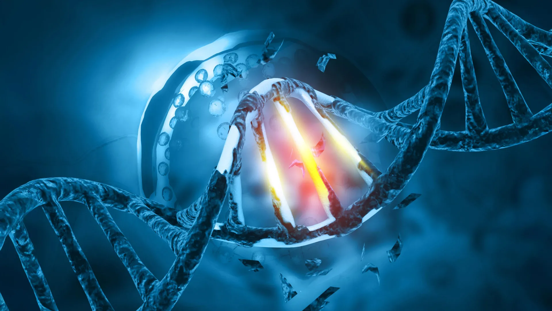
Their analyses revealed that TRF2 does not operate exclusively at the ends of chromosomes. Rather, it binds directly to specific regulatory regions—such as promoters and enhancers—spread throughout the entire genome. Crucially, many of these non-telomeric genomic sites are enriched with secondary DNA structures known as G-quadruplexes (G4s).
G-quadruplexes are stable, four-stranded nucleic acid secondary structures formed in guanine-rich nucleic acid sequences. While historically viewed as potential genomic hazards capable of impeding replication and transcription, contemporary molecular biology recognizes G4s as critical epigenetic and transcriptional regulatory nodes. Intriguingly, these same secondary structures are currently under intensive investigation by the broader scientific community as novel therapeutic targets in oncology, given their involvement in regulating oncogene expression and telomerase activity in cancer cells.
The Penn Medicine study demonstrated that TRF2 interacts directly with these genomic G-quadruplex formations to modulate the transcriptional networks required to preserve muscle stem cell identity. By stabilizing or interacting with G4 structures at specific genetic loci, TRF2 ensures that satellite cells retain the molecular memory and operational capacity needed to execute muscle repair.
"We found that TRF2 works through these secondary DNA structures to preserve the identity of muscle stem cells and keep them capable of repairing damaged muscle," Dr. Mourkioti noted. "That was completely unexpected, as it bridges two entirely different realms of molecular biology: telomere maintenance and genome-wide transcriptional regulation via non-B-form DNA structures."
Official Responses and Academic Implications
The academic and clinical communities have responded to the publication with considerable enthusiasm, viewing the discovery as a conceptual bridge between disparate fields of biomedical research. Dr. Mourkioti and her laboratory are currently expanding their investigations to evaluate whether manipulating TRF2 pathways or targeting the associated G-quadruplex structures could yield translational therapies for muscular dystrophies and other degenerative myopathies.
From a therapeutic standpoint, enhancing TRF2-mediated pathways in dystrophic muscle could theoretically prolong the functional lifespan of endogenous satellite cells, mitigating the exhaustion phase that characterizes late-stage muscular dystrophy. By preserving stem cell identity and preventing the fibrotic conversion of regenerating tissue, clinicians might slow the inexorable decline observed in patients with conditions like Duchenne muscular dystrophy.
Furthermore, the discovery offers intriguing clues regarding a long-standing paradox in cancer biology: while skeletal muscle represents one of the largest tissue masses in the human body and experiences continuous mechanical stress and micro-injury requiring high rates of cellular turnover, primary skeletal muscle cancers (such as soft-tissue sarcomas like rhabdomyosarcoma) are relatively rare compared to carcinomas originating in epithelial tissues.
The specialized regulatory mechanisms by which muscle stem cells utilize proteins like TRF2 to manage regeneration without inducing oncogenic transformation may illuminate protective pathways inherent to striated muscle. Understanding how satellite cells maintain tight control over their proliferative and identity-preserving networks could inform broader cancer research, offering insights into how tissues balance the imperative for tissue repair against the risk of runaway cellular proliferation.
Broader Impact and Future Horizons
As the scientific community digests these findings, the research underscores the growing sophistication of modern genomics, where proteins long pigeonholed into singular functions are increasingly recognized as multi-tasking master regulators. The identification of TRF2 as a steward of muscle stem cell identity redefines the boundaries of telomere biology and opens up new methodologies for addressing tissue degeneration.
The study received primary financial backing and support from competitive grants awarded by the National Institutes of Health (NIH), specifically through the National Institute of Arthritis and Musculoskeletal and Skin Diseases (NIAMS), under award identifiers R01 DK123356, R01 CA174904, GM101149, and FDN-143330. With these sustained resources, the Mourkioti laboratory and collaborating institutions are poised to advance from foundational discovery toward preclinical drug development, aiming to translate these molecular insights into viable clinical interventions for patients afflicted with debilitating muscle-wasting disorders.