Scientists at the Johns Hopkins Kimmel Cancer Center and the Johns Hopkins Bloomberg School of Public Health have unlocked a critical biological mechanism that explains how a rare and aggressive form of kidney cancer progresses. In a study published on April 22 in the scientific journal Cell Reports, researchers detailed how specific genetic mutations create abnormal proteins that condense into microscopic liquid droplets within cells. These droplets act as command centers, aberrantly activating genes responsible for driving tumor growth and metastasis.
The findings offer a profound leap forward in understanding translocation renal cell carcinoma (tRCC), a devastating malignancy that predominantly affects children and young adults and currently lacks standard, highly effective treatment protocols. Supported substantially by funding from the National Institutes of Health (NIH), the breakthrough not only illuminates the pathology of tRCC but also highlights potential therapeutic vulnerabilities that could reshape treatment paradigms for multiple fusion gene-driven cancers.
Decoding the Mechanics of Translocation Renal Cell Carcinoma
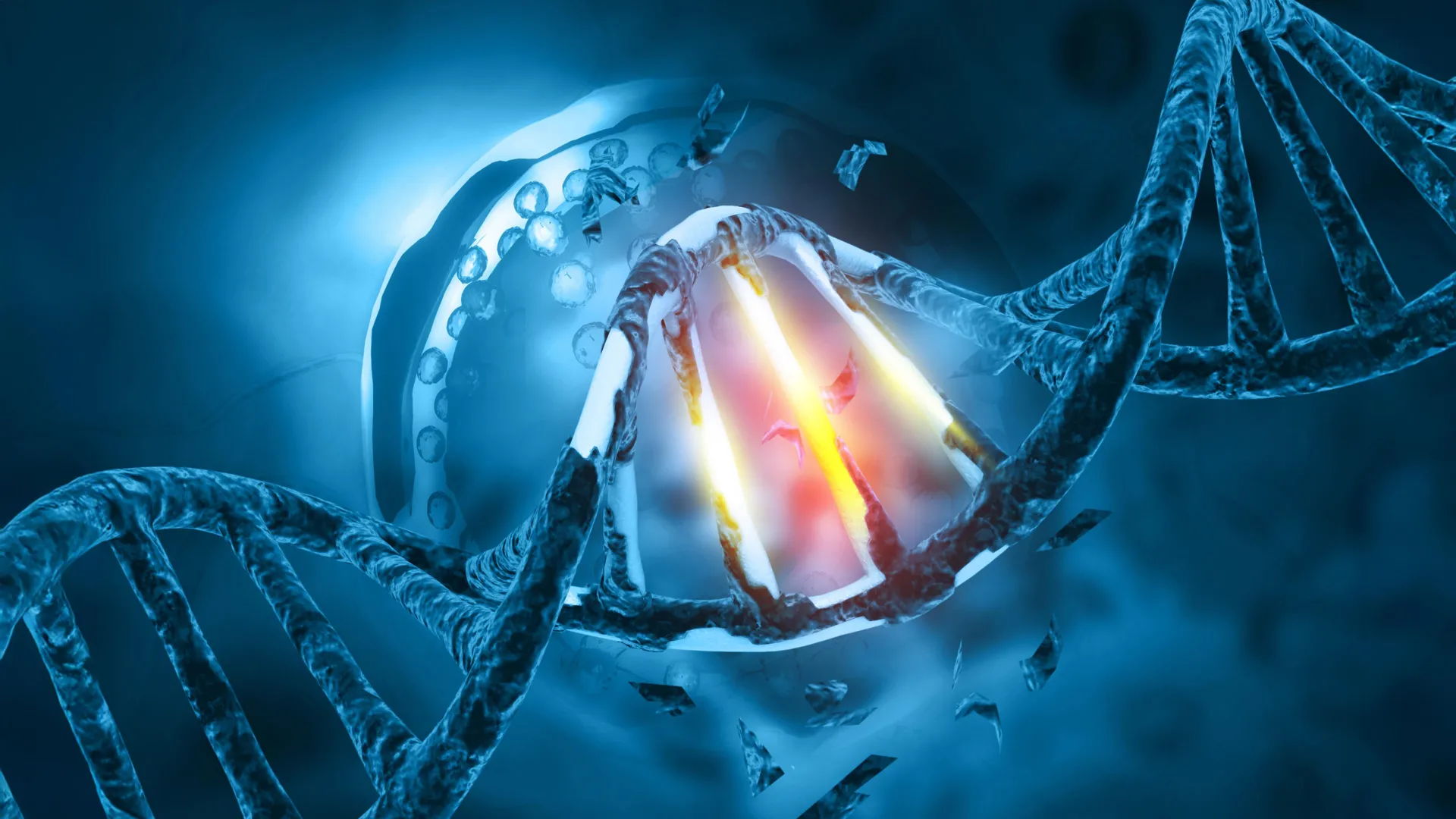
Translocation renal cell carcinoma is triggered by chromosomal rearrangements—genetic accidents in which segments of chromosomes break off and fuse improperly. In the case of tRCC, the tail end of the TFE3 gene becomes fused with the beginning of one of roughly 20 different partner genes, most commonly PRCC, NONO, and SFPQ. These genetic hybrids direct the cell to manufacture abnormal TFE3 fusion proteins that do not exist in healthy human tissue.
While oncologists and molecular biologists have long recognized that these chimeric proteins are the root cause of tRCC, the precise physical mechanisms by which they drove oncogenesis remained elusive until now.
To investigate, senior author Danfeng "Dani" Cai, Ph.D., an assistant professor of biochemistry and molecular biology at the Johns Hopkins Bloomberg School of Public Health, led a team that introduced fluorescent tags to TFE3 fusion proteins within patient-derived cancer cells. Utilizing advanced microscopy, the investigators observed something remarkable: rather than diffusing evenly throughout the cell, the fusion proteins coalesced into distinct, glowing dots within the cell nucleus—the compartment housing human DNA.
These formations are known scientifically as liquid-liquid phase separated condensates. Similar to how oil droplets form and separate in vinegar, these cellular condensates concentrate specific molecules within a localized microenvironment to drive localized biochemical reactions. Cai’s laboratory specializes in studying how such condensates regulate cellular processes.
Within these specific droplets, the research team discovered the presence of key regulatory markers: a protein typically associated with active transcription, and another protein dedicated to switching genes on. This convergence strongly indicated that the TFE3 fusion proteins were not merely present in the nucleus, but were actively interacting with chromatin to alter genetic expression.
Mapping the Epigenetic Landscape and Chromatin Remodeling
To understand precisely how these condensates reprogram cellular behavior, Cai partnered with Eneda Toska, Ph.D., an assistant professor of oncology at the Johns Hopkins Kimmel Cancer Center. Together, the researchers analyzed the interaction between the TFE3 fusion proteins and the cellular genome.
DNA inside human cells is tightly packaged and wound around histone proteins into structures called chromatin, often visualized as beads on a string. When chromatin is densely packed, access to genes is blocked, effectively silencing them. Conversely, when chromatin loops open up, transcription machinery can easily access the underlying DNA sequence to transcribe genes into proteins.
Through genomic mapping and molecular analysis, Toska and her team discovered that the TFE3 fusion condensates directly manipulate this architecture.
"We found that these fusion proteins open and close different sites on the chromatin by making chemical modifications," Toska explained. "They bind, regulate and redesign the chromosome landscape, interacting with target genes that promote cell proliferation and movement—functions that cancer needs to grow and spread."
By actively reshaping the epigenetic and chromosomal landscape, the fusion proteins unleash a cascade of transcription that continuously signals the cancer cells to multiply, migrate, and invade surrounding tissues.
Pinpointing the Vulnerability: The Coiled-Coil Domain
A crucial breakthrough in the study occurred when the research team set out to determine which structural components of the fusion proteins were responsible for forming the liquid droplets in the first place.
By employing targeted genetic engineering, the researchers systematically deleted various segments of the TFE3 fusion proteins. Their experiments revealed that removing a minute structural segment—specifically, a "coiled-coil" domain located within the junction connecting the TFE3 tail to its fusion partner—completely altered the protein’s physical properties.
Without the coiled-coil domain, the modified fusion proteins lost their ability to form liquid condensates. Crucially, once the droplets failed to form, the proteins also lost their capacity to activate the oncogenic genes responsible for cancer progression.
"Individually, all the protein components found in the TFE3 fusions, including full-length TFE3, NONO, and SFPQ, are typically involved in the cell machinery that turns on genes to make proteins," noted Cai. "However, we found when in the form of these fusion proteins, they acquire an even stronger ability to control what genes get turned on."
This structural dependency represents an ideal therapeutic target. Because the formation of liquid condensates is an absolute prerequisite for the cancer-promoting activity of the fusion proteins, disrupting these droplets pharmacologically could halt disease progression.
Broader Implications for Oncology and Fusion-Driven Cancers
While tRCC is classified as a rare malignancy, the broader implications of this research extend far across the oncology landscape. Chromosomal translocations and resulting fusion genes are hallmark drivers of numerous other difficult-to-treat cancers, including Ewing sarcoma, various forms of leukemia, and certain types of sarcomas.
"Other cancers, such as Ewing sarcoma and leukemia, are caused by fusion genes as well," Cai emphasized. "It’s possible that these fusion genes form similar droplets, or condensates, that regulate genes in these cancers and could react to similar treatment strategies."
By establishing a universal paradigm regarding how chimeric transcription factors utilize phase separation to drive oncogenesis, the Johns Hopkins team has provided a blueprint that researchers studying other fusion-driven malignancies can readily apply.
Following the publication of the study, the scientific community has responded with cautious optimism. Although translating these bench-science findings into clinical therapeutics will require extensive development, the identification of a physical structure—the liquid condensate—provides pharmaceutical researchers with a tangible physical entity to target using small molecules or specialized drugs.
Future Directions and Clinical Development
Armed with these insights, the research team is already looking toward the next phase of investigation. Their immediate goal is to identify all additional protein components and molecular partners trapped within or recruited by the TFE3 liquid condensates.
Mapping the complete molecular census of these droplets will allow investigators to design high-throughput drug screens. By testing libraries of small molecules and existing pharmacological compounds, scientists hope to discover agents capable of selectively dissolving or preventing the formation of these oncogenic condensates without disrupting normal cellular housekeeping functions.
Such therapeutic agents could ultimately fill a glaring void in modern medicine: a targeted, standardized treatment protocol for patients diagnosed with translocation renal cell carcinoma.
The research was made possible through collaborative efforts spanning multiple institutions and departments. Additional co-authors from the Johns Hopkins Bloomberg School of Public Health included Choon Leng So, Ye Jin Lee, Wanlu Chen, Binglin Huang, Emily De Sousa, Yangzhengyu Gao, Marie Elena Portuallo, Sumaiya Begum, Kasturee Jagirdar, Vito Rebecca, and Hongkai Ji. They were joined by Bujamin Vokshi of the Johns Hopkins University School of Medicine and W. Marston Linehan of the National Cancer Institute.
Financial backing for the extensive multi-year project was provided by several grants from the National Institutes of Health, including the National Institute of General Medical Sciences (grant R35GM142837); the National Cancer Institute (grants K22CA245487, R01CA276187, and K01CA245124); and the National Human Genome Research Institute (grants R01HG013409 and R01HG010889). Additional support was provided by a Department of Defense Kidney Cancer Idea Development Award (grant W81XWH2210900), a Jayne Koskinas Ted Giovanis grant, and a Johns Hopkins Provost Catalyst Award.
In accordance with institutional transparency policies, study co-author Eneda Toska disclosed that she has received research grant funding from AstraZeneca, alongside consulting fees from both AstraZeneca and Menarini.