Researchers at the University of Hong Kong (HKUMed) have successfully identified the biological mechanism that translates physical activity into skeletal strength, unveiling a crucial cellular pathway that could revolutionize the treatment of osteoporosis and age-related bone degradation. Published in the peer-reviewed journal Signal Transduction and Targeted Therapy, the study centers on a mechanoreceptor protein known as Piezo1, which acts as the body’s internal physical activity sensor. This landmark discovery not only clarifies how mechanical forces preserve bone mass but also lays the scientific groundwork for "exercise mimetics"—pharmacological agents designed to replicate the osteoprotective benefits of physical movement for individuals who are physically incapable of exercising.
Global Implications of Age-Related Bone Loss
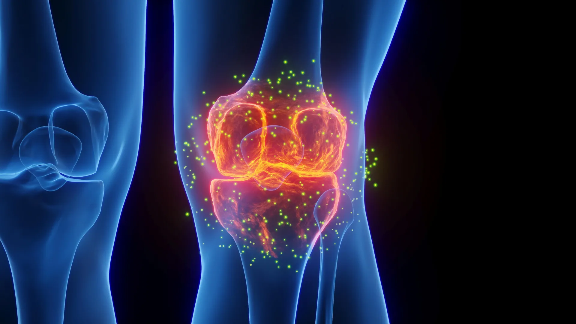
The implications of this research extend far into the global healthcare landscape, addressing a silent epidemic that disproportionately affects aging populations. According to epidemiological data from the World Health Organization (WHO), roughly one in three women and one in five men over the age of fifty will suffer an osteoporotic fracture during their lifetime. These fractures frequently trigger a cascade of negative health outcomes, including chronic pain, permanent loss of mobility, diminished autonomy, and elevated mortality rates, particularly following hip fractures in elderly demographics.
In regions with rapidly aging populations, the strain is acutely felt. In Hong Kong, for instance, demographic aging has driven the prevalence of osteoporosis to staggering levels, impacting approximately 45 percent of women and 13 percent of men aged 65 and older. Beyond the individual suffering, these fractures impose a formidable economic and infrastructural burden on healthcare systems worldwide, consuming billions of dollars annually in acute care, long-term rehabilitation, and institutional support.
Understanding the mechanics of skeletal aging requires examining the microscopic environment within human bones. Bone tissue is a dynamic, living structure in a constant state of remodeling, regulated by osteoblasts (bone-forming cells) and osteoclasts (bone-resorbing cells). Deep within the bone marrow reside mesenchymal stem cells, multipotent progenitor cells capable of differentiating into either bone tissue or adipocytes (fat cells). Throughout youth and early adulthood, mechanical loading from daily movement and weight-bearing exercise stimulates these stem cells to favor osteogenesis, maintaining dense and robust skeletal structures.
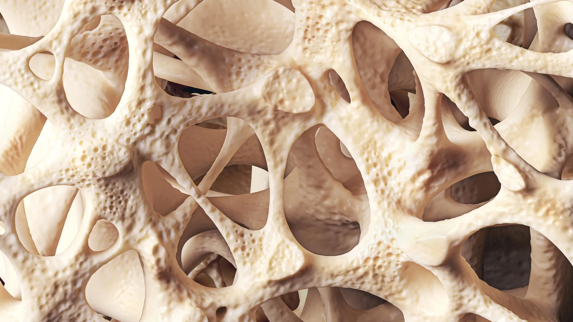
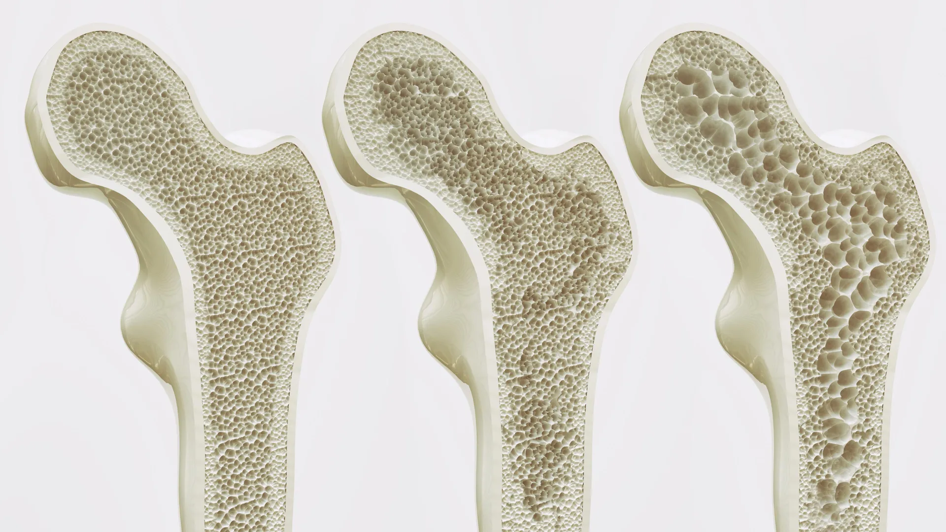
However, the natural aging process alters this delicate cellular equilibrium. As individuals grow older, cellular senescence and systemic shifts promote adipogenesis within the bone marrow. Mesenchymal stem cells increasingly differentiate into fat cells rather than bone cells. This progressive accumulation of bone marrow adipose tissue crowds out healthy osseous matrix, reducing overall bone mineral density, increasing porosity, and establishing a self-perpetuating cycle of structural deterioration that standard osteoporotic medications struggle to reverse once advanced.
The Chronology of the Discovery: From Mechanics to Molecules
The breakthrough by the HKUMed team represents the culmination of years of targeted investigation into mechanobiology—the study of how physical forces influence biological cells. Led by Professor Xu Aimin, Director of the State Key Laboratory of Pharmaceutical Biotechnology and Chair Professor in the Department of Medicine at HKUMed, alongside Research Assistant Professor Dr. Wang Baile and international collaborators including Professor Éric Honore of the French National Centre for Scientific Research (CNRS), the research initiative systematically traced how physical forces translate into cellular instructions.
The investigation began with the hypothesis that specific transmembrane proteins must exist to convert physical forces—such as tension, compression, and shear stress generated by muscle contractions and gravity—into biochemical signals. Utilizing advanced murine models combined with human bone marrow stem cell cultures, the research consortium zeroed in on Piezo1. Known broadly as an ion channel responsive to mechanical stretch, its specific role in bone marrow stem cell lineage allocation during physical loading had remained poorly understood until now.
Through a series of rigorous in vivo and in vitro experiments, the researchers demonstrated that activating Piezo1 via mechanical simulation or pharmacological agonists significantly suppressed adipogenesis within the bone marrow while concurrently upregulating pathways responsible for new bone formation. Conversely, the experimental deletion or silencing of the Piezo1 gene in mouse models led to accelerated bone loss. The absence of the protein triggered the pathological secretion of specific inflammatory signaling molecules, namely Ccl2 and lipocalin-2. These pro-inflammatory cytokines further drove stem cells down the adipogenic pathway while actively inhibiting osteogenic differentiation, thereby accelerating the osteoporotic phenotype. Crucially, the team discovered that chemically blocking these inflammatory signals partially rescued bone health, highlighting multiple points for therapeutic intervention.
Official Responses and Expert Analysis
The significance of identifying the Piezo1 pathway has drawn praise from the international scientific community, highlighting the collaborative nature of the project which united top-tier institutions in Hong Kong and France.
"Osteoporosis and age-related bone loss affect millions worldwide, often leaving elderly and bedridden patients vulnerable to fractures and loss of independence," stated Professor Xu Aimin during the release of the findings. "Current treatments rely heavily on physical activity, which many patients simply cannot perform. We need to understand how our bones get stronger when we move or exercise before we can find a way to replicate the benefits of exercise at the molecular level. This study is a critical step towards that goal."
Elaborating on the therapeutic potential, Professor Xu noted the core achievement of the study: "We have essentially decoded how the body converts movement into stronger bones. We have identified the molecular exercise sensor, Piezo1, and the signalling pathways it controls. This gives us a clear target for intervention. By activating the Piezo1 pathway, we can mimic the benefits of exercise, effectively tricking the body into thinking it is exercising, even in the absence of movement."
Dr. Wang Baile emphasized the immediate clinical relevance for vulnerable populations who are routinely excluded from traditional physical therapy regimens. "This discovery is especially meaningful for older individuals and patients who cannot exercise due to frailty, injury, or chronic illness," Dr. Wang remarked. "Our findings open the door to developing ‘exercise mimetics’—drugs that chemically activate the Piezo1 pathway to help maintain bone mass and support independence."
Adding a European perspective, Professor Éric Honoré of the Institute of Molecular and Cellular Pharmacology at CNRS, UniCA, and Inserm, who also serves as a Visiting Professor in the Department of Pharmacology and Pharmacy at HKUMed, underscored the broader preventive implications of the discovery. "This offers a promising strategy beyond traditional physical therapy," Professor Honoré stated. "In the future, we could potentially provide the biological benefits of exercise through targeted treatments, thereby slowing bone loss in vulnerable groups such as bedridden patients or those with limited mobility, and substantially reducing their risk of fractures."
Translating Science into Clinical Reality
The successful identification of Piezo1 as the mechanical transducer of exercise opens an entirely new therapeutic frontier in endocrinology and orthopedics. Traditional pharmacological treatments for osteoporosis largely fall into two categories: antiresorptive agents (such as bisphosphonates), which slow down bone breakdown, and anabolic agents (such as teriparatide), which stimulate bone formation. However, neither approach addresses the underlying root cause of age-related bone marrow adiposity or replicates the systemic, multi-tissue benefits of physical conditioning.
Exercise mimetics offer a paradigm shift. By designing small molecules or biologics that selectively target and activate the Piezo1 pathway within mesenchymal stem cells, future therapeutics could trick the bone marrow into maintaining an osteogenic profile regardless of the patient’s actual physical activity levels. This would prove invaluable not only for the elderly and bedridden but also for individuals undergoing prolonged immobilization due to severe trauma, neurological disorders, or spaceflight, where microgravity induces rapid skeletal degeneration.
The collaborative research team is already pivoting toward the next phase of development: translational clinical applications. Funding and infrastructural support for these ongoing investigations have been secured through a robust coalition of grants, including the Areas of Excellence Scheme and the General Research Fund of the Research Grants Council; the Health and Medical Research Fund under the Health Bureau of the Government of the Hong Kong Special Administrative Region; the National Key R&D Program of China; the National Natural Science Foundation of China; the Human Frontier Science Program; the French National Research Agency; Fondation de France; Fondation pour la Recherche Médicale; and the Macau Science and Technology Development Fund.
As the research moves from murine models toward human clinical trials, the medical community maintains cautious optimism. While developing safe and effective exercise mimetics will require rigorous pharmacokinetic and pharmacodynamic optimization, the HKUMed study provides the foundational map required to navigate this complex biological terrain. By bridging the gap between mechanical force and molecular signaling, researchers have brought humanity one step closer to preserving skeletal integrity across the entire lifespan, offering renewed hope to millions facing the specter of frailty and bone loss.