Every second of every day, the human body undergoes a silent, staggering feat of biological engineering. Deep within the spongy interiors of the skeletal system, the bone marrow acts as a relentless production factory, generating millions of fresh blood cells and immune defenders to maintain systemic health. This nonstop renewal relies on a meticulously calibrated ecosystem: a delicate, interdependent relationship between hematopoietic stem cells (HSCs), supportive stromal cells, and an intricate network of biochemical and immune signals. Yet, this vital regenerative engine is not immune to the relentless pressures of time. As individuals age, this biological balance inevitably frays.
For years, medical science viewed the deterioration of bone marrow primarily as a consequence of intrinsic genetic errors accumulating within stem cells themselves. However, a landmark international study co-led by researchers from the European Molecular Biology Laboratory (EMBL), the University of Basel, and the University Medical Center (UMC) Mainz has fundamentally shifted this paradigm. By conducting an unprecedented molecular and spatial mapping of the human bone marrow microenvironment, the research team has revealed that chronic inflammation and structural remodeling within the marrow’s ecosystem act as primary drivers of pre-leukemic states and aggressive blood cancers—often long before any clinical symptoms manifest in the patient.
The findings, published in Nature Communications alongside a complementary study led by Marc Raaijmakers from the Erasmus MC Cancer Institute in Rotterdam, offer a transformative view of how blood disorders begin. By redirecting scientific focus from mutated stem cells alone to the surrounding cellular niche that nourishes them, the research opens entirely new frontiers for early diagnostics, preventative therapies, and interventions targeting age-related chronic inflammation.
The Silent Onset of Clonal Hematopoiesis and Myelodysplastic Syndrome
To understand the gravity of these findings, one must examine the precarious trajectory of aging hematopoiesis. Over decades of life, the delicate equilibrium of the bone marrow becomes vulnerable to multiple stressors, including natural aging, chronic systemic inflammation, and somatic mutations. While the human body possesses remarkable DNA repair mechanisms, occasional mutations in HSCs slip through unchecked. When a single mutated stem cell gains a slight survival or replication advantage over its healthy neighbors, it begins to outpace them, slowly expanding its clone throughout the marrow.
This phenomenon is known as clonal hematopoiesis of indeterminate potential, commonly referred to as CHIP. Clinically silent in its early stages, CHIP is remarkably prevalent in older demographics. Epidemiological data indicates that CHIP appears in approximately 10 to 20 percent of adults over the age of 60, and its prevalence surges to nearly 30 percent in individuals over 80. Although people living with CHIP typically experience no overt symptoms, the condition carries profound long-term health risks. Individuals with CHIP face a tenfold increase in the incidence of blood cancers and a doubled likelihood of cardiovascular disease and premature mortality.
When CHIP progresses or when distinct genetic lesions accumulate further, it can evolve into more severe disorders, such as myelodysplastic syndrome (MDS). MDS is a related clonal bone marrow disorder characterized by inefficient blood-cell production, progressive cytopenias, and eventual bone marrow failure. The condition predominantly affects older populations, striking up to 20 in every 100,000 adults over the age of 70. Most alarming is MDS’s propensity for malignant transformation: approximately 30 percent of MDS cases ultimately advance to acute myeloid leukemia (AML), an aggressive, rapidly progressing, and frequently fatal cancer of the blood and bone marrow.
Despite the severe morbidity and mortality associated with these conditions, the precise contribution of the bone marrow microenvironment—the physical and biochemical niche housing these stem cells—to disease initiation and progression has remained obscure until now.
Mapping the Hidden Architectural Shifts of the Bone Marrow Niche
To uncover how mutated HSC clones successfully seize dominance within the marrow, an international research consortium launched an exhaustive molecular and spatial analysis of human bone marrow samples. The biological specimens were meticulously gathered through the BoHemE cohort study, a collaborative effort involving hematology experts, notably Uwe Platzbecker at the National Center for Tumor Diseases (NCT) Dresden.
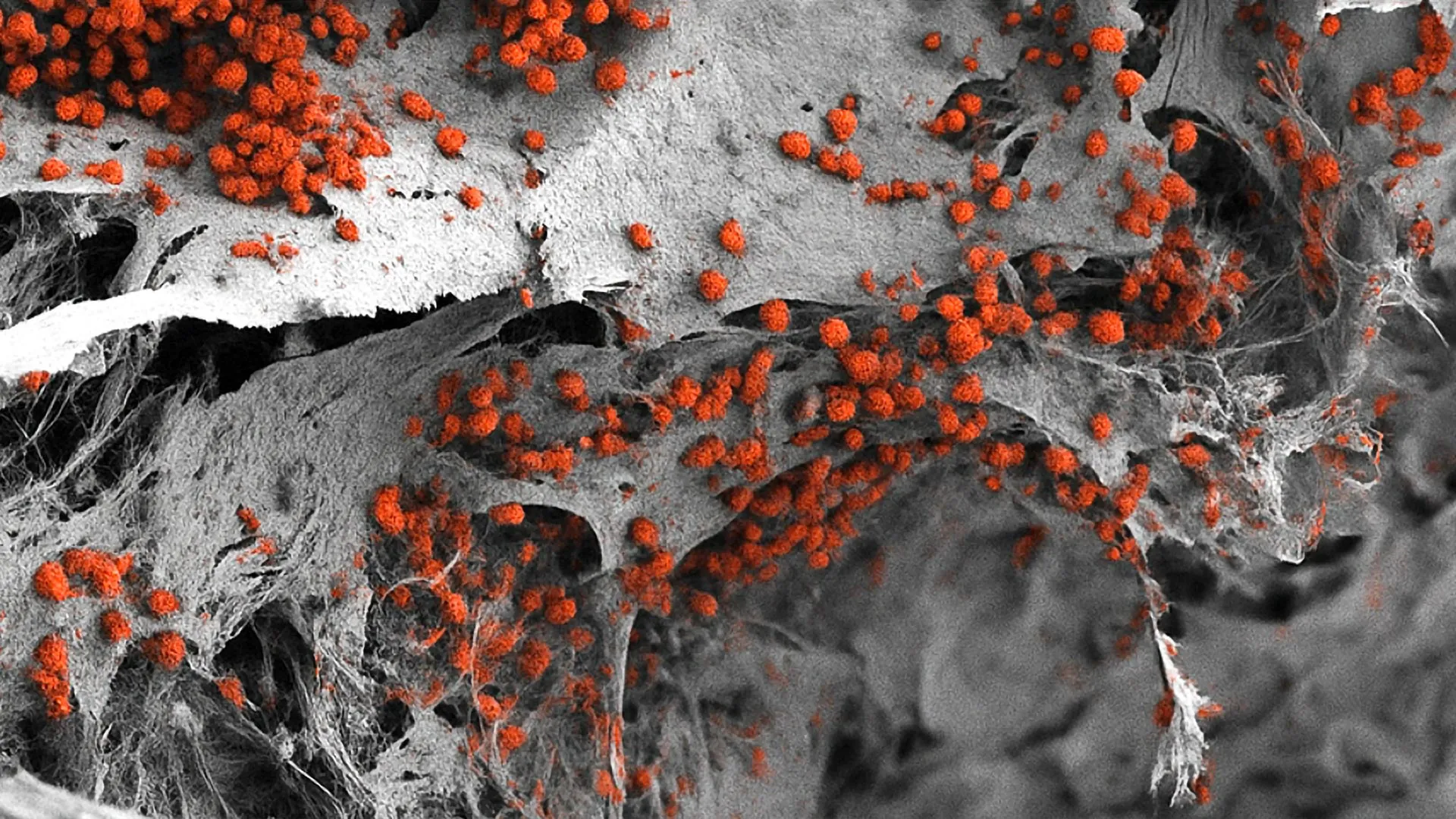
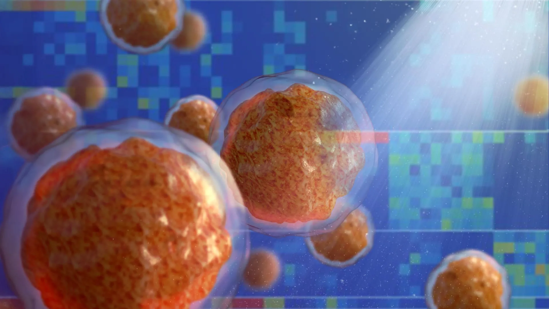
Deploying a powerful suite of cutting-edge technologies, the research team combined single-cell RNA sequencing, high-resolution biopsy imaging, advanced proteomics, and complex co-culture models. This multi-layered approach allowed the scientists to construct a high-resolution, comprehensive map of the bone marrow microenvironment across three distinct cohorts: healthy donors, individuals with incidental CHIP, and patients diagnosed with advanced MDS.
The resulting data revealed an unexpected and dramatic cellular shift that initiates long before any clinical pathology becomes apparent. The researchers discovered that as individuals age and develop CHIP or MDS, a specialized population of inflammatory stromal cells gradually replaces the normal mesenchymal stromal cells (MSCs) that typically provide structural and biochemical support to healthy stem cells.
"I was surprised to observe such pronounced remodeling of the bone marrow microenvironment already in individuals with CHIP, although the underlying cause-and-effect relationships remain unclear," noted Judith Zaugg, co-senior author of the study, EMBL Group Leader, and Professor at the University of Basel.
Unlike their healthy counterparts, these newly identified inflammatory mesenchymal stromal cells (iMSCs) undergo a profound functional transformation. They begin synthesizing and secreting massive quantities of interferon-induced cytokines and chemokines. These inflammatory signaling molecules act as beacons, actively recruiting and hyperactivating interferon-responsive T cells within the marrow niche.
Once activated, these T cells further amplify the local inflammatory output, establishing a self-sustaining, feed-forward loop. This chronic inflammatory cascade not only disrupts normal hematopoiesis—suppressing the production of healthy red blood cells, white blood cells, and platelets—but also induces pathological vascular changes within the bone marrow architecture.
Dissecting the Drivers of Marrow Inflammation
One of the most perplexing questions facing the research team was whether the mutated hematopoietic cells themselves directly triggered this aggressive inflammatory response within the surrounding stromal cells. To answer this, the investigators needed a reliable method to distinguish mutated cells from their healthy, non-mutated neighbors within the complex single-cell datasets.
To achieve this, the team utilized SpliceUp, an innovative computational method developed by co-lead author and EMBL alumnus Maksim Kholmatov, working in close collaboration with Pedro Moura and Eva Hellström-Lindberg from the Karolinska Institute. SpliceUp operates by scanning single-cell RNA sequencing data to identify abnormal RNA-splicing patterns—a hallmark of mutated cells in disorders like MDS.
By applying this tool, the researchers made a counterintuitive discovery: the mutated hematopoietic cells themselves did not appear to be the direct, primary triggers of the inflammatory storm within the stroma. Instead, the inflammatory network within the microenvironment appears to take on a life of its own, eventually becoming dominant and systematically dismantling the marrow’s normal regenerative architecture.
Furthermore, the study illuminated specific mechanical failures within the diseased niche. "Another striking observation was that MDS stem cells couldn’t trigger stromal cells to produce CXCL12, an important signal that triggers blood cells to settle in the bone marrow," explained Karin Prummel, co-lead author and EMBL postdoctoral researcher. "This failure may help explain why the bone marrow stops working properly."
Maksim Kholmatov echoed the significance of these findings. "It was quite surprising to see the lack of a direct inflammatory effect that we could attribute to the mutant cells," Kholmatov stated. "However, when viewed in the context of changes in the T cell and stromal compartments, it underlines the importance of the bone marrow microenvironment in shaping disease progression."
Inflammation as an Early Catalyst for Malignant Evolution
The implications of these discoveries stretch far beyond basic cell biology, fundamentally redefining how modern hematology must approach the prevention and treatment of age-related blood disorders. By demonstrating that localized chronic inflammation plays a central role in the earliest phases of disease development, the research elevates the bone marrow microenvironment—often referred to simply as the bone marrow niche—to a premier therapeutic target.
Traditional oncology has long focused almost exclusively on eradicating mutated malignant cells through cytotoxic chemotherapy, targeted molecular inhibitors, or stem cell transplantation. However, these new findings suggest that targeting the mutated clones alone may be insufficient if the supportive ecosystem that fostered their growth remains corrupted and inflamed.
By directing therapeutic strategies toward the microenvironment, clinicians may soon be able to intercept disease progression at its inception. For older adults diagnosed with CHIP, future interventions could involve the use of targeted anti-inflammatory drugs or novel therapies designed to modulate aberrant interferon signaling, thereby preserving normal bone marrow function and preventing the transition to full-blown MDS or AML.
Moreover, the unique molecular signatures identifying iMSCs and interferon-responsive T cells could soon serve as reliable early prognostic biomarkers. Clinicians could utilize these markers to screen aging populations, identifying individuals at the highest risk of malignant progression long before clinical cytopenias or leukemia manifest.
"Our findings reveal that the bone marrow microenvironment actively shapes the earliest stages of malignant evolution," emphasized Borhane Guezguez, Principal Investigator in the Department of Hematology at UMC Mainz and co-senior author of the study. "As advances in molecular profiling allow us to detect pre-leukemic states years before clinical onset, understanding how stromal and immune cells interact provides a foundation for preventive therapies that intercept disease progression before leukemia develops."
Inflammaging, Systemic Aging, and Future Horizons
Beyond the specific context of hematological malignancies, this research contributes vital pieces to the broader scientific understanding of "inflammaging"—a term used by biogerontologists to describe the low-grade, chronic, systemic inflammation that accompanies aging and underpins a vast spectrum of age-related diseases, including cardiovascular disorders, type 2 diabetes, neurodegeneration, and solid-tumor cancers.
Historically, the bone marrow was perceived merely as a passive manufacturing plant for blood cells, shielded within the skeletal structure. This study firmly repositions the marrow as a central player in systemic aging—an organ that is both profoundly affected by and actively responsible for driving systemic inflammatory aging. By mapping how complex cross-talk between immune cells and stromal components drives pathological remodeling, the research establishes a robust mechanistic model that can now be applied to investigate inflammatory remodeling in other myeloid malignancies and advanced leukemias.
Despite these monumental breakthroughs, the researchers emphasize that much work remains. "It will be crucial to study these processes over time; our current findings are based on cross-sectional data," noted Professor Zaugg.
This temporal dimension carries profound implications for existing medical procedures, such as allogeneic hematopoietic stem cell transplantation. Currently, transplants replace diseased or malignant hematopoietic cells with healthy donor stem cells, but they typically leave the patient’s original bone marrow microenvironment intact. Zaugg and her colleagues are now actively investigating whether the bone marrow niche retains a biological "memory" of past disease—a cellular and molecular imprint that might adversely influence how the niche interacts with and supports newly introduced, healthy donor stem cells.
The publication of this study, appearing alongside complementary insights from Erasmus MC Cancer Institute, marks a watershed moment in hematology. By bridging spatial biology, single-cell genomics, and clinical medicine, researchers have illuminated the hidden architecture of the aging marrow. As the medical community digests these revelations, the path toward intercepting blood cancers at their earliest whispers—by healing the microenvironment rather than just targeting the mutated seed—is finally coming into focus.
The international research effort involved a broad consortium of collaborating institutions, including the University Medical Center Mainz, the University of Basel, University Hospital Dresden, the Karolinska Institute in Sweden, The Jackson Laboratory in the United States, Sorbonne University in France, and German Consortium for Translational Cancer Research (DKTK) partner institutions, including the German Cancer Research Center (DKFZ) and NCT Dresden. Financial support for the research was provided by the DKTK-CHOICE program, the European Research Council (ERC) under the grant EpiNicheAML awarded to Judith Zaugg, the Marie Skłodowska-Curie Actions (MSCA)-funded ITN ENHPATHY, EMBO, the Swiss National Science Foundation, and the José Carreras Leukämie-Stiftung.