
Nearly a century ago, a monumental shift in optical microscopy forever changed how scientists examine biological samples. Developed in the 1930s by Dutch physicist Frits Zernike, the phase-contrast technique addressed a fundamental limitation of light microscopy: biological cells, composed largely of water, are nearly transparent. When light passes through them, cellular structures such as nuclei and mitochondria cause negligible shifts in brightness, rendering them virtually indistinguishable without toxic chemical stains that destroy living tissue. Zernike discovered that light passing through a specimen experiences a minute spatial delay, altering its phase. By artificially shifting the phase of unscattered light by 90 degrees, his innovation translated these invisible phase shifts into stark, visible differences in brightness. The profound impact of this discovery was officially recognized with the Nobel Prize in Physics in 1953, setting a precedent that phase manipulation is essential for high-contrast biological imaging.
Now, a team of physicists at the University of California, Berkeley, alongside collaborators at the Chan Zuckerberg Biohub, has successfully translated this Nobel Prize-winning principle into the realm of modern electron microscopy. By developing an unprecedentedly intense laser phase plate, these researchers have overcome a critical half-century bottleneck in structural biology. This breakthrough significantly enhances cryoelectron microscopy (cryo-EM) and cryoelectron tomography (cryo-ET), pushing the technological boundaries to visualize biological macromolecules and intricate cellular landscapes previously obscured by insurmountable signal-to-noise limitations.
The Main Facts and Technological Innovation
At the heart of this technological leap is a device capable of generating what is widely considered the world’s most intense focused continuous-wave laser. Traditional electron microscopes utilize a beam of electrons to magnify objects up to 10,000 times more powerfully than conventional light microscopes, providing atomic-scale details of macromolecular structures. However, achieving high contrast without damaging fragile biological specimens has remained an ongoing challenge.
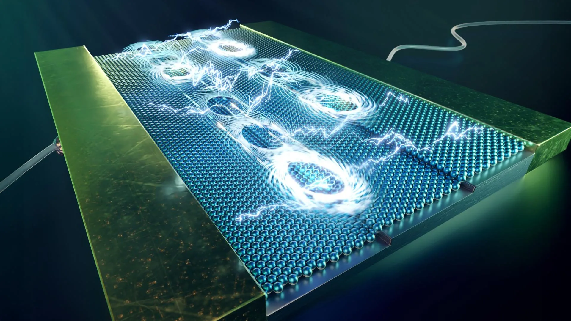
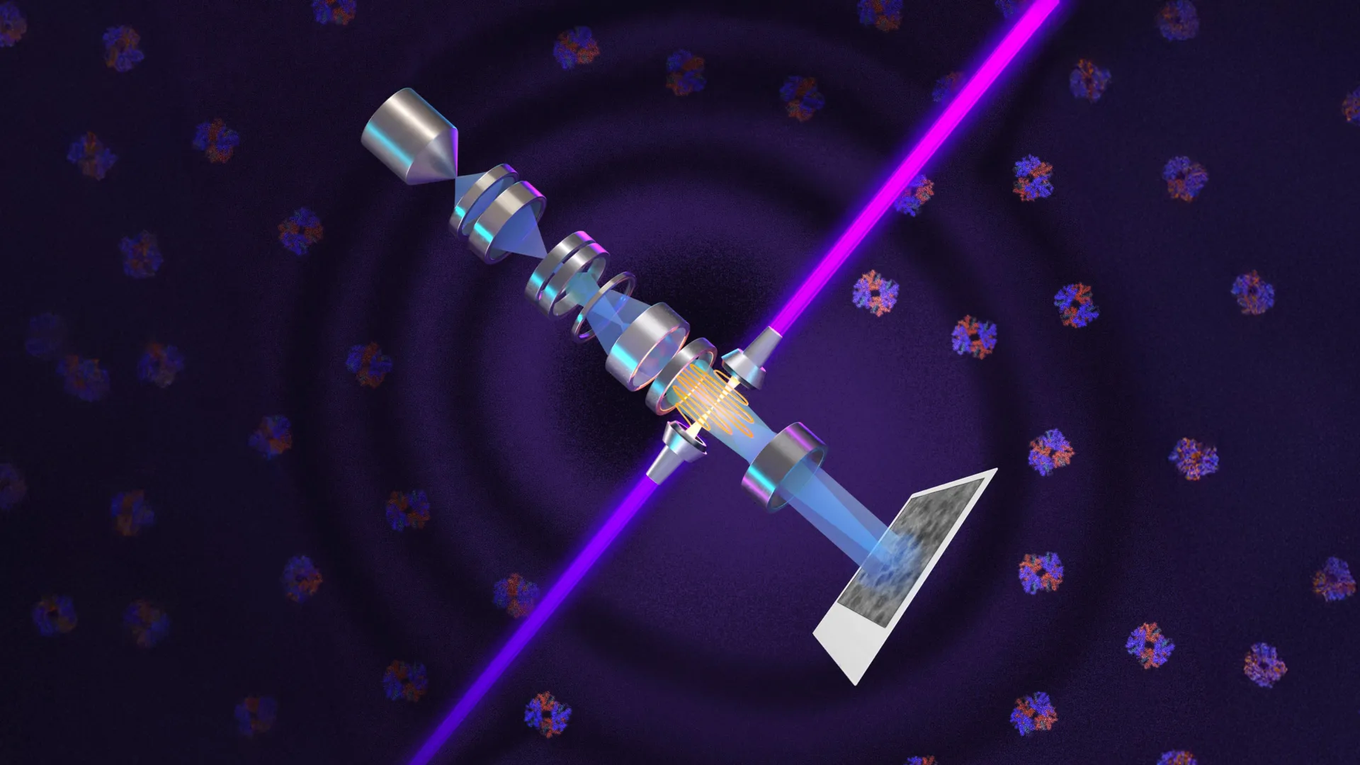
The UC Berkeley research team’s laser phase plate does not merely illuminate a specimen; instead, it interacts directly with the microscope’s electron beam. By confining a continuous laser beam inside a specialized spherical optical cavity lined with mirrors, the light reflects more than 10,000 times. This optical accumulation concentrates roughly 75 kilowatts of power—surpassing the intensity of industrial welding lasers and military-grade directed-energy systems—into a microscopic space measuring just a few microns across.
When the microscope’s electron beam intersects this high-intensity laser focus, its phase is precisely altered by 90 degrees, perfectly mirroring Zernike’s optical phase-contrast concept. This direct phase shift drastically enhances the signal-to-noise ratio, making faint, low-contrast specimens stand out sharply against background noise. To house and test this advanced engineering, UC Berkeley physics professor Holger Müller and his team acquired a state-of-the-art Thermo Fisher Krios cryo-EM instrument, christening the customized microscope Theia, named after the ancient Greek Titaness of light and radiance. Concurrently, a parallel engineering effort directed by Bridget Carragher at Biohub in Redwood City, California, has pioneered a dual-laser configuration operating at right angles to minimize optical distortions and component wear.
A 15-Year Chronology of Scientific Persistence
The realization of the laser phase plate is the culmination of a rigorous 15-year scientific endeavor marked by theoretical milestones and persistent engineering hurdles.
The chronological development of the technology spans several key periods:
- 1930: Frits Zernike invents phase-contrast microscopy, establishing the mathematical and physical foundations for phase manipulation, later earning him the 1953 Nobel Prize in Physics.
- Early 1940s to 2000s: Early attempts to apply phase-contrast principles to electron microscopy repeatedly stall due to severe technical roadblocks, including excessive beam attenuation, severe image instability, and compromised resolution.
- 2010: Holger Müller and Robert Glaeser, a UC Berkeley professor emeritus of molecular and cell biology, publish a foundational theoretical paper proposing the use of an intense laser field to impart the necessary phase shift on electron beams without sacrificing intensity.
- 2010–2025: Backed by funding from the National Institutes of Health and later the Chan Zuckerberg Biohub, Müller spends a decade and a half engineering the ultra-intense continuous-wave optical cavity required to generate the necessary laser focus.
- Recent Months: The Berkeley team successfully integrates the laser phase plate into a customized electron microscope, testing it across multiple biological samples and publishing their findings in the journal Science, while Biohub releases a complementary preprint detailing their dual-laser architecture.
Supporting Data and Molecular Scale Limitations
To contextualize the importance of this breakthrough, one must examine the dimensional constraints that have historically crippled conventional cryo-EM and cryo-cytology. Protein mass is measured in daltons, where one dalton equals one-twelfth the mass of a carbon-12 atom. Standard cryo-EM instruments encounter severe analytical walls when attempting to image macromolecules smaller than approximately 70 kilodaltons (kDa).
This threshold represents a profound structural blind spot in modern biochemistry. Approximately 90% of the entire human proteome—the complete catalog of proteins encoded by the human genome—consists of molecules falling below this 70 kilodalton limit. Vital regulatory proteins, signaling molecules, and numerous human and animal disease targets are simply too small or generate too little scattering contrast to be successfully resolved by standard cryo-EM protocols.
By introducing the laser phase plate, researchers have demonstrated the ability to clearly resolve proteins as small as 50 kilodaltons, testing samples such as muscle-derived aldolase and oxygen-carrying hemoglobin. The performance gains are particularly pronounced for smaller, structurally complex targets. Furthermore, Müller’s theoretical frameworks suggest that subsequent optimizations could push this limit down to 17 kilodaltons—the approximate molecular weight of myoglobin—by implementing a tightly focused electron beam rather than the defocused illumination currently required.
Official Responses and Expert Perspectives
The academic and industrial communities have responded to the breakthrough with considerable enthusiasm, viewing the development as a foundational paradigm shift for structural biology and biomedical research.
"Cryo-EM has become the new, fastest-growing method for resolving the structure of biological macromolecules, and cryo-ET is expected to show how these molecules work together in their natural, cellular context," stated Holger Müller, who led the UC Berkeley development effort. "But because of signal-to-noise limitations, the majority of human and animal proteins are too small to be analyzed by these methods. The increase in signal-to-noise ratio provided by this laser phase plate is expected to overcome these important limitations."
Bridget Carragher, founding technical director of imaging at Biohub, emphasized the chaotic microenvironment inside living cells that cryo-tomography aims to decode. "With cryo-ET, we’re looking at small, very complicated cellular material that’s incredibly crowded inside the cell," Carragher noted. "It’s like a forest of trees, and you’re trying to find one leaf on one tree in there. Cryo-ET needs a dramatic step forward in contrast, so we can start to see what’s going on inside the cell. That’s what the laser phase plate promises to give us."
Echoing these sentiments, Stephani Otte, Biohub’s Vice President of Imaging Science, highlighted the broader biomedical implications of rendering the invisible visible. "This technology is a step function change for biology," Otte remarked. "We are going to be able to see how molecular machines operate inside the living cell, in context, for the first time. What was once invisible will become visible—and that changes everything about how we understand disease."
Broader Impact and Future Implications
The integration of laser phase plates into cryo-EM and cryo-electron tomography carries sweeping implications for foundational science, pharmaceutical design, and translational medicine. Over the past decade, cryo-EM has transformed drug discovery by enabling researchers to map the three-dimensional atomic structures of large therapeutic targets without the painstaking process of crystallization required by traditional X-ray crystallography.
However, the inability to image smaller proteins has left vast pharmaceutical landscapes unexplored. By unlocking access to the sub-70 kilodalton proteome, the laser phase plate enables scientists to study critical drug targets that were previously invisible. In the realm of cryo-ET—which constructs three-dimensional reconstructions of intact cellular environments by compiling tilt-series images—the signal amplification provided by the laser phase plate promises to reveal the precise choreography of macromolecular complexes interacting within their native cellular matrix.
As research teams at UC Berkeley and Biohub continue refining their optical hardware in partnership with leading instrument manufacturer Thermo Fisher Scientific, the scientific community stands on the precipice of a new observational epoch. By bridging a century-old optical theory with state-of-the-art quantum and laser engineering, this innovation transforms the fundamental capabilities of microscopy, opening unprecedented avenues for decoding the molecular mechanisms of human health and disease.