
Mitochondria, the indispensable powerhouses of eukaryotic cells, are responsible for generating the vast majority of the energy required for cellular functions through the process of oxidative phosphorylation. This critical role is supported by their unique possession of their own distinct genetic material, mitochondrial DNA (mtDNA). Within each cell, hundreds to thousands of copies of mtDNA are meticulously organized into compact structures known as nucleoids. For decades, scientists have observed a striking regularity in the spatial arrangement of these nucleoids within the mitochondrial network, a phenomenon crucial for ensuring reliable mtDNA transmission during cell division and for maintaining equitable gene expression across the organelle. However, the precise mechanism by which this precise spatial organization is achieved has remained an enduring enigma in cell biology.
Unraveling a Cellular Enigma: The Mystery of Nucleoid Spacing
The functional integrity of mitochondria and their DNA is paramount to cellular health. Aberrations in mitochondrial function are not isolated incidents; they are intricately linked to a spectrum of severe human diseases. These range from debilitating metabolic and neurological disorders, such as liver failure and encephalopathy, to age-related degenerative conditions like Alzheimer’s and Parkinson’s disease. The profound implications of mitochondrial dysfunction underscore the importance of understanding the fundamental processes that govern their operation, including the organized distribution of their genetic material.
Prior to the recent breakthrough, proposed explanations for nucleoid spacing relied on mechanisms such as mitochondrial fusion and fission, or molecular tethering. However, as Suliana Manley, professor at the Laboratory of Experimental Biophysics (LEB) at EPFL, noted, these hypotheses failed to fully account for the observed phenomenon. "Proposed mechanisms related to mitochondrial fusion, fission, or molecular tethering cannot explain it, since nucleoid spacing is maintained even when they are disrupted," Manley stated. This observation highlighted the inadequacy of existing models and pointed towards a more fundamental, perhaps overlooked, biophysical process.
A Rediscovered Phenomenon: Mitochondrial Pearling Takes Center Stage
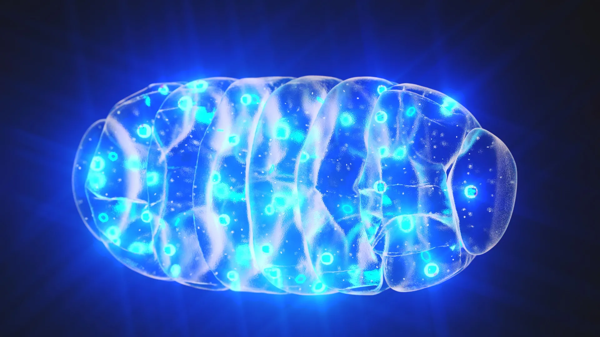
The research conducted by Professor Suliana Manley and her postdoctoral fellow Juan Landoni at EPFL has now illuminated the long-sought mechanism responsible for maintaining the regular spacing of mitochondrial nucleoids. Their groundbreaking work identifies and elevates a previously underappreciated process termed "mitochondrial pearling." This dynamic transformation involves temporary shape changes in mitochondria, causing them to adopt a characteristic "beads on a string" appearance. During these transient phases, clusters of mtDNA are effectively separated and redistributed, leading to a more even spread of nucleoids throughout the mitochondrial network.
Visualizing the Unseen: Advanced Microscopy Techniques Illuminate Mitochondrial Dynamics
To unravel the intricacies of mitochondrial pearling, the research team employed a sophisticated arsenal of advanced imaging techniques. These included cutting-edge super-resolution microscopy, which allows for visualization of cellular structures with unprecedented detail, correlated light and electron microscopy (CLEM) for combining the strengths of both optical and electron imaging, and phase contrast microscopy for observing living cells in real-time.
The integration of these powerful tools enabled the scientists to meticulously track individual nucleoids, capture the rapid and ephemeral changes in mitochondrial morphology during pearling events, and gain a deeper understanding of the internal organizational architecture of these vital organelles. The ability to observe these dynamic processes within living cells provided crucial insights that were previously unattainable.
The Mechanics of Pearling: A Bead-like Transformation
Live-cell imaging experiments revealed that mitochondrial pearling is a remarkably dynamic process, capable of occurring multiple times within a single minute. During these events, mitochondria transiently form a series of evenly spaced constrictions along their elongated structures, creating the distinct "pearls." Crucially, the inter-pearl distances observed during these transformations closely mirror the typical spacing found between nucleoids in a resting mitochondrion.
Further detailed observation confirmed that most of these pearl-like segments contain a nucleoid positioned near their center. Interestingly, the formation of these structures can also occur even in the absence of mtDNA, suggesting that the physical scaffolding of the mitochondrion plays a significant role in guiding this process. As the pearling event progresses, larger aggregations of nucleoids often fragment into smaller units. These smaller nucleoid clusters then migrate and settle into adjacent pearls. When the mitochondrion subsequently reverts to its more typical tubular shape, the nucleoids remain distributed, thus preserving their even spacing and ensuring a consistent genetic landscape within the organelle.
Unraveling the Control Mechanisms: Calcium and Internal Membranes
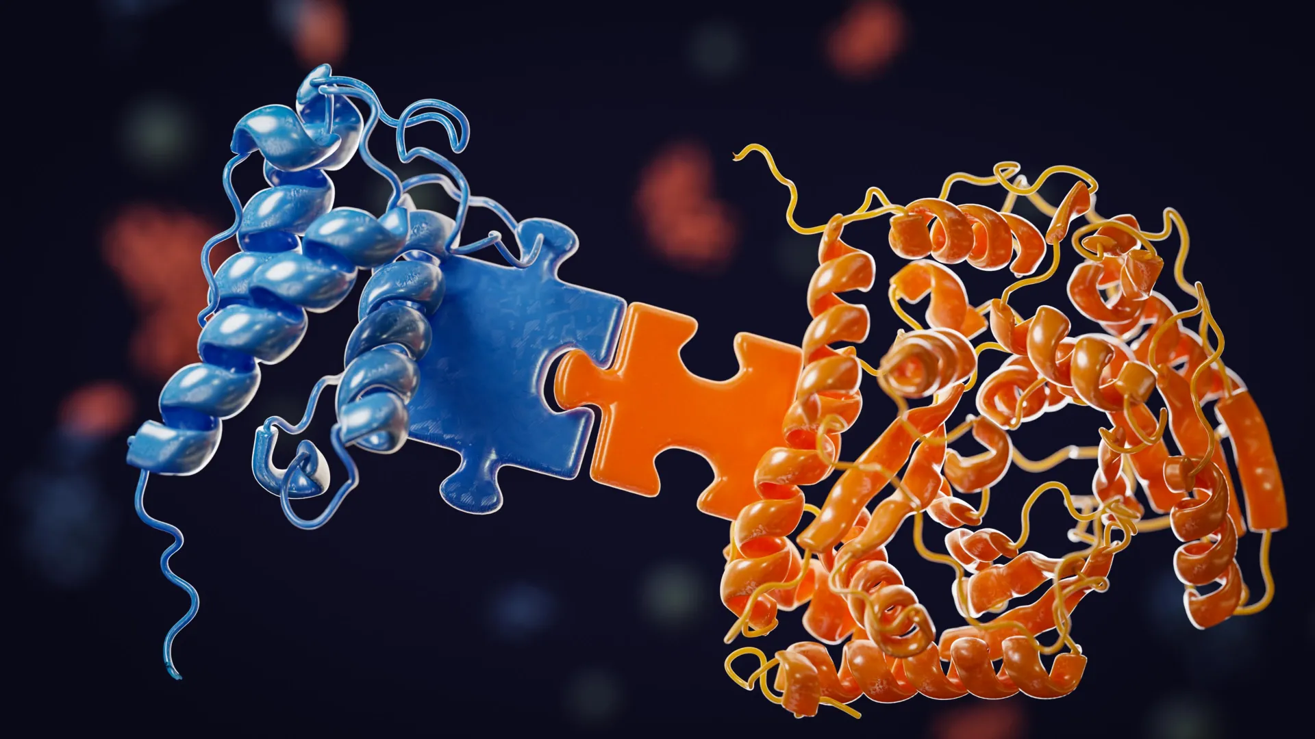
Beyond describing the physical process, the researchers delved into the regulatory factors that govern mitochondrial pearling. Through a series of targeted genetic and pharmacological experiments, they identified key elements that trigger and maintain this organization. Notably, the influx of calcium ions into the mitochondria serves as a potent trigger for initiating the pearling process. Additionally, the internal membrane structures within the mitochondria play a crucial role in facilitating and sustaining the separation of nucleoids during these transformations.
Conversely, when these regulatory pathways are disrupted, the study observed a tendency for nucleoids to aggregate rather than maintain their characteristic even distribution. This finding provides critical insight into how disruptions in calcium signaling or mitochondrial membrane integrity could lead to the pathogenic clumping of mtDNA, a phenomenon observed in various diseases.
A Century-Old Observation Re-evaluated: From Anomaly to Essential Mechanism
The historical context of mitochondrial pearling adds a fascinating dimension to this discovery. Juan Landoni pointed out the long-standing underestimation of this phenomenon. "Since Margaret Reed Lewis first sketched mitochondrial pearling in 1915, it has largely been dismissed as an anomaly linked to cellular stress," Landoni remarked. "Over a century later, it is emerging as an elegantly conserved mechanism at the heart of mitochondrial biology. This biophysical process offers a simple and energy efficient means to distribute the mitochondrial genome."
Margaret Reed Lewis, a pioneering cytologist, documented these "beaded" mitochondrial forms in the early 20th century. However, her observations were largely relegated to the realm of artifacts or responses to cellular distress, failing to be recognized as a fundamental cellular process. The current research effectively reclaims this observation, elevating it from a mere curiosity to a cornerstone of mitochondrial organization.
Broader Implications: Towards New Therapeutic Avenues
The implications of this discovery extend far beyond academic curiosity. The findings underscore a fundamental principle in cellular biology: that intricate organization is not solely achieved through complex molecular machinery but also through elegant biophysical processes. Understanding the precise mechanics of mitochondrial pearling and its regulatory pathways opens up new avenues for investigating diseases associated with mitochondrial dysfunction.
The current understanding of conditions like Alzheimer’s, Parkinson’s, and various metabolic disorders often implicates impaired mitochondrial function. If abnormal pearling or nucleoid clumping contributes to the pathogenesis of these diseases, then targeting these specific biophysical processes could lead to novel therapeutic strategies. For instance, interventions aimed at promoting healthy pearling or preventing nucleoid aggregation might offer a way to restore mitochondrial integrity and function, potentially slowing disease progression or even reversing certain aspects of cellular damage.
Supporting Data and Future Research Directions
While the current study provides a robust framework, further research is warranted to fully elucidate the nuances of mitochondrial pearling. Quantification of pearling frequency across different cell types and under varying physiological conditions would provide valuable comparative data. Investigating the specific protein interactions and molecular signals that mediate calcium influx and internal membrane dynamics during pearling could reveal new therapeutic targets. Furthermore, exploring the evolutionary conservation of this mechanism across diverse organisms would shed light on its fundamental importance in eukaryotic life.
The discovery also highlights the limitations of static cellular models. The dynamic nature of mitochondrial pearling underscores the necessity of employing live-cell imaging and advanced biophysical techniques to truly understand cellular processes. The ability to observe events that occur on timescales of seconds to minutes within living cells is revolutionizing our understanding of cellular biology.
A Paradigm Shift in Understanding Cellular Energetics
The identification of mitochondrial pearling as the primary mechanism for nucleoid spacing represents a significant paradigm shift in our understanding of cellular energetics and genetic organization. It moves beyond a purely molecular view to incorporate the crucial role of physical forces and dynamic shape changes in maintaining cellular order. This elegantly simple yet profoundly effective biophysical process ensures the faithful transmission and equitable expression of the mitochondrial genome, thereby safeguarding cellular energy production and overall organismal health. The century-long journey from a sketched anomaly to a fundamental biological mechanism exemplifies the iterative and often surprising nature of scientific discovery. The ongoing research in this area promises to unlock further secrets of mitochondrial function and pave the way for innovative therapeutic interventions.


Leave a Reply