
In a landmark discovery that challenges long-held anatomical assumptions, researchers at the Medical University of South Carolina (MUSC) have identified a critical control point within the brain’s waste-clearance system. The study, published in the journal iScience, provides the first direct evidence in humans that the middle meningeal artery (MMA) serves as a primary conduit for the drainage of cerebrospinal and interstitial fluids. This finding, made possible through high-resolution imaging technology originally developed for space exploration, marks a significant leap in our understanding of how the human brain maintains its internal environment and sheds light on the mechanisms behind neurodegenerative diseases.
For decades, the scientific community operated under the premise that the brain was immunologically privileged and physically isolated from the body’s lymphatic system. However, the work led by Onder Albayram, Ph.D., an associate professor in the Department of Pathology and Laboratory Medicine at MUSC, suggests that the brain relies on a sophisticated and highly regulated network of vessels to expel metabolic waste. The identification of the MMA as a functional part of this "cleanup" system represents a paradigm shift in neuroanatomy, offering new targets for the treatment of conditions ranging from Alzheimer’s disease to traumatic brain injury.
A New Era in Neuro-Lymphatic Research
The concept of a brain-specific lymphatic system is a relatively recent development in the field of neuroscience. For over a century, the meninges—the three protective membranes (dura mater, arachnoid mater, and pia mater) surrounding the brain and spinal cord—were viewed primarily as a mechanical barrier. It was not until the mid-2010s that researchers rediscovered lymphatic vessels within these membranes, sparking a global effort to map the pathways through which the brain clears out "trash" such as misfolded proteins.
The MUSC study builds upon this foundation by pinpointing the exact location and behavior of these drainage channels in living humans. By focusing on the middle meningeal artery, which resides in the dura mater, the team has found what appears to be a major exit route. While the MMA was previously known only for supplying blood to the meninges and the skull, Albayram’s research demonstrates that it is surrounded by a lymphatic structure that facilitates the outward flow of fluid from the central nervous system.
The NASA Connection: From Spaceflight to Brain Health
One of the most remarkable aspects of this research is the technology employed to capture the movement of fluid in real-time. The MUSC team utilized advanced magnetic resonance imaging (MRI) sequences developed through a collaboration with NASA. These tools were not originally intended for clinical pathology but were designed to solve a specific problem facing astronauts: Spaceflight-Associated Neuro-ocular Syndrome (SANS).
Astronauts spending extended periods in microgravity often experience vision changes and physical alterations to the brain’s shape, thought to be caused by fluid shifts toward the head. NASA developed specialized real-time MRI tools to monitor these fluid dynamics with unprecedented precision. By applying this technology to terrestrial medicine, Albayram’s team was able to observe the brain’s drainage system with a level of detail that standard clinical MRI scans cannot achieve.
In the study, five healthy volunteers underwent six hours of continuous monitoring. The researchers tracked the movement of cerebrospinal fluid (CSF) and interstitial fluid (ISF) along the pathway of the MMA. The results were starkly different from typical circulatory observations. While blood moves through arteries in a fast, pulsatile manner driven by the heart, the fluid observed around the MMA moved at a slow, steady pace. This "slow-flow" signature is a hallmark of lymphatic drainage rather than arterial circulation, providing the team with visual confirmation of a secondary system operating alongside the blood vessels.
Validating the Findings: Biological and Histological Evidence
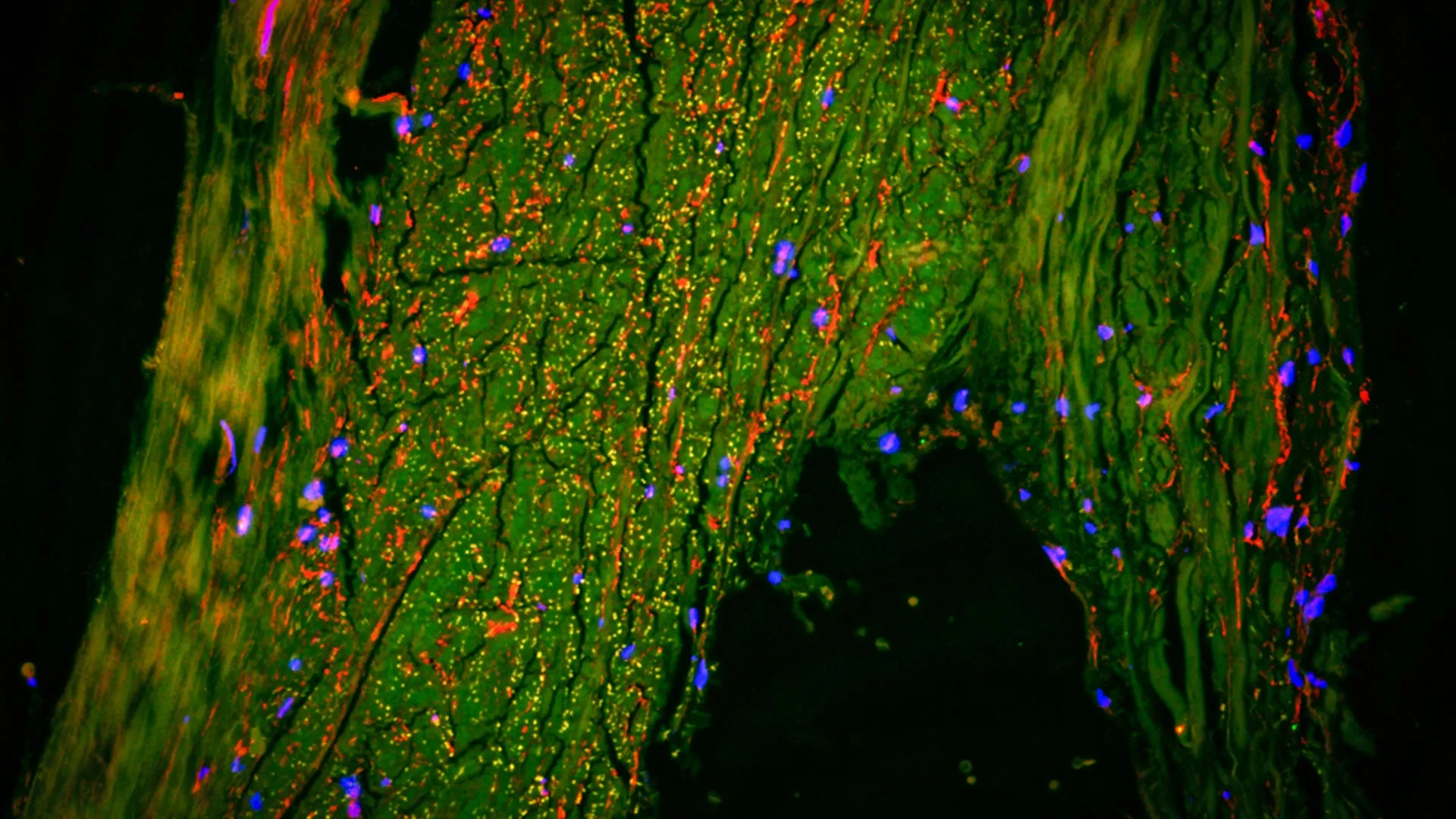
To ensure that the imaging data reflected biological reality, the MUSC researchers collaborated with experts at Cornell University to perform ultra-high-resolution histological analysis on human brain tissue. Using multiplexed imaging—a technique that allows for the simultaneous visualization of various cell types—the team examined the area surrounding the middle meningeal artery at the microscopic level.
The analysis confirmed the presence of lymphatic endothelial cells (LECs) lining the channels adjacent to the MMA. These cells are the building blocks of lymphatic vessels throughout the rest of the human body, responsible for absorbing fluid and transporting it to lymph nodes. The presence of these specific cellular markers in the meninges, coupled with the real-time MRI data showing slow-moving fluid, provides a "smoking gun" for the existence of a functional lymphatic exit point at the MMA.
"The correlation between the live imaging and the tissue samples is what makes this study so robust," Albayram noted during the presentation of the findings. "We aren’t just seeing a pattern on a screen; we are seeing the actual cellular architecture that makes this drainage possible."
A Chronology of Discovery: Redefining the Meninges
The discovery of the MMA’s role is the latest chapter in a rapidly evolving timeline of neuro-lymphatic research.
- Pre-2012: The brain is widely believed to lack a traditional lymphatic system. It is thought that waste simply diffuses into the CSF and is absorbed into the bloodstream.
- 2012: Researchers at the University of Rochester identify the "glymphatic system," a waste-clearance pathway in the brain’s parenchyma managed by glial cells.
- 2015: Independent teams at the University of Virginia and the University of Helsinki rediscover lymphatic vessels in the dural sinuses of mice, and later in humans.
- 2022: Dr. Albayram’s team at MUSC publishes research in Nature Communications providing visual evidence of meningeal lymphatic vessels in humans using specialized MRI techniques.
- 2024: The current study in iScience identifies the MMA as a specific, major control point for this drainage, utilizing NASA-derived technology to monitor flow in real-time.
This timeline illustrates a shift from viewing the brain as an isolated organ to seeing it as deeply integrated into the body’s systemic waste-management infrastructure.
Supporting Data: Flow Dynamics and Waste Clearance
The data gathered during the six-hour MRI sessions provided critical metrics on how the brain’s "plumbing" operates. The researchers measured the velocity and directionality of fluid movement, noting that the clearance along the MMA was most active during periods of rest. This aligns with existing theories that the brain’s waste-removal system is significantly more active during sleep.
Furthermore, the study highlighted the importance of the "perivascular space"—the gap between the blood vessel wall and the surrounding tissue. In the case of the MMA, this space acts as a highway for lymphatic fluid. If this highway becomes narrowed or blocked, metabolic byproducts can accumulate. In healthy subjects, the flow was found to be consistent and unobstructed, providing a baseline for what "normal" drainage looks like.
Implications for Neurodegenerative Disease and Aging
The discovery of a specific control point like the MMA has profound implications for the study of Alzheimer’s disease and other forms of dementia. One of the defining characteristics of Alzheimer’s is the accumulation of amyloid-beta and tau proteins in the brain. If the MMA serves as a primary exit for these proteins, then a "clogged" or dysfunctional drainage system could be a contributing factor to the onset of the disease.
"As we age, our vessels lose elasticity and our lymphatic systems become less efficient," Albayram explained. "By identifying the MMA as a key exit point, we can now investigate whether age-related changes in this specific vessel contribute to the buildup of toxic proteins."
Beyond Alzheimer’s, the study also opens new doors for understanding:
- Traumatic Brain Injury (TBI): After a head injury, the brain often experiences swelling and an influx of inflammatory markers. Effective drainage is crucial for recovery.
- Psychiatric Conditions: Emerging research suggests that neuro-inflammation plays a role in depression and anxiety. A better understanding of how the brain clears inflammatory signals could lead to new therapeutic approaches.
- Autoimmune Disorders: The connection between the brain and the body’s immune system via the lymphatics may explain how systemic inflammation affects cognitive function.
Future Research and Clinical Applications
Following the success of this study, the MUSC team is already moving toward clinical applications. The next phase of research involves comparing the drainage patterns of healthy individuals with those of patients diagnosed with early-stage neurodegenerative diseases. By using the NASA-developed MRI tools, clinicians may eventually be able to diagnose "lymphatic failure" in the brain before physical symptoms of cognitive decline appear.
The long-term goal is the development of treatments that can "flush" the system or improve the efficiency of the MMA drainage pathway. This could involve pharmacological interventions to dilate lymphatic vessels or lifestyle modifications designed to optimize the brain’s natural cleanup cycle.
The scientific community has reacted with cautious optimism. While the study size was small—limited to five healthy individuals for the intensive real-time monitoring—the combination of advanced imaging and histological verification provides a compelling case. Independent experts suggest that if these findings are replicated in larger cohorts, the MMA could become as central to neurology as the major heart valves are to cardiology.
Conclusion: A New Frontier in Human Anatomy
The work of Dr. Albayram and his colleagues at MUSC, supported by the technological prowess of NASA and the analytical depth of Cornell University, has mapped a previously invisible frontier of human anatomy. By proving that the middle meningeal artery is a functional component of the brain’s lymphatic system, the study provides a new lens through which to view human health and disease.
As the research moves forward, the focus will remain on understanding the "normal" functioning of the brain to better identify the "abnormal." In the words of Albayram, "We are finally beginning to see the full picture of how the brain maintains itself. Once we understand the mechanics of the drain, we can learn how to fix it when it breaks." This discovery marks not just an end to a long-standing anatomical mystery, but the beginning of a new era in the prevention and treatment of brain disorders.


Leave a Reply