
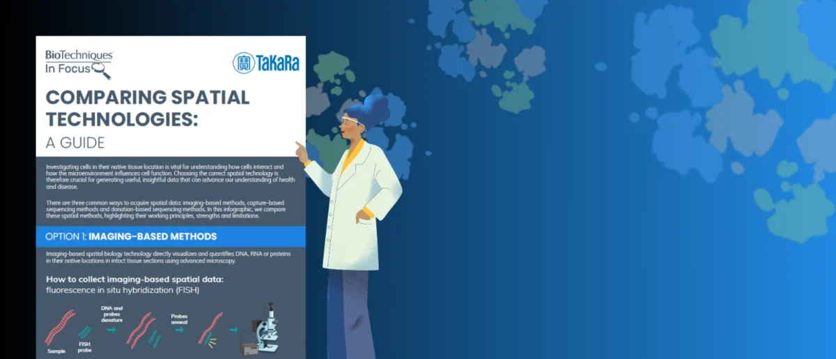
The landscape of biological research is undergoing a profound transformation, driven by an increasing demand to understand molecular processes within their native tissue context. A newly released infographic, featured as part of BioTechniques‘ "In Focus" series on spatial technologies and produced in association with Takara Bio, offers a crucial comparative analysis of three primary methodologies for collecting spatial data: imaging-based methods, capture-based sequencing techniques, and an emerging class termed "donation-based sequencing methods." This comprehensive resource details the working principles, inherent strengths, and critical limitations of each approach, providing researchers with an essential guide to navigating this rapidly evolving field. The infographic is designed to demonstrate how the latest advancements, particularly within the novel donation-based techniques, are poised to overcome long-standing challenges in spatial data acquisition, promising to unlock deeper insights into biological systems and disease mechanisms.
The Imperative of Spatial Biology: Moving Beyond Bulk Analysis
For decades, molecular biology relied heavily on "bulk" sequencing approaches, which involve homogenizing tissue samples and then analyzing the average molecular profile of millions of cells. While invaluable for identifying general trends and molecular signatures, these methods inherently strip away crucial spatial information—the precise location of cells, their interactions, and the heterogeneous distribution of molecules within a tissue. This loss of context is a significant limitation, as biological functions, disease progression, and therapeutic responses are profoundly influenced by the spatial organization of cells and molecules. Understanding the spatial dimension is critical for deciphering complex biological phenomena, such as tumor microenvironments, neuronal circuits, embryonic development, and immune responses.
Spatial biology has emerged as a field dedicated to preserving and analyzing this invaluable locational data. It seeks to map the distribution of RNA, proteins, metabolites, and other biomolecules directly within tissue sections, providing an unprecedented view of cellular architecture and molecular interactions in their natural state. This ability to integrate molecular data with anatomical context is revolutionizing our understanding of health and disease, paving the way for more precise diagnostics and targeted therapies.
A Chronological Overview of Spatial Technology Development
The journey towards robust spatial biology tools has been progressive, marked by incremental innovations building upon foundational molecular techniques.
-
Early Forays (Pre-2000s): Initial attempts to localize molecules included techniques like immunohistochemistry (IHC) for proteins and in situ hybridization (ISH) for RNA. While groundbreaking for their time, these methods offered limited multiplexing capabilities, typically allowing the detection of only a few targets simultaneously, and provided semi-quantitative rather than precise quantitative data. Fluorescence in situ hybridization (FISH) improved multiplexing but still faced challenges in scalability and high-throughput application across entire transcriptomes.
-
First-Generation Spatial Transcriptomics (Mid-2010s): The mid-2010s witnessed a significant leap with the introduction of technologies like 10x Genomics Visium. This method, often considered a hybrid, combined elements of both imaging and capture. It involved placing tissue sections onto slides containing spatially barcoded oligonucleotides. mRNA molecules diffusing from cells within specific spots would bind to these barcodes, allowing for subsequent sequencing and spatial reconstruction of gene expression patterns. While revolutionary, these early platforms typically offered resolution at the level of 50-100 µm spots, meaning each spot often contained multiple cells, thus obscuring single-cell spatial resolution. Other innovations like Slide-seq also contributed to this era, pushing the boundaries of spatial resolution.
-
Diversification and Refinement (Late 2010s – Early 2020s): The field rapidly diversified, with researchers developing distinct methodological paradigms to address specific challenges related to resolution, throughput, and multiplexing. This period saw the maturation of both imaging-based and capture-based sequencing methods, each with unique advantages and limitations.
Exploring the Methodologies: Imaging, Capture, and Donation-Based Sequencing
The BioTechniques infographic provides a critical comparison of the three major technological streams, offering a valuable resource for researchers selecting the most appropriate tool for their specific scientific questions.
1. Imaging-Based Methods: Precision Through Visualization
Imaging-based spatial techniques rely on directly visualizing and quantifying molecules (RNA, proteins) within a tissue section using microscopy. These methods typically employ fluorescent probes that bind to specific targets, with sophisticated imaging systems capturing their precise locations.
-
Working Principles: These techniques often involve cycles of hybridization, imaging, and probe removal (or inactivation) to detect a large number of different RNA or protein targets within the same tissue section. Examples include Multiplexed Error-Robust FISH (MERFISH), Sequential Fluorescence In Situ Hybridization (seqFISH), and Spatial Transcriptomics with A Real-time Readout (STARmap). Technologies like Vizgen MERSCOPE and Akoya Biosciences’ PhenoCycler/Fusion (formerly CODEX and Phenoptics) and GeoMx Digital Spatial Profiler (DSP) also fall into this category, albeit with different molecular readout strategies. GeoMx DSP, for instance, uses UV light to cleave photocleavable probes from specific regions of interest, which are then collected and sequenced.
-
Strengths:
- High Spatial Resolution: Imaging-based methods can achieve subcellular resolution, allowing researchers to pinpoint the exact location of molecules within individual cells and organelles. This level of detail is crucial for understanding cell compartmentalization and fine-grained molecular interactions.
- High Multiplexing: Advanced iterative imaging techniques can detect hundreds to thousands of RNA transcripts or proteins simultaneously within a single tissue section, providing rich molecular profiles.
- Direct Visualization: The ability to visually confirm the location of molecules within the tissue context offers intuitive data interpretation and validation.
- Preservation of Tissue Integrity: These methods generally preserve the tissue architecture, allowing for subsequent histological analysis or re-analysis.
-
Limitations:
- Limited Field of View and Throughput: While offering high resolution, many imaging-based methods are limited by the microscope’s field of view, making it challenging to analyze large tissue areas or many samples simultaneously. Scaling up can be time-consuming and labor-intensive.
- Targeted Panels: Many imaging techniques rely on pre-designed probe panels, meaning they are typically not whole-transcriptome or whole-proteome methods. While multiplexing is high, it’s often not exhaustive.
- Data Analysis Complexity: The sheer volume and complexity of image data generated require sophisticated computational tools and expertise for processing, segmentation, and quantification.
- Equipment Cost and Expertise: High-end microscopy systems and specialized reagents can be expensive, and operating them often requires significant technical expertise.
2. Capture-Based Sequencing Methods: Unbiased Transcriptomic Profiling
Capture-based sequencing methods combine spatial information with the power of next-generation sequencing to provide comprehensive molecular profiles across entire tissue sections.

-
Working Principles: These techniques typically involve placing a tissue section onto a specialized glass slide containing an array of spatially barcoded oligonucleotides. When the tissue is permeabilized, mRNA molecules diffuse from the cells and bind to the unique spatial barcodes in their vicinity. These mRNA-barcode complexes are then reverse transcribed into cDNA, collected, amplified, and sequenced. The spatial coordinates are subsequently reconstructed computationally based on the unique barcodes. The 10x Genomics Visium platform is the most prominent example in this category.
-
Strengths:
- Whole-Transcriptome Analysis: A major advantage is the ability to profile nearly all detectable mRNA transcripts within the tissue, providing an unbiased view of gene expression without the need for pre-selected gene panels.
- Relatively Straightforward Workflow: Compared to some iterative imaging methods, the experimental workflow for capture-based sequencing can be more streamlined, especially for researchers familiar with standard sequencing protocols.
- Scalability: These methods can analyze larger tissue areas than many high-resolution imaging techniques, making them suitable for broader tissue mapping studies.
- Quantitative Data: Provides quantitative gene expression levels, enabling detailed differential expression analysis.
-
Limitations:
- Lower Spatial Resolution: The primary drawback of many capture-based methods is their reliance on "spots" or "pixels" that are typically larger than a single cell (e.g., 50-100 µm diameter spots, often containing 5-30 cells). This limits the ability to resolve single-cell spatial heterogeneity.
- RNA Diffusion Artifacts: During permeabilization and capture, mRNA molecules can diffuse away from their original cellular location, leading to a blurring of spatial resolution and potential misattribution of transcripts.
- Requires Tissue Dissociation for Single-Cell Context: While providing spatial context, if single-cell resolution is ultimately desired, researchers often need to integrate these data with single-cell RNA sequencing data, which involves tissue dissociation.
- Cost of Sequencing: Generating whole-transcriptome data for multiple samples can incur significant sequencing costs.
3. Donation-Based Sequencing Methods: A Novel Class for Enhanced Spatial Precision
The term "donation-based sequencing methods" represents a newer, potentially more advanced class of spatial biology techniques. While not yet a universally standardized nomenclature across the entire field, its inclusion in the BioTechniques infographic suggests a conceptual grouping of innovative approaches designed to overcome the specific limitations of imaging and capture-based methods, especially concerning resolution and targeted molecular profiling. This class likely encompasses techniques where molecular material is actively and precisely "donated" or extracted from defined spatial locations for subsequent sequencing, moving beyond passive diffusion or purely optical readouts.
-
Working Principles (Inferred): This category likely includes methods that combine highly targeted physical or molecular extraction with sequencing. Examples might include advanced forms of laser capture microdissection (LCM) coupled with highly sensitive sequencing, or novel microfluidic/nanofluidic devices that precisely sample subcellular or single-cell regions within a tissue. It could also refer to in situ sequencing approaches where molecules are directly sequenced within the tissue, but with a refined method of identifying and "donating" their sequence information to a readout system. The key differentiating factor is a more active, precise, and potentially less destructive sampling or information transfer mechanism compared to broad diffusion capture. Given Takara Bio’s support, it could also hint at proprietary technologies that leverage their expertise in molecular biology and sequencing. These methods aim to achieve high spatial resolution while retaining the comprehensive molecular profiling benefits of sequencing.
-
Strengths (Inferred):
- Potentially Superior Spatial Resolution: By employing more precise molecular extraction or in situ sequencing techniques, these methods aim to achieve single-cell or even subcellular resolution with high fidelity, minimizing diffusion artifacts.
- Enhanced Molecular Specificity: The ability to precisely target and extract molecules from specific cellular compartments or microenvironments could lead to more accurate and specific molecular profiles.
- Flexibility in Target Selection: Depending on the specific technology, these methods could offer a balance between targeted (like imaging) and unbiased (like capture) molecular profiling, allowing researchers to focus on specific regions or molecules while still having the capacity for broader analysis.
- Overcoming Diffusion Limitations: The "donation" aspect implies a more controlled transfer of molecular information, mitigating the spatial blurring often seen in capture-based methods.
- Integration Potential: These methods may be designed for easier integration with downstream bioinformatics pipelines and multi-omics analysis.
-
Limitations (Inferred):
- Technological Complexity: Novel methods often entail intricate experimental setups and require specialized expertise.
- Throughput Challenges: Achieving high resolution often comes with a trade-off in throughput, making large-scale studies potentially more laborious or costly.
- Early Stage Development: As a "new class," these methods may still be in earlier stages of optimization and broader adoption compared to established techniques.
- Cost: Advanced, novel technologies can be expensive to develop, implement, and run.
Overcoming Common Challenges in Spatial Data Collection
The scientific community continually grapples with several inherent challenges in spatial biology:
- The Resolution-Throughput Trade-off: High spatial resolution often comes at the expense of throughput (ability to process many samples or large areas), and vice-versa.
- Data Complexity: Spatial datasets are inherently multi-dimensional, combining image data with molecular readouts, requiring advanced computational and statistical tools for analysis and visualization.
- Accessibility and Cost: Specialized equipment, reagents, and bioinformatics expertise can be prohibitive for many research labs.
- Standardization and Reproducibility: Ensuring consistent and comparable results across different platforms, labs, and experimental conditions remains a critical goal.
The emergence of "donation-based sequencing methods" is explicitly highlighted by BioTechniques and Takara Bio as a promising avenue for overcoming these challenges. By potentially offering a more balanced approach that combines high resolution with comprehensive molecular profiling, these new techniques could democratize spatial biology, making it more accessible and robust for a wider range of research applications.
The Role of Takara Bio and BioTechniques in Advancing the Field
This collaborative initiative underscores the critical efforts by industry leaders and scientific publishers to accelerate research. Takara Bio, a global leader in life science research tools, plays a pivotal role in innovating and commercializing cutting-edge technologies. Their support for this infographic signifies their commitment to the spatial biology domain, likely reflecting their ongoing research and development in next-generation sequencing, molecular biology reagents, and potentially novel spatial platforms that align with the "donation-based" concept. Their involvement ensures that researchers have access to the latest tools and insights to push the boundaries of discovery.
BioTechniques, as a peer-reviewed journal and scientific publisher, serves as an essential conduit for disseminating critical information and fostering scientific discourse. Its "In Focus" series is specifically designed to provide in-depth analysis and educational resources on rapidly evolving fields. By curating content like this infographic, animated videos, and expert interviews, BioTechniques empowers researchers to understand complex technologies, compare methodologies, and make informed decisions about experimental design. The inclusion of an illustration by Tobias Dumbraveanu further highlights the effort to make complex scientific information visually engaging and digestible.
Market Growth and Broader Implications of Spatial Technologies
The spatial biology market is experiencing explosive growth, reflecting the scientific community’s recognition of its transformative potential. Industry reports indicate that the global spatial transcriptomics market, valued at approximately USD 250 million in 2023, is projected to reach over USD 1.5 billion by 2030, exhibiting a compound annual growth rate (CAGR) exceeding 25%. This growth is fueled by increasing investments in genomics research, the rising prevalence of chronic diseases (especially cancer and neurodegenerative disorders), and technological advancements leading to more accessible and powerful platforms. Venture capital funding for companies specializing in spatial omics has also seen a significant uptick, signaling strong investor confidence in the sector’s future.
The implications of these advancements are far-reaching:
- Drug Discovery and Development: Spatial biology provides an unparalleled view of drug-target interactions within the tissue context, enabling more precise target identification, biomarker discovery, and efficacy testing. It can help explain why drugs work in some patients but not others, guiding the development of more effective and safer therapeutics.
- Precision Medicine: By mapping the unique molecular landscape of a patient’s disease (e.g., a tumor), spatial technologies can inform personalized treatment strategies, moving beyond generalized approaches to highly tailored interventions based on the specific spatial heterogeneity of a disease.
- Fundamental Biological Understanding: Spatial analysis is crucial for dissecting complex biological processes like embryonic development, organogenesis, and the intricate cell-cell communication networks in healthy and diseased tissues. It helps researchers understand how tissue architecture influences cellular function and vice versa.
- Disease Diagnostics: High-resolution spatial profiling could lead to novel diagnostic tools that identify disease signatures earlier and with greater specificity, improving patient outcomes.
The continued innovation, particularly in developing new classes of techniques like "donation-based sequencing," promises to address existing bottlenecks and expand the applicability of spatial biology across diverse research areas. As these technologies mature and become more integrated with artificial intelligence and machine learning for data analysis, they are set to unlock even deeper insights into the fundamental mechanisms of life and disease.
The BioTechniques infographic, supported by Takara Bio, serves as a vital tool for researchers navigating this dynamic field. By clearly delineating the current state-of-the-art and highlighting the potential of emerging methodologies, it equips the scientific community with the knowledge necessary to harness the full power of spatial biology in their pursuit of groundbreaking discoveries. Researchers are encouraged to download the infographic and explore the accompanying animated video and interview to fully grasp how these latest techniques are poised to overcome common challenges and accelerate scientific understanding.


Leave a Reply