
For decades, the medical community has recognized a strong correlation between physical exercise and the maintenance of cognitive function, yet the precise mechanical triggers for this relationship have remained partially obscured. A groundbreaking study published on April 27 in the journal Nature Neuroscience has unveiled a previously unknown physical link between the body’s core and the brain’s internal environment. Researchers at Pennsylvania State University have demonstrated that simple physical movements, specifically the contraction of abdominal muscles, act as a hydraulic pump that physically shifts the brain and facilitates the circulation of cerebrospinal fluid (CSF). This movement is critical for the "cleansing" of the brain, as it helps flush out metabolic waste products that are often implicated in the development of neurodegenerative disorders such as Alzheimer’s and Parkinson’s disease.
The Mechanical Foundation of Brain Health
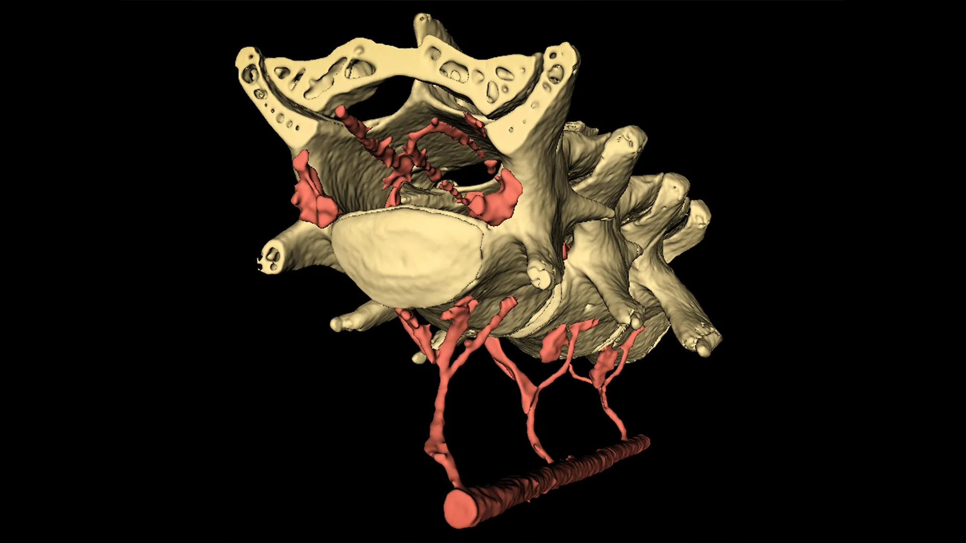
The human brain does not sit static within the skull; rather, it exists in a dynamic environment where it is constantly bathed in cerebrospinal fluid. The Penn State research team, led by Patrick Drew, a professor of engineering science and mechanics, neurosurgery, biology, and biomedical engineering, has identified a direct mechanical pathway that connects the abdomen to the cranial cavity. When an individual engages their core—whether through a deliberate exercise or a routine action like standing up—the resulting pressure in the abdominal cavity is transmitted through a complex network of veins known as the vertebral venous plexus.
This network of veins acts as a conduit. When abdominal muscles tighten, they exert pressure on the blood vessels in the torso, forcing blood upward toward the spinal cord and the brain. This sudden influx of pressure causes a minute but significant physical displacement of the brain within the skull. This shifting motion is not merely an incidental byproduct of movement; rather, it appears to be a fundamental physiological mechanism designed to drive the flow of CSF through the brain’s intricate channels.
"Our research explains how just moving around might serve as an important physiological mechanism promoting brain health," stated Professor Drew, who also serves as the associate director of the Huck Institutes of the Life Sciences. He compared the process to a hydraulic system where the abdominal muscles serve as the primary pump. The study suggests that even the smallest movements, such as bracing the core to take a step or adjusting one’s posture, are enough to trigger this hydraulic effect.
Advanced Imaging and the Mouse Model Experiments
To reach these conclusions, the interdisciplinary team employed a sophisticated experimental design involving moving mice and high-resolution imaging technology. The researchers utilized two primary methods to capture the real-time effects of muscle contraction on brain physiology: two-photon microscopy and microcomputed tomography (micro-CT).
Two-photon microscopy allowed the team to view living tissue at a cellular level with extreme precision, while micro-CT provided high-resolution, three-dimensional visualizations of the entire organ structure. By observing mice in motion, the researchers noted a consistent pattern: the brain would shift just milliseconds after the abdominal muscles tightened, but immediately before the animal initiated a full limb movement. This timing indicated that the preparation for movement itself—the tensing of the core—was the catalyst for the brain’s displacement.
To isolate the variable of abdominal pressure, the researchers conducted a secondary experiment on lightly anesthetized mice. In this controlled environment, they applied gentle pressure to the mice’s abdomens, mimicking the force of a muscle contraction without any other bodily movement. The pressure applied was remarkably low—less than the pressure a human experiences during a standard blood pressure cuff test. Despite the minimal force, the results were definitive: the brain shifted in response to the abdominal pressure and returned to its original baseline position the moment the pressure was released. This confirmed that the brain’s movement is a direct, rapid, and reversible response to internal pressure changes in the torso.
Computational Modeling: The Brain as a Porous Sponge
Understanding that the brain moves was only the first step. The researchers then needed to determine how this movement influenced the flow of cerebrospinal fluid. Given the limitations of current imaging technology in capturing the rapid, microscopic movement of fluids deep within the brain, the team turned to advanced computer simulations.
Francesco Costanzo, a professor of engineering science and mechanics, biomedical engineering, mechanical engineering, and mathematics at Penn State, led the modeling efforts. Costanzo and his team faced the significant challenge of accounting for the "special physics" that occur when fluid particles cross the various membranes within the brain. To overcome this complexity, they conceptualized the brain not as a solid mass, but as a biological sponge.
"The brain has a structure similar to a sponge, in the sense that you have a soft skeleton and fluid can move through it," Costanzo explained. Using this sponge-like model, the team simulated how physical displacement forces fluid through spaces of varying sizes, from large folds to microscopic pores. The simulations revealed that the gentle "squeezing" and shifting caused by abdominal contractions effectively drove fluid across the brain’s surfaces and through its tissue.
Costanzo extended the sponge analogy to describe the waste-clearing process. "How do you clean a dirty sponge? You run it under a tap and squeeze it out," he noted. In this context, the abdominal contractions provide the "squeeze" that helps circulate the "water" (CSF), flushing away the "dirt" (metabolic waste).
Historical Context and the Glymphatic System
This study builds upon a growing body of evidence regarding the brain’s waste-management systems. Historically, the brain was thought to be somewhat isolated from the body’s lymphatic system. However, the discovery of the "glymphatic system" over a decade ago revolutionized neuroscience by showing that the brain has its own dedicated plumbing system for waste removal, primarily active during sleep.
Previous research by Professor Drew and others had focused on how sleep and the loss of certain neurons affected the timing of CSF flow. While sleep remains a critical period for brain detoxification, the new findings suggest that wakeful physical activity provides a complementary, mechanical boost to this process. This discovery fills a significant gap in our understanding of why sedentary lifestyles are often linked to cognitive decline, whereas active lifestyles are protective.
Broader Implications for Neurodegenerative Disease
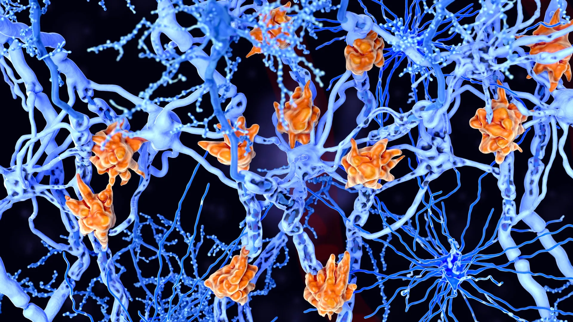
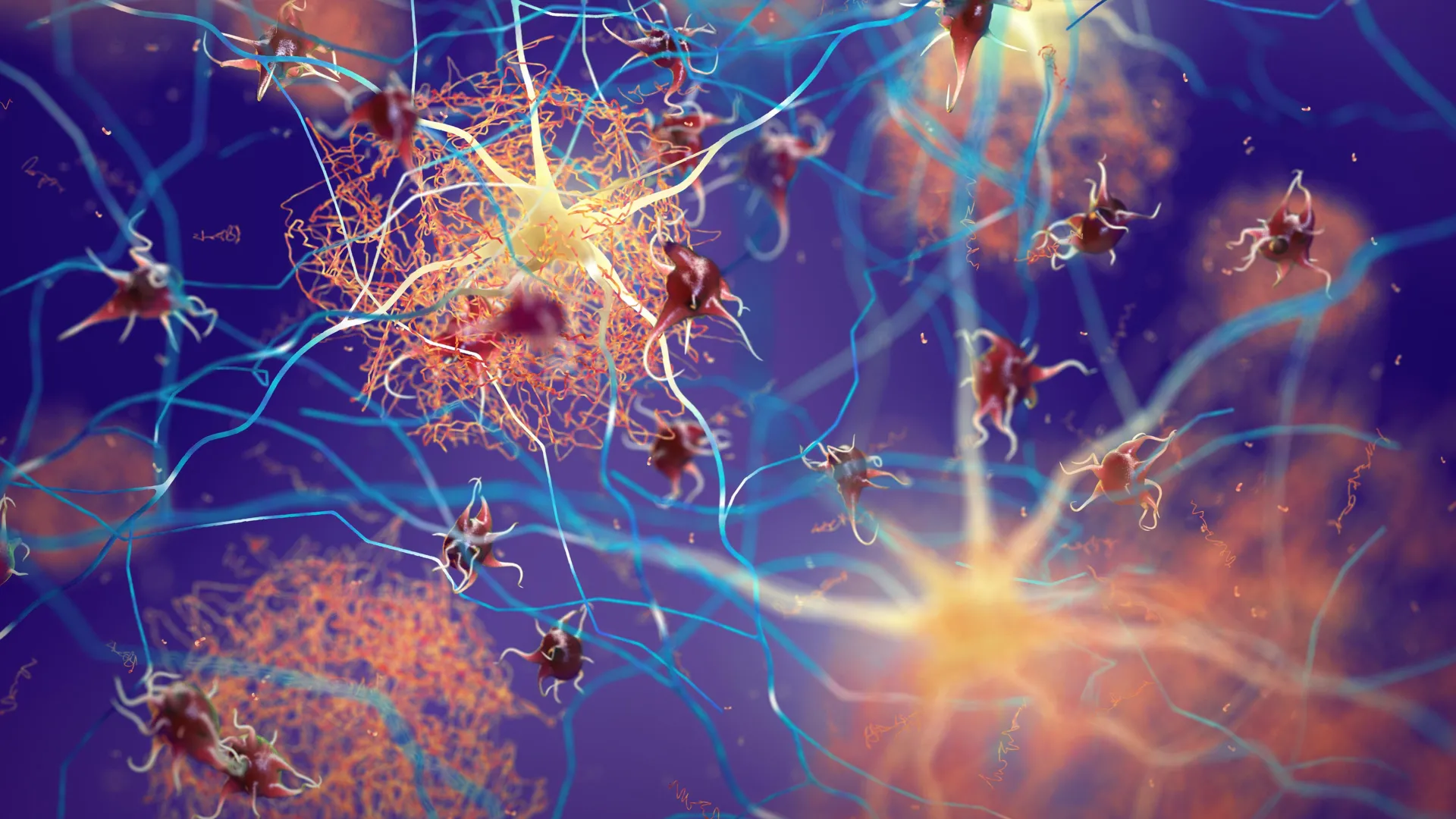
The implications of this research are far-reaching, particularly in the context of an aging global population. Neurodegenerative diseases like Alzheimer’s are characterized by the accumulation of toxic proteins, such as beta-amyloid and tau, which form plaques and tangles that disrupt neural communication. If regular movement helps "flush" these proteins out of the brain daily, then physical activity is not just a general health recommendation—it is a specific mechanical requirement for neural maintenance.
The study suggests that the "dose" of movement required to trigger this cleaning mechanism is surprisingly low. Because even small contractions of the abdominal muscles can induce brain movement, the findings provide hope for individuals with limited mobility. It implies that even light core-engagement exercises or assisted movements could potentially offer some level of neuroprotective benefit by maintaining CSF circulation.
Furthermore, this research opens the door for new therapeutic interventions. Understanding the hydraulic link between the abdomen and the brain could lead to the development of medical devices or physical therapy techniques designed to optimize CSF flow in patients who are unable to exercise vigorously.
An Interdisciplinary Effort
The success of the study was predicated on its interdisciplinary nature, combining the fields of neurosurgery, biology, mechanical engineering, and mathematics. The research team included a diverse group of scholars, including postdoctoral researchers C. Spencer Garborg and Beatrice Ghitti, and former Penn State faculty such as Qingguang Zhang. The project also involved significant contributions from graduate and undergraduate students, highlighting the collaborative environment at Penn State’s Center for Quantitative Imaging.
The study received support from several major institutions, including the National Institutes of Health (NIH), the Pennsylvania Department of Health, and the American Heart Association. These organizations have a vested interest in the intersection of cardiovascular health and brain function, a link that this research has made more tangible.
Future Research Directions
While the results in mice are compelling, Professor Drew emphasized that the next critical step is to validate these findings in human subjects. "This kind of motion is so small… it could make such a difference for your brain health," Drew said, noting that more research is needed to quantify exactly how much movement is necessary for optimal waste clearance in humans.
Future studies will likely utilize advanced fMRI techniques and other non-invasive imaging to observe the hydraulic relationship between the human abdomen and the cranium. Researchers will also look to see if specific types of exercise—such as yoga, weightlifting, or walking—provide different levels of CSF circulation.
As the scientific community continues to unravel the complexities of the brain-body connection, this study stands as a pivotal reminder that the health of the mind is physically anchored in the movement of the body. The simple act of engaging the core may be one of the most effective, accessible tools humans have for preserving their cognitive longevity.


Leave a Reply