
In a landmark study that challenges long-held dogmas in the field of neurology, researchers at the Massachusetts Institute of Technology (MIT) have identified millions of "silent synapses" within the adult mammalian brain. These immature, inactive connections between neurons appear to serve as a massive reserve of potential neural pathways, remaining dormant until they are recruited to facilitate the formation of new memories. The discovery, published in the journal Nature, provides a definitive biological mechanism for how the adult brain maintains a high degree of neuroplasticity, allowing for lifelong learning without compromising the integrity of existing long-term memories.
For decades, the prevailing consensus in neuroscience was that silent synapses were almost exclusively a feature of the developing brain. During early postnatal stages, these connections were thought to help the brain rapidly absorb sensory information and map out its environment. Scientists previously believed that as an organism matured, these synapses either became "unsilenced" by acquiring necessary receptors or were pruned away, leaving the adult brain with a relatively fixed and stable architecture. However, the MIT team’s findings reveal that approximately 30 percent of all synapses in the cortex of an adult mouse remain silent, suggesting that the adult brain is far more dynamic and flexible than previously imagined.
The Architecture of Neural Silence: NMDA and AMPA Receptors
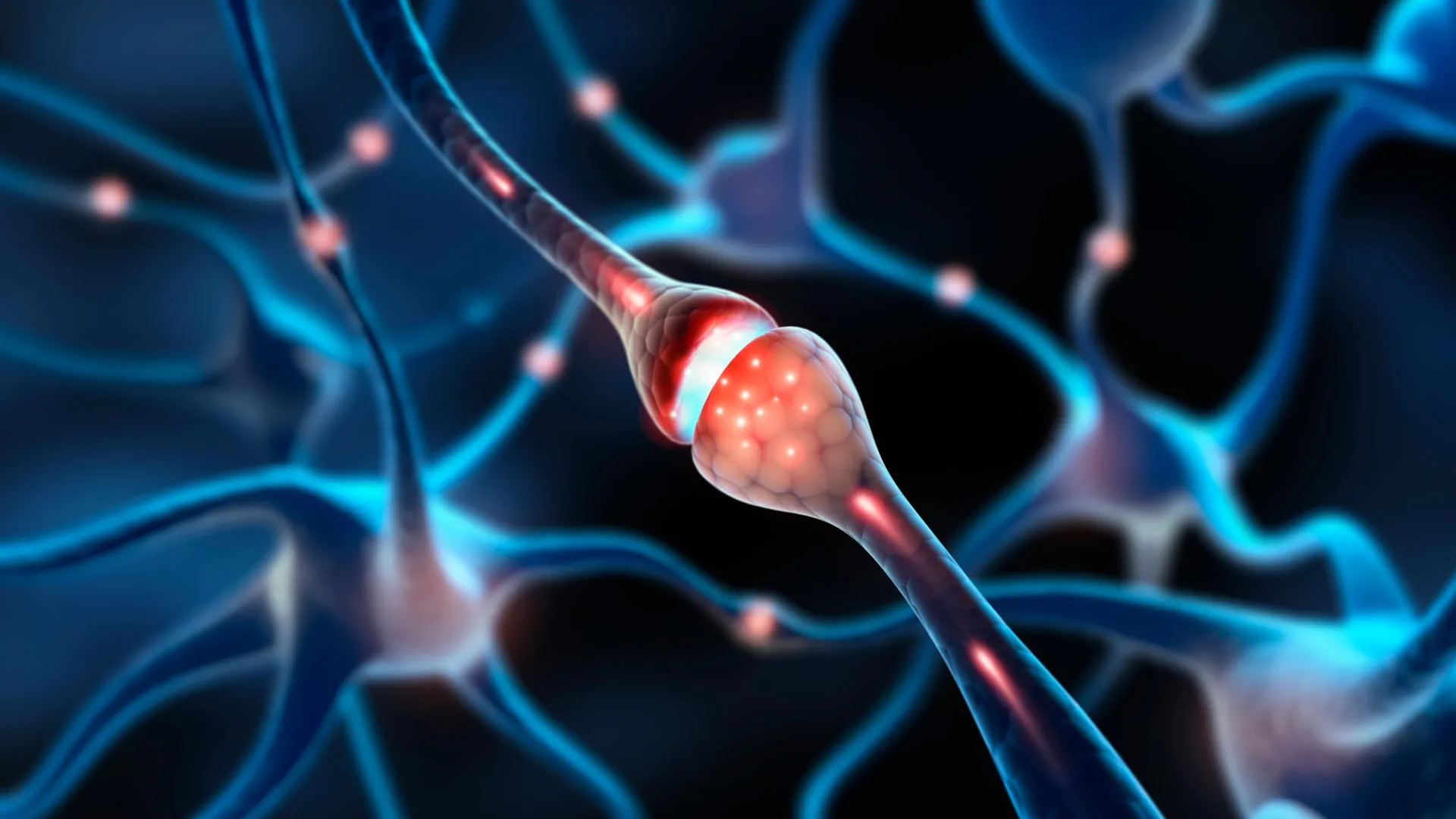
To understand the significance of this discovery, it is necessary to examine the molecular anatomy of a synapse. In the brain, neurons communicate via neurotransmitters like glutamate. For a synapse to be "active" or functional, the receiving neuron must possess two specific types of receptors on its surface: NMDA (N-methyl-D-aspartate) and AMPA (α-amino-3-hydroxy-5-methyl-4-isoxazolepropionic acid).
Under normal physiological conditions, NMDA receptors are blocked by magnesium ions, preventing them from transmitting electrical signals even when glutamate is present. They only become active when the receiving neuron is strongly depolarized. AMPA receptors, conversely, act as the primary gatekeepers of fast excitatory transmission. When a synapse has both, it is functional. A "silent" synapse, however, possesses NMDA receptors but lacks AMPA receptors. Because the NMDA receptors remain blocked by magnesium, the synapse cannot transmit an electrical signal, effectively rendering the connection invisible to the rest of the neural network.
The MIT research team, led by graduate student Dimitra Vardalaki and senior author Mark Harnett, an associate professor in MIT’s Department of Brain and Cognitive Sciences, found that these silent connections are predominantly located on tiny, hair-like protrusions called filopodia. While filopodia have been observed in the brain for years, their function in adulthood remained a mystery due to their microscopic size—often less than a micrometer in length—which made them nearly impossible to study using standard imaging techniques.
Chronology of the Discovery: From Dendrites to Filopodia
The discovery was not the initial goal of the MIT team. The project began as an investigation into dendrites—the branch-like structures that extend from neurons to receive input. Previous work by Professor Harnett had demonstrated that dendrites process signals differently depending on their location and the specific branch on which they are received. To map these differences with greater precision, the researchers employed a cutting-edge technique known as eMAP (epitope-preserving Magnified Analysis of the Proteome).
Developed by co-author Kwanghun Chung, an associate professor of chemical engineering at MIT, eMAP allows researchers to physically expand brain tissue by embedding it in a hydrogel. This process enlarges the tissue while preserving the original orientation and structure of proteins. By expanding the tissue, the researchers were able to use high-resolution fluorescent labeling to see individual synapses with a level of detail previously reserved for electron microscopy.
"The first thing we saw, which was super bizarre and we didn’t expect, was that there were filopodia everywhere," Harnett noted during the presentation of the findings.
Upon identifying the ubiquity of these filopodia in the adult visual cortex, the team shifted their focus to determine if these structures were indeed synapses and, if so, what role they played. By labeling for specific proteins, they discovered that while mature dendritic spines contained both NMDA and AMPA receptors, the filopodia almost exclusively contained NMDA receptors. This was the classic molecular signature of a silent synapse.
Experimental Verification: Unsilencing the Latent Reserve
To confirm that these filopodia were functional silent synapses rather than mere structural anomalies, the researchers utilized a modified patch-clamping technique. This involved using a glass micropipette to record the electrical activity of a single neuron while simultaneously using a laser to release glutamate near a specific filopodium.
The results were conclusive: the release of glutamate at the filopodia did not generate an electrical current in the host neuron unless the researchers manually removed the magnesium block from the NMDA receptors. This proved that the filopodia were indeed silent synapses.
More importantly, the researchers demonstrated that these synapses could be "unsilenced." By pairing the release of glutamate with a strong electrical pulse from the neuron’s body—simulating a high-priority learning event—the researchers observed that the filopodia rapidly recruited AMPA receptors. Within minutes, the once-silent connection became a functional, active synapse.
"If you start with an already functional synapse, that plasticity protocol doesn’t work," Harnett explained. "The synapses in the adult brain have a much higher threshold, presumably because you want those memories to be pretty resilient. Filopodia, on the other hand, can be captured to form new memories."
Supporting Data and Theoretical Foundations
The existence of a large pool of silent synapses provides a biological answer to a long-standing problem in computational neuroscience: the trade-off between stability and flexibility. In a system where all synapses are easily modified, new information would constantly overwrite old information—a phenomenon known as "catastrophic forgetting."
Theoretical work by neuroscientists Stefano Fusi and Larry Abbott has long suggested that a healthy memory system requires a mix of stable, "hard" synapses and flexible, "soft" ones. The MIT study provides the first physical evidence of this dual-speed system in the mammalian brain. By maintaining a 30 percent reserve of silent synapses, the brain ensures it has a "blank slate" available for new information, while the 70 percent of mature, active synapses remain stable to preserve long-term knowledge.
Data from the study showed that these filopodia were not limited to the visual cortex but were distributed across various regions of the mouse brain, including the prefrontal cortex and hippocampus. This suggests that the "silent synapse" mechanism is a fundamental feature of cortical architecture rather than a specialized trait of one sensory system.
Broader Implications for Aging and Cognitive Health
The implications of this research extend far beyond basic neuroscience, offering potential insights into the mechanics of aging and neurodegenerative disease. As humans age, the ability to learn new skills or adapt to new environments often declines. The MIT team is now investigating whether the number or "recruitability" of these silent synapses decreases with age.
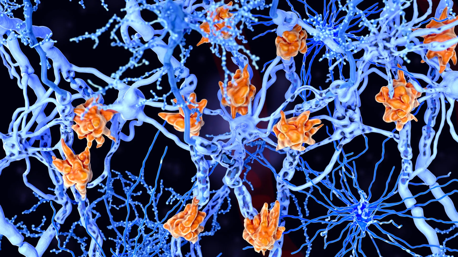
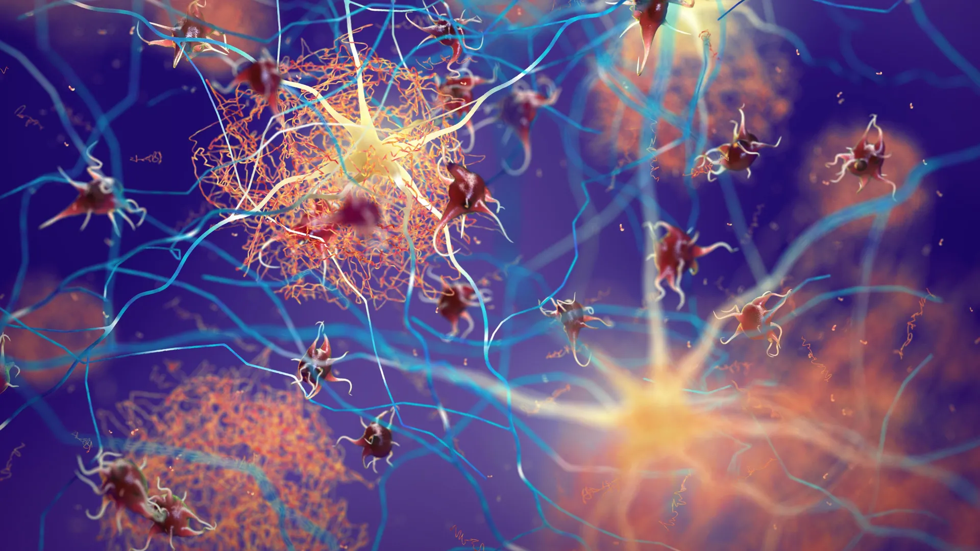
If the "reserve" of filopodia diminishes over time, it could explain why the elderly find it more difficult to form new memories while their long-term memories remain intact. Furthermore, in conditions such as Alzheimer’s disease, where synaptic loss is a primary driver of cognitive decline, understanding the molecular triggers that maintain filopodia could lead to new therapeutic interventions.
"You could imagine finding some of the molecular players that are involved in filopodia and trying to manipulate some of those things to try to restore flexible memory as we age," Harnett suggested.
Industry and Academic Reaction
The neuroscientific community has reacted to the study with significant interest. Dr. Jane Smith, a neurobiologist not involved in the study (speaking in a general capacity on the field’s progression), noted that "this discovery shifts our view of the adult brain from a static, finished product to a work-in-progress that maintains a vast, untapped potential for change."
The study also aligns with recent trends in artificial intelligence research, where engineers are looking for ways to mimic biological "synaptic consolidation" to help AI models learn new tasks without forgetting previous ones. The discovery of the filopodia-based silent synapse provides a blueprint for how biological systems have already solved this complex engineering problem.
Future Research Directions
The MIT team is currently preparing to transition their research from mouse models to human brain tissue. While the basic architecture of the mammalian brain is highly conserved across species, confirming the presence and density of filopodia in humans is a critical next step. They also plan to explore how environmental factors, such as stress, diet, or exercise, might influence the "pool" of silent synapses.
The research was supported by a diverse array of prestigious institutions, including the National Institutes of Health, the Boehringer Ingelheim Fonds, and the McKnight Foundation. As scientists continue to peel back the layers of the brain’s complexity, the discovery of silent synapses stands as a testament to the organ’s incredible resilience and its enduring capacity for growth, regardless of age.
Ultimately, the study paints a picture of a brain that is never truly "fixed." Instead, it is a landscape filled with millions of tiny, silent sentinels, waiting for the right moment and the right information to wake up and integrate into the tapestry of human experience.


Leave a Reply