
The human brain has long been perceived as a highly shielded organ, protected by the blood-brain barrier and floating in a shock-absorbing reservoir of cerebrospinal fluid. However, a groundbreaking study published on April 27 in Nature Neuroscience reveals that the brain is far more physically integrated with the body’s mechanical movements than previously understood. Researchers at Pennsylvania State University have identified a direct hydraulic link between the contraction of abdominal muscles and the movement of fluid within the brain, offering a profound mechanical explanation for why physical activity is essential for cognitive longevity and the prevention of neurodegenerative diseases.
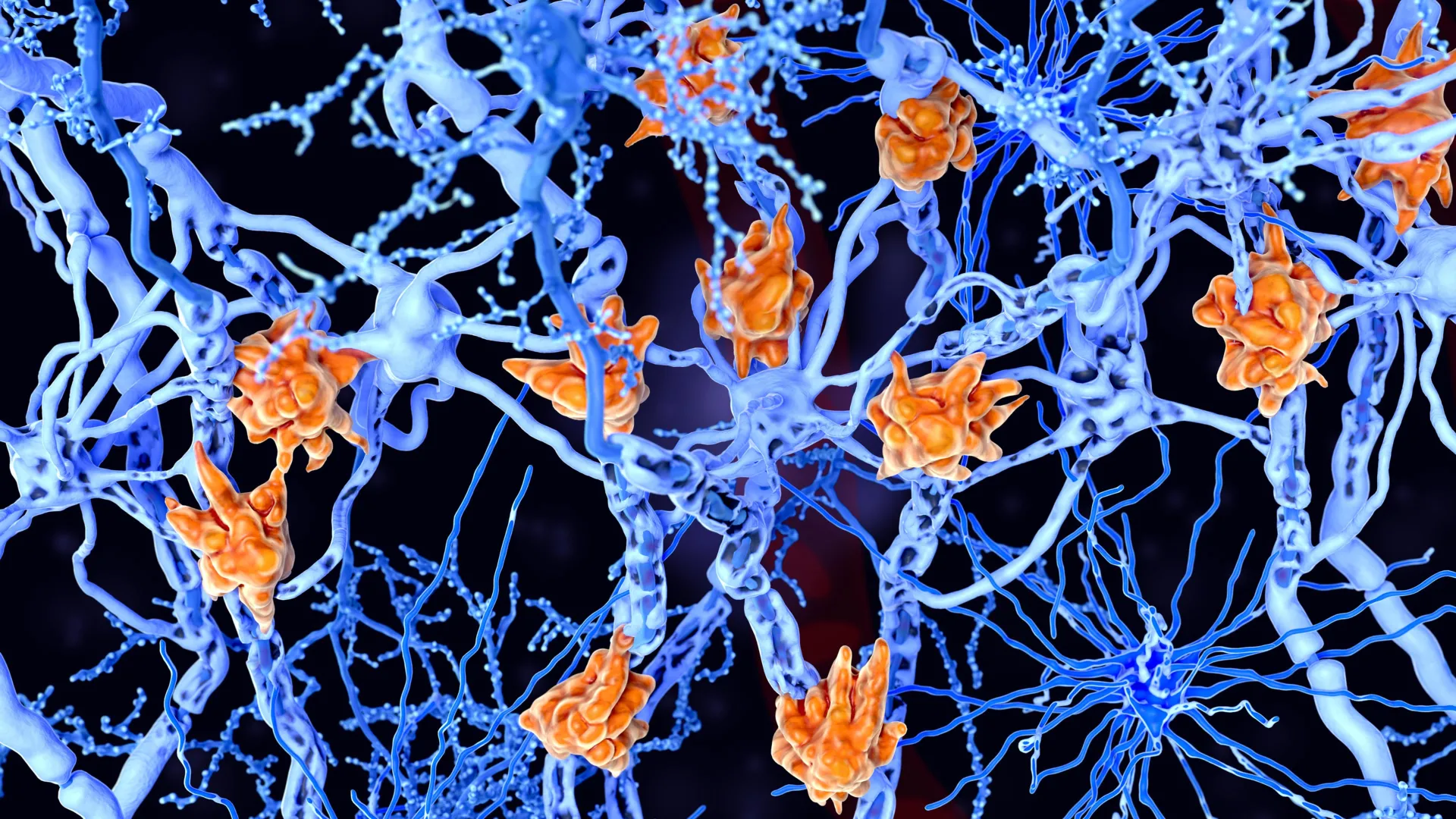
By utilizing a combination of high-resolution imaging in animal models and complex computer simulations, the interdisciplinary team demonstrated that even minor physical actions—such as bracing the core to stand or taking a single step—trigger a cascade of pressure changes. These changes physically shift the brain within the skull, acting as a pump that facilitates the circulation of cerebrospinal fluid (CSF). This circulation is critical for the "glymphatic" system, the brain’s waste-removal pathway, which flushes out metabolic byproducts that are otherwise linked to conditions such as Alzheimer’s and Parkinson’s disease.
The Hydraulic Mechanism: From Core to Cranium
The central finding of the research revolves around a sophisticated hydraulic interaction. Patrick Drew, a professor of engineering science and mechanics, neurosurgery, biology, and biomedical engineering at Penn State, served as the corresponding author. He explained that the process begins in the torso. When a subject engages their abdominal muscles, the resulting internal pressure does not remain localized. Instead, it compresses blood vessels within the abdomen that are part of a larger network known as the vertebral venous plexus.
This plexus is a valveless network of veins that runs along the spinal column, providing a direct conduit between the thoracic/abdominal cavities and the cranial vault. When the abdominal muscles tighten, they force blood out of the abdominal compartment and into this spinal network. Because the system is hydraulic, the increased volume in the spinal veins transmits pressure upward into the skull. This pressure causes the brain to shift slightly—a subtle but significant physical displacement.
"In this study, we found that when the abdominal muscles contract, they push blood from the abdomen into the spinal cord, just like in a hydraulic system, applying pressure to the brain and making it move," Drew said. This movement is not merely a byproduct of exertion but appears to be a functional necessity for brain maintenance. The gentle "sloshing" effect caused by the brain’s displacement drives the flow of CSF through the brain’s intricate channels, effectively "washing" the neural tissue.
Advanced Imaging: Capturing the Brain in Motion
To validate this theory, the researchers employed two distinct and highly advanced imaging technologies to observe the phenomenon in live mice. The use of multiple modalities allowed the team to see both the micro-level cellular changes and the macro-level organ displacement.
The first technique, two-photon microscopy, allowed the researchers to look deep into the living tissue of the mice. This provided real-time data on how individual vessels and fluid compartments reacted during movement. The second technique, microcomputed tomography (micro-CT), offered a high-resolution, three-dimensional view of the entire anatomy. This allowed the team to track the movement of the brain as a whole relative to the skull.
The data revealed a striking temporal correlation: the brain began to shift just milliseconds before the mice actually initiated physical locomotion. This timing corresponds with the "pre-movement" contraction of the abdominal muscles, which animals (and humans) use to stabilize their posture before moving. To isolate the role of abdominal pressure from other factors like heart rate or neural activity, the team conducted a controlled experiment on lightly anesthetized mice. They applied gentle pressure to the animals’ abdomens—less pressure than a human would feel during a standard blood pressure cuff test. Even without any voluntary movement from the mice, this external abdominal pressure caused the same brain displacement and fluid movement observed during active exercise.
"Importantly, the brain began moving back to its baseline position immediately upon relief of the abdominal pressure," Drew noted. "This suggests that abdominal pressure can rapidly and significantly alter the position of the brain within the skull."
The "Dirty Sponge" Analogy: Modeling Fluid Dynamics
While imaging confirmed the movement of the brain, the rapid and complex nature of CSF flow proved difficult to capture through traditional photography alone. To bridge this gap, the team turned to computational modeling. Francesco Costanzo, a professor of engineering science and mechanics at Penn State, led the effort to simulate how these mechanical shifts influence fluid dynamics.
Costanzo described the brain’s structure as being analogous to a sponge—a porous, soft skeleton through which fluid can permeate. However, modeling this is exceptionally difficult because the brain involves independent movements of tissue and fluid, as well as movements that are "coupled" or dependent on one another. To solve this, the team accounted for the unique physics that occur when fluid particles cross the various membranes within the brain.
"Keeping with the idea of the brain as a sponge, we also thought of it as a dirty sponge—how do you clean a dirty sponge?" Costanzo asked. "You run it under a tap and squeeze it out."
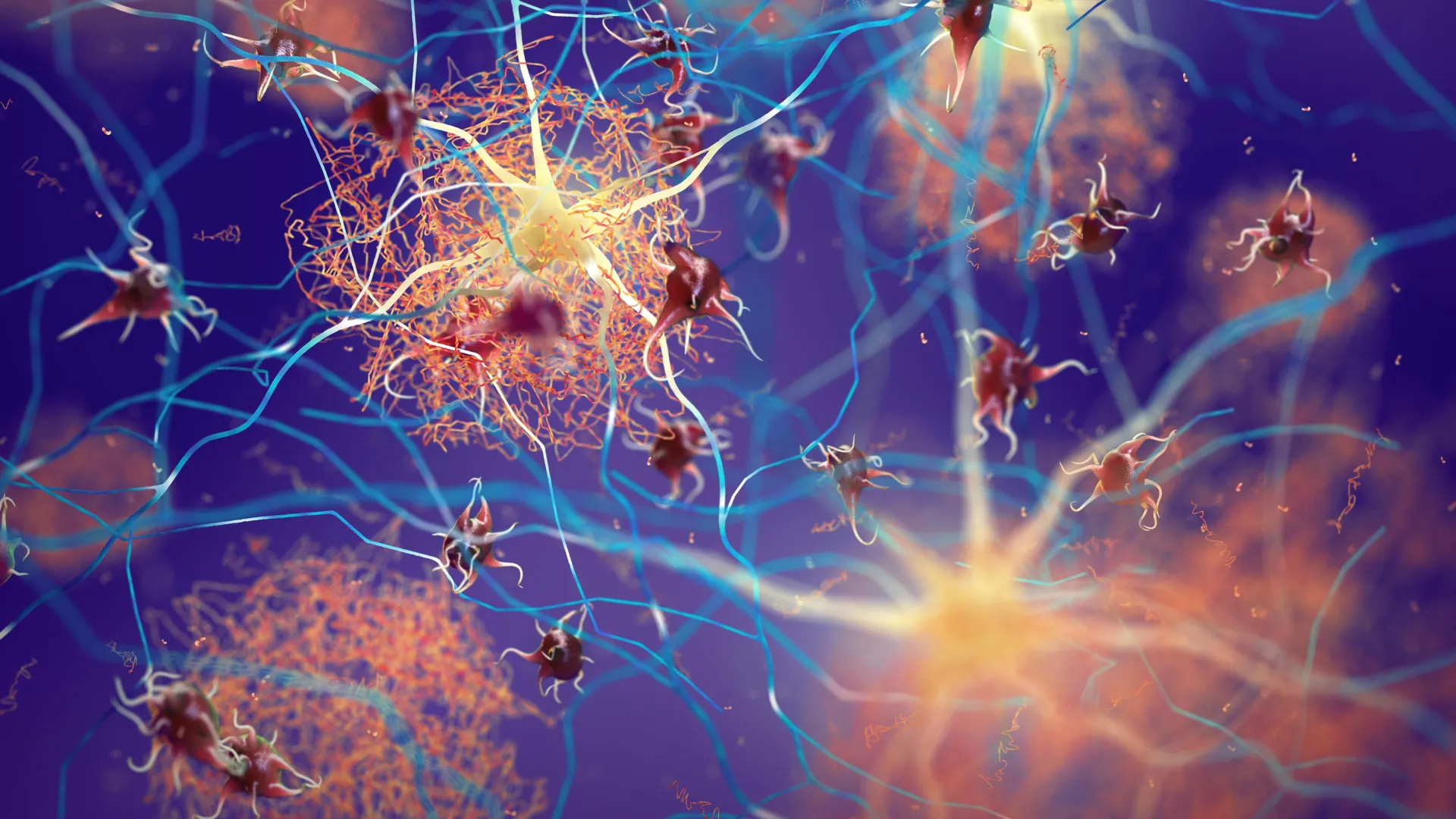
The simulations demonstrated that the physical displacement of the brain acts as the "squeeze." When the brain shifts due to abdominal pressure, it forces CSF through the "pores" of the brain tissue. This motion facilitates the transit of fluid into the perivascular spaces, where it can pick up waste products like amyloid-beta—the protein fragments that form plaques in the brains of Alzheimer’s patients—and carry them away into the lymphatic system.
Historical Context and the Evolution of Glymphatic Theory
This study builds upon a decade of research into the glymphatic system, a term coined in 2012 by researcher Maiken Nedergaard. Prior to this discovery, it was largely believed that the brain recycled its own waste internally or that the process was extremely slow. Nedergaard’s work showed that the brain has a dedicated "plumbing system" that becomes highly active during sleep.
However, the Penn State study adds a critical new dimension to this understanding. While sleep is a passive period of waste clearance, physical movement appears to be an active, mechanical driver of the same system. Previously, it was thought that the pulsing of arteries was the primary pump for CSF. This new data suggests that the "abdominal pump" may be an equally, if not more, significant contributor to fluid circulation during waking hours.
The timeline of this research reflects an increasing move toward interdisciplinary science. Patrick Drew’s background in engineering and neurosurgery, combined with Costanzo’s expertise in mathematics and mechanics, allowed the team to view the brain not just as a biological organ, but as a mechanical system subject to the laws of fluid dynamics and hydraulics.
Implications for Public Health and Neurodegenerative Disease
The implications of these findings for human health are vast. As global populations age, the incidence of neurodegenerative diseases is rising, and sedentary lifestyles are increasingly recognized as a major risk factor. This research provides a specific, mechanical reason why "sitting is the new smoking" in the context of brain health.
If everyday movements—walking, standing up, or even adjusting one’s posture—are necessary to "squeeze the sponge" of the brain, then prolonged inactivity could lead to a stagnation of CSF. This stagnation may allow metabolic waste to accumulate, potentially accelerating the onset of cognitive decline.
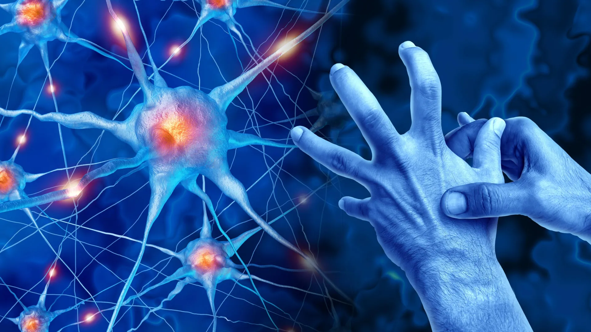
"This kind of motion is so small. It’s what’s generated when you walk or just contract your abdominal muscles, which you do when you engage in any physical behavior," Drew said. "It could make such a difference for your brain health."
The study also opens new avenues for therapeutic interventions. For individuals with limited mobility, such as those with paralysis or the elderly, it may be possible to develop devices or physical therapy routines that mimic the effects of abdominal contractions to ensure their brains continue to receive the benefits of fluid circulation. Furthermore, it highlights the importance of core strength, not just for spinal stability, but as a fundamental component of the brain’s waste-management infrastructure.
Analysis: A New Frontier in Biomechanics
The Penn State study represents a shift in how neurobiology is studied. By focusing on the vertebral venous plexus and the hydraulic nature of the spinal-cranial connection, the researchers have identified a "hidden" highway between the gut and the brain. While the "gut-brain axis" is a popular topic in microbiology regarding the microbiome, this study establishes a literal, physical "gut-brain axis" based on pressure and fluid.
The fact that the brain shifts anticipatorily—before the limb actually moves—suggests that the central nervous system has evolved to integrate these pressure changes into its basic operational framework. This suggests that the brain is not a passive passenger in the body but an active participant in the mechanical stresses of life.
However, the researchers caution that more work is needed to translate these findings from mice to humans. Human anatomy is larger and more complex, and our upright posture creates different gravitational pressures than those experienced by four-legged mice. Nevertheless, the fundamental structures—the abdominal muscles, the vertebral venous plexus, and the CSF-filled cranial cavity—are conserved across species, making the hydraulic model highly plausible for humans.
Research Team, Funding, and Institutional Support
The study was a massive collaborative effort involving experts across multiple departments at Penn State and other institutions. Co-authors included C. Spencer Garborg, Beatrice Ghitti, Qingguang Zhang, Joseph M. Ricotta, Noah Frank, Sara J. Mueller, Denver L. Greenawalt, Hyunseok Lee, Kevin L. Turner, Ravi T. Kedarasetti, and Marceline Mostafa.
The imaging work was largely conducted at the Penn State Center for Quantitative Imaging, a facility of the Institute of the Energy and the Environment. The project received significant financial backing from major health organizations, including the National Institutes of Health (NIH), the Pennsylvania Department of Health, and the American Heart Association.
As the scientific community continues to explore the link between physical activity and cognitive function, this study provides a vital piece of the puzzle. It moves the conversation beyond the vague benefits of "exercise" and into the realm of specific, quantifiable mechanical processes that keep the brain clean, healthy, and functional. By understanding the brain as a sponge that needs to be squeezed, science is one step closer to developing more effective strategies for maintaining mental acuity throughout the human lifespan.


Leave a Reply