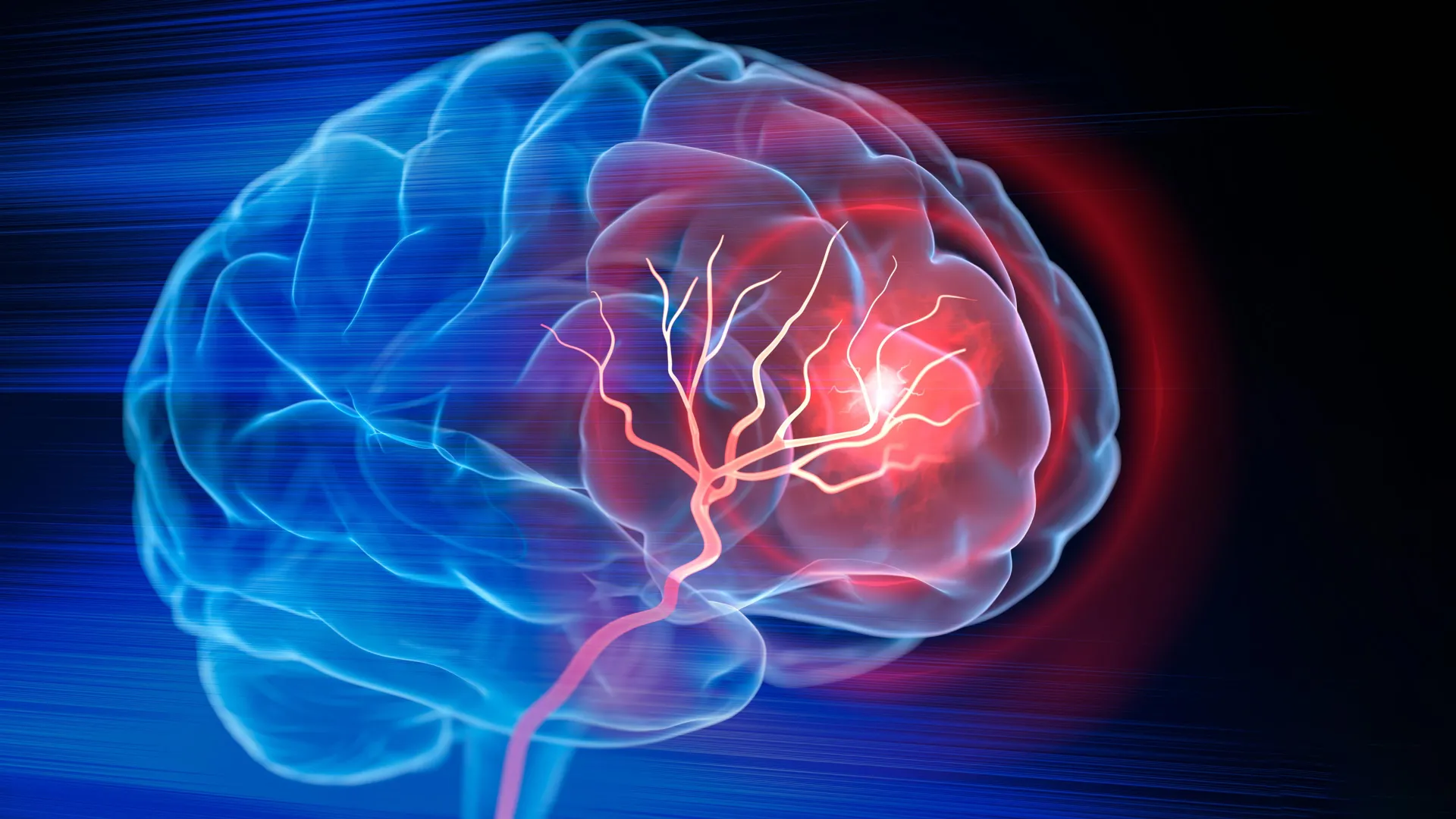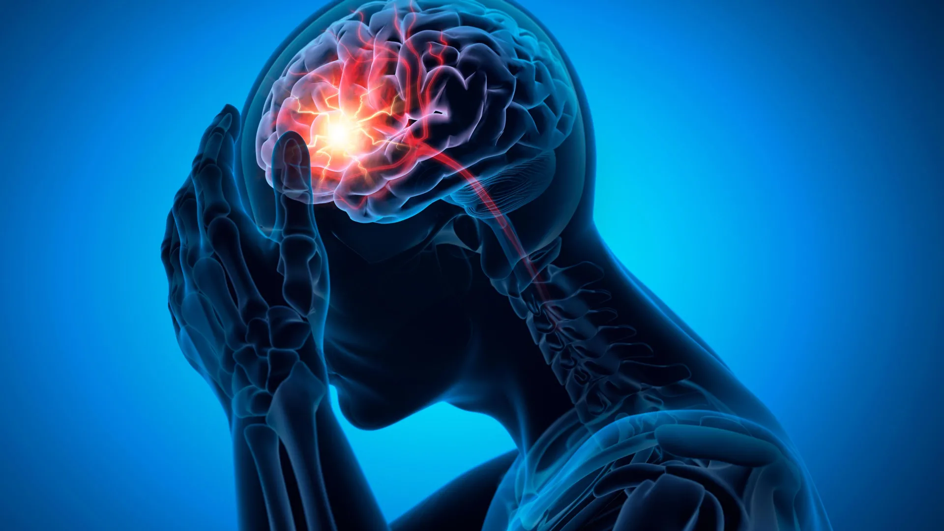
A landmark study led by researchers at the University of Edinburgh and the UK Dementia Research Institute has challenged decades of medical assumptions regarding the pathophysiology of lacunar ischemic strokes. For years, the medical community has largely treated lacunar strokes—a common form of stroke that accounts for approximately 25% of all ischemic cases—as a byproduct of atherosclerosis, the buildup of fatty plaques within the arteries. However, new evidence suggests that the primary driver of these strokes is not arterial blockage from plaque, but rather a fundamental change in the structural integrity of the brain’s blood vessels, specifically characterized by the widening and elongation of the arteries.
This shift in understanding provides a long-sought explanation for why traditional stroke prevention strategies, such as the administration of aspirin and other antiplatelet medications, have frequently proven ineffective for patients suffering from lacunar infarcts. The findings, recently published in the prestigious journal Circulation, are expected to catalyze a significant overhaul in clinical protocols and the development of targeted pharmaceutical interventions.
The Pathophysiology of Lacunar Stroke and Small Vessel Disease
Lacunar strokes occur deep within the brain when one of the small, penetrating arteries becomes obstructed. While individually small, these strokes are collectively a leading cause of vascular cognitive impairment and physical disability. They are the most common cause of "silent" strokes—brain injuries that do not present immediate, obvious symptoms but cumulatively lead to dementia, gait instability, and mood disorders.
The underlying condition responsible for these strokes is known as Cerebral Small Vessel Disease (SVD). SVD is a systemic condition affecting the microvasculature of the brain. Until now, the precise mechanisms through which SVD led to lacunar infarction remained a subject of intense debate. Many clinicians operated under the "atheroma" hypothesis, which posited that small plaques formed at the openings of these tiny vessels, similar to how they form in the larger carotid or coronary arteries.
The new research suggests a different mechanism: "arterial remodeling." Instead of narrowing due to fat deposits, the vessels appear to undergo a process of dilation and distortion. This suggests that the problem is not a simple plumbing issue involving a "clog," but rather a biological failure of the vessel wall itself, likely involving the endothelial lining—the thin layer of cells that regulates blood flow and protects brain tissue from harmful substances in the blood.
Study Methodology and Comparative Analysis
To investigate the divergence between large-vessel disease and lacunar stroke, the international research team, which included collaborators from China and Mexico, conducted a longitudinal study involving 229 participants. These individuals had recently experienced either a lacunar stroke or a mild non-lacunar stroke (a stroke caused by larger artery disease).
The methodology was rigorous, utilizing advanced magnetic resonance imaging (MRI) to track changes in the brain’s architecture over time. Participants underwent baseline scans immediately following their initial event and were brought back for follow-up imaging one year later. This allowed the researchers to observe not only the damage caused by the initial stroke but also the progression of SVD and the emergence of new, "silent" brain lesions.
The researchers specifically compared two distinct vascular markers:
- Arterial Narrowing (Stenosis): The traditional marker of atherosclerosis, where fatty deposits restrict blood flow.
- Arterial Widening (Ectasia): The enlargement and lengthening of the arteries, indicating a weakening of the vessel’s structural integrity.
The results were stark. The analysis revealed that narrowing of the large arteries showed no significant correlation with the incidence of lacunar stroke or the severity of small vessel disease. Conversely, patients with widened arteries were more than four times more likely to have suffered a lacunar stroke. Furthermore, those with significant arterial widening exhibited a much higher rate of "white matter hyperintensities"—areas of the brain that appear bright on MRI scans and signify chronic damage to the brain’s wiring.
The Phenomenon of "Silent" Strokes and Ongoing Risk
One of the most concerning findings of the study was the high rate of recurrence despite standard medical care. Over the course of the one-year follow-up period, more than 25% of the participants developed new "silent" strokes. These are small areas of tissue death caused by interrupted blood supply that, while not causing a "stroke syndrome" (such as paralysis or loss of speech), contribute to long-term cognitive decline.
Crucially, these patients were already receiving "gold standard" preventive treatments, including antiplatelet drugs and cholesterol-lowering statins. The fact that a quarter of the patients continued to suffer brain damage despite these medications underscores the limitations of the current plaque-focused treatment model. It suggests that the traditional "one size fits all" approach to ischemic stroke prevention is failing a significant portion of the population.
Shifting Clinical Focus: The LACI-3 Trial
The implications of this research are already being translated into clinical trials. The researchers emphasized that if the problem is vessel wall dysfunction rather than plaque, then the solution must focus on stabilizing the microvasculature.
The LACunar Intervention Trial 3 (LACI-3) is currently at the forefront of this effort. This trial is evaluating the efficacy of two existing drugs—cilostazol and isosorbide mononitrate—specifically for the treatment of lacunar strokes.
- Cilostazol: A drug that inhibits platelet aggregation but also promotes vasodilation and protects the endothelial lining.
- Isosorbide Mononitrate: Often used for chest pain, this drug releases nitric oxide, which helps maintain the health and flexibility of blood vessels.
By focusing on drugs that support the health of the small vessel walls, researchers hope to reduce the progression of SVD and prevent the cognitive decline that so often follows lacunar stroke. This represents a pivot toward "precision medicine" in the field of neurology, where the specific subtype of stroke dictates the pharmacological response.
Expert Reactions and Global Implications
Professor Joanna Wardlaw, a world-leading expert in neuroimaging and the study’s lead investigator, highlighted the necessity of this paradigm shift. "This study provides strong evidence that lacunar stroke is not caused by fatty blockage of larger arteries, but by disease of the small vessels within the brain itself," Wardlaw stated. "Recognizing this distinction is crucial, because it explains why conventional treatments like antiplatelet drugs are not as effective for this type of stroke and highlights the urgent need to develop new therapies that target the underlying microvascular damage."
The medical community has reacted with cautious optimism. Dr. Richard Isaacson, a neurologist not involved in the study, noted that these findings could change the way physicians counsel patients. "For years, we’ve told patients that a stroke is a stroke, and we treat them all with the same blood thinners and statins. This research tells us we need to be more nuanced. We are looking at a different disease process entirely."
From an economic perspective, the impact of these findings is substantial. Stroke is a leading cause of long-term healthcare expenditure globally. By identifying a more effective way to prevent the "silent" damage associated with SVD, healthcare systems could potentially reduce the burden of dementia and late-life disability, which are often the end-stage results of untreated small vessel disease.
Chronology of Research Milestones
The path to this discovery has been decades in the making:
- 1960s: Pathologist C. Miller Fisher first described "lacunes" (small holes) in the brain during autopsies, hypothesizing they were caused by small vessel disease.
- 1990s-2000s: The advent of high-resolution MRI allowed doctors to see these strokes in living patients, though the assumption remained that plaque was the culprit.
- 2010s: Large-scale observational studies began to suggest that antiplatelet therapy had a higher risk-to-benefit ratio in lacunar stroke patients compared to other stroke types.
- 2020-2023: The LACI-1 and LACI-2 trials established the safety and potential efficacy of targeting vessel health rather than plaque.
- Current Study (2024): The publication in Circulation provides the definitive link between arterial widening (rather than narrowing) and lacunar stroke risk.
Conclusion and Future Outlook
The University of Edinburgh’s findings mark a turning point in vascular neurology. By decoupling lacunar stroke from the traditional model of atherosclerosis, the research opens the door to a new era of treatment focused on microvascular health.
Future research is expected to delve deeper into why these vessels widen in the first place. Potential culprits include chronic inflammation, genetic predispositions affecting the extracellular matrix of the vessel walls, and the long-term effects of hypertension on the brain’s autoregulatory mechanisms.
As the LACI-3 trial progresses, the medical community remains hopeful that a new standard of care is on the horizon—one that will finally offer effective protection for the millions of people worldwide at risk of lacunar stroke and the cognitive decline that accompanies it. The study was supported by a broad coalition of funders, including the UK Medical Research Council, Alzheimer’s Society, and the British Heart Foundation, reflecting the cross-disciplinary importance of this microvascular breakthrough.