
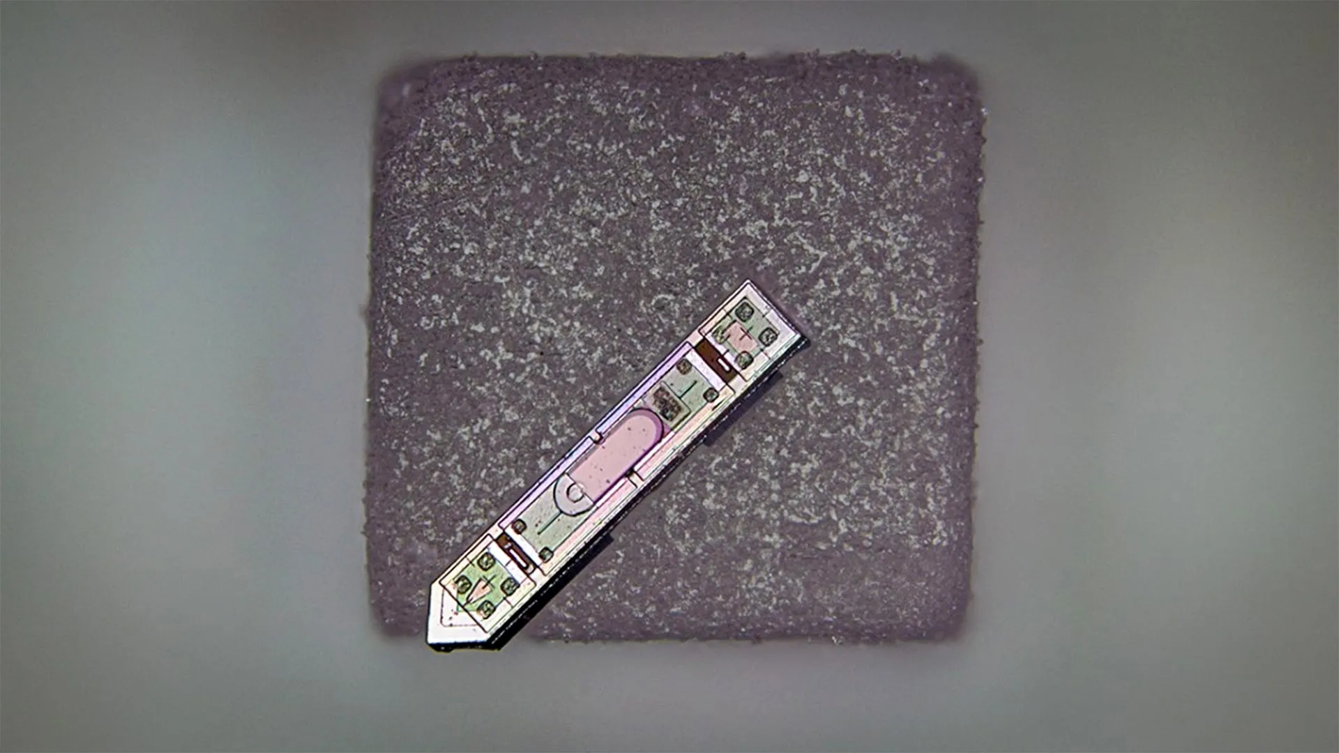
In a landmark achievement for the field of neurotechnology, a multidisciplinary team of researchers at Cornell University has engineered a microscale neural implant so small it can rest on a single grain of salt. This device, known as a microscale optoelectronic tetherless electrode (MOTE), represents a significant leap forward in the quest to interface electronics with biological systems. Measuring approximately 300 microns in length and 70 microns in width, the MOTE is capable of wirelessly transmitting high-fidelity brain activity data from a living subject for over a year, overcoming the traditional barriers of size, longevity, and connectivity that have long hindered the development of permanent brain-computer interfaces (BCIs).
The research, recently published in the journal Nature Electronics, highlights a paradigm shift in how scientists monitor the central nervous system. Led by Alyosha Molnar, a professor in the School of Electrical and Computer Engineering at Cornell, and Sunwoo Lee, an assistant professor at Nanyang Technological University (NTU), the project demonstrates that microelectronic systems can be scaled down to nearly invisible proportions without sacrificing the robustness required for long-term medical applications.
The Engineering Behind the MOTE Device
The primary challenge in developing neural implants has always been the trade-off between size and functionality. Traditional implants, such as the Utah Array or the flexible "threads" utilized by companies like Neuralink, often require physical wires (tethers) to provide power and extract data. These wires create significant risks, including the potential for infection, mechanical strain on delicate brain tissue, and the gradual buildup of scar tissue, known as gliosis, which eventually insulates the electrode from the neurons it is meant to monitor.
The MOTE addresses these issues by eliminating physical connections entirely. At its core, the device is an integrated optoelectronic system. It utilizes a semiconductor diode made from aluminum gallium arsenide (AlGaAs), a high-performance material often used in high-speed electronics and photonics. This specific material was chosen for its dual-functionality: it acts as a photovoltaic cell to harvest energy from incoming light and as a light-emitting diode (LED) to transmit data back to an external receiver.
To operate the device, researchers use red and infrared laser beams. These specific wavelengths are chosen because they can safely penetrate biological tissue with minimal absorption or scattering, allowing the light to reach the implant situated beneath the surface of the brain. The MOTE captures this light to power its internal circuitry, which includes a low-noise amplifier and an optical encoder. When the device detects electrical signals from nearby neurons, it converts those signals into pulses of infrared light.
Advanced Data Encoding and Power Efficiency
One of the most innovative aspects of the MOTE is its communication protocol. To maximize power efficiency, the research team implemented Pulse Position Modulation (PPM). This is the same encoding technique used in deep-space optical communications, such as satellite-to-ground transmissions.
"By using pulse position modulation for the code, we can use very, very little power to communicate and still successfully get the data back out optically," Professor Molnar explained. In PPM, the information is encoded not by the intensity or frequency of the light, but by the precise timing of the pulses. This allows the MOTE to transmit high-resolution data while consuming only a fraction of the energy required by traditional radio-frequency (RF) wireless systems.
The integration of all these components—the power harvester, the amplifier, the encoder, and the transmitter—into a footprint of 0.021 square millimeters is a feat of micro-fabrication. The device is manufactured using techniques similar to those used in the production of commercial microchips, suggesting that if the technology is validated for human use, it could eventually be mass-produced at a relatively low cost.
A Chronology of Development and Testing
The journey toward the MOTE began several years ago in Professor Molnar’s lab at Cornell. Sunwoo Lee, then a postdoctoral researcher, spearheaded the initial design and fabrication phases. The development timeline involved several critical milestones:
- Conceptualization (2018-2019): The team identified the limitations of RF-based wireless implants, specifically the "antenna problem." RF antennas must be a certain size relative to the wavelength of the signal, which sets a hard floor on how small an RF-based implant can be. Light, having much shorter wavelengths, allows for much smaller "antennas" (the diodes).
- Fabrication and Bench Testing (2020): The researchers perfected the AlGaAs manufacturing process, ensuring the diodes could efficiently switch between power-reception and data-transmission modes.
- In Vivo Integration (2021-2022): The devices were implanted in animal models. Unlike previous micro-implants that often failed within weeks due to moisture ingress or biological rejection, the MOTE was designed with specialized encapsulation layers to protect the delicate electronics from the saline environment of the brain.
- Long-term Monitoring (2023): The researchers successfully demonstrated that the MOTE could remain functional and provide clear neural recordings for a period exceeding 12 months.
The success of the year-long trial is particularly noteworthy. In the field of neuroprosthetics, "chronic stability" is the gold standard. Many experimental implants provide excellent data for the first month, only to see signal quality degrade as the body’s immune system reacts to the foreign object. The MOTE’s tiny displacement—meaning it displaces very little brain matter—appears to significantly reduce the inflammatory response, contributing to its longevity.
Implications for Medical Science and Diagnostics
The potential applications for the MOTE extend far beyond basic neuroscience research. Because the device is constructed without the large metallic components typically found in wireless electronics, it offers a unique advantage: compatibility with Magnetic Resonance Imaging (MRI).
Standard neural implants often contain significant amounts of metal, which can cause dangerous heating or create massive signal interference (artifacts) during an MRI scan. This has historically forced patients with neural implants to avoid MRIs, depriving them of a critical diagnostic tool. The MOTE’s semiconductor-based architecture and optical communication method could allow patients to undergo high-resolution imaging without risk or data loss.
Furthermore, the technology is not limited to the brain. Researchers suggest the MOTE could be adapted for use in the spinal cord to monitor recovery from trauma or to manage chronic pain. Its small size makes it an ideal candidate for "bio-integrated" sensors that could be placed in peripheral nerves throughout the body, providing a real-time map of the nervous system’s activity.
Future Outlook: The Smart Skull and Beyond
As the research moves into its next phase, the Cornell team is looking at how to integrate these sensors into a broader medical ecosystem. One proposed innovation involves the use of "smart" artificial skull plates. For patients who require a craniotomy (the removal of a portion of the skull) due to injury or surgery, an artificial plate embedded with optoelectronics could be installed. This plate would serve as a relay station, providing the laser light needed to power MOTEs deeper in the brain and collecting the infrared data pulses to be sent to an external computer.
While the current results are promising, the path to human clinical trials involves several more years of rigorous testing. Future iterations of the MOTE will likely aim to increase the number of recording channels. Currently, a single MOTE records from a small cluster of neurons; however, by deploying an "array" of dozens or even hundreds of these salt-grain-sized devices, researchers could theoretically map large-scale neural networks with unprecedented resolution.
The ethical and regulatory implications of such technology are also being considered. As neural implants become smaller and less invasive, the barrier to entry for brain-monitoring technology lowers. Experts in the field of neuroethics emphasize the need for robust data privacy and "neural rights" frameworks to ensure that the data harvested by such devices remains the sole property of the individual.
Conclusion
The development of the MOTE by Cornell University and its collaborators marks a pivotal moment in the evolution of microelectronics. By successfully shrinking a fully functional, wireless, and long-lasting neural interface to the size of a grain of salt, the team has proven that the "tetherless" future of neurology is not just a theoretical possibility, but a tangible reality. As this technology matures, it promises to provide deeper insights into the mysteries of the human brain, offering new hope for the treatment of neurological disorders and the advancement of human-machine integration.


Leave a Reply