The map of the human genome, once thought to be a thoroughly cataloged blueprint of life, continues to yield astonishing secrets. Modern genomic sequencing technologies and advanced mass spectrometry are currently driving a revolution in molecular biology, shedding light on the so-called "hidden proteome." For decades, vast stretches of human DNA were dismissed as evolutionary junk or transcribed into non-coding RNA molecules with supposedly negligible biological utility. Today, that dogma is crumbling. Researchers are discovering that regions of the genome long believed to be silent are actually bustling hubs of molecular activity, encoding functional micro-proteins and regulatory RNAs that quietly orchestrate the fundamental mechanics of human cells.
A landmark study conducted by a team of molecular biologists at the University of Gothenburg in Sweden has delivered a striking contribution to this emerging field. Investigators at the university’s prestigious Sahlgrenska Academy have successfully identified a previously unknown protein—designated as MSEP—alongside the specific RNA molecule that encodes it, known as ARHGEF17-AS1. Traditionally categorized as a non-coding RNA, ARHGEF17-AS1 has now been unmasked as a dual-threat genomic actor. Not only does it help regulate cellular operations in its native RNA form, but it also translates into the novel MSEP protein. Together, these two distinct entities play an indispensable role in maintaining the cellular machinery responsible for pulling chromosomes apart during cell division.
The implications of this discovery extend far beyond basic cell biology. Because uncontrolled and rapid cell division is the defining hallmark of oncology, scientists immediately turned their attention to how MSEP and its parent RNA behave in malignant environments. Their findings reveal that MSEP is abnormally upregulated in various cancer cell lines compared to normal, healthy cells. When the researchers experimentally suppressed the production of either the ARHGEF17-AS1 RNA or the MSEP protein in cultured cervical cancer cells, the malignant cells experienced a significant drop in growth and proliferation. While clinical applications remain distant, this revelation opens a compelling new window into the vulnerabilities of tumors and offers fresh avenues for targeted cancer research.
The Anatomy of the Hidden Proteome and the Evolution of Genomics
To understand the magnitude of the Gothenburg discovery, one must examine the technological evolution that made it possible. When the Human Genome Project was declared essentially complete in the early 2000s, scientists were surprised to find that only about two percent of the human genome actually codes for classical, well-characterized proteins. The remaining ninety-eight percent was largely sidelined as non-coding DNA. Over the subsequent decade, however, the launch of major international consortia like the ENCODE (Encyclopedia of DNA Elements) project revealed a far more complex reality: vast portions of this "dark matter" genome are actively transcribed into RNA molecules.
While many of these transcripts were found to regulate gene expression, epigenetics, and chromatin structure without ever becoming proteins, a lingering suspicion remained among pioneering biochemists. They hypothesized that sensitive ribosome-profiling techniques and high-resolution mass spectrometry might uncover tiny, overlooked open reading frames (ORFs) embedded within what were thought to be non-coding transcripts. This gave rise to the concept of the hidden proteome—micro-proteins encoded by short, unconventional sequences that standard automated gene-annotation software routinely filtered out as statistical noise.
The scientific community has spent the past several years systematically mining these overlooked regions. The identification of MSEP by the University of Gothenburg research team fits directly into this global scientific movement. By employing cutting-edge transcriptomic and proteomic pipelines, the Swedish researchers were able to look past historical annotations and spot a hidden translation product that traditional models had missed entirely.
Unraveling MSEP and ARHGEF17-AS1: A Dual-Action Cellular Mechanism
The discovery of the MSEP protein and its relationship with the ARHGEF17-AS1 RNA molecule challenges simplistic views of genetic architecture. In biological textbooks, the central dogma dictates a linear flow of information: DNA is transcribed into messenger RNA, which is subsequently translated into a protein. ARHGEF17-AS1 blurs these clean lines by functioning simultaneously as a regulatory RNA transcript and as the physical template for the MSEP protein.
To dissect the individual contributions of these two molecular components, the research team designed a series of precise intervention experiments using cultured cells. By utilizing molecular tools to selectively target and degrade the ARHGEF17-AS1 RNA molecule without halting the production of the MSEP protein—and conversely, by selectively disrupting the translation of MSEP while leaving the RNA intact—the scientists were able to isolate their respective functions.
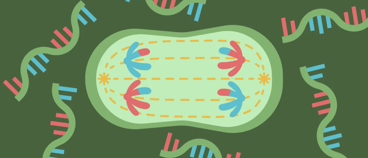
The results were unexpected. Sagar Mahale, a researcher at Sahlgrenska Academy and the first author of the published studies, noted that the RNA molecule and the protein do not operate through identical pathways. Instead, they act independently via distinct molecular cascades, yet they ultimately converge on the same vital objective: maintaining the structural integrity of the mitotic apparatus. During mitosis—the phase of the cell cycle where a parent cell divides into two genetically identical daughter cells—the mitotic spindle must form correctly to segregate chromosomes evenly. If this machinery fails, cells can suffer from aneuploidy, a condition characterized by an abnormal number of chromosomes, which is a frequent driver of tumorigenesis. Both the ARHGEF17-AS1 RNA and the MSEP protein act as critical stabilization factors for this delicate cellular scaffolding, ensuring that division proceeds without error.
Implications for Oncology: Curbing Cancer Cell Proliferation
Given that successful mitosis is the absolute prerequisite for tumor expansion, investigators immediately hypothesized that abnormalities in the MSEP pathway might be exploited by cancer cells. To test this hypothesis, the Gothenburg researchers analyzed the expression profiles of MSEP across a variety of established cancer cell lines and compared them against normal, non-malignant human cell lines.
The data revealed a clear pattern: MSEP levels were consistently elevated in the cancer cells examined. This overexpression suggests that rapidly dividing malignancies may rely heavily on elevated levels of MSEP and its associated RNA to sustain their hyperactive mitotic schedules without suffering catastrophic chromosomal missegregation.
To evaluate the functional dependency of cancer cells on these factors, the research team performed targeted knockdown experiments. When the expression of ARHGEF17-AS1 RNA was silenced or MSEP production was artificially blocked in cultured cervical cancer cells, the proliferation rate of the cancer cells dropped markedly. Deprived of the structural reinforcement normally provided by the MSEP network, the malignant cells struggled to complete successful divisions, leading to growth arrest or cell death.
Chronology and Collaborative Scope of the Research

The path leading to these breakthroughs spanned multiple years of meticulous biochemical analysis, bridging transcriptomics, proteomics, and cellular imaging.
The initial phases of the investigation began with bioinformatic screenings of human genomic databases at the University of Gothenburg, where researchers scanned for unannotated translation products hidden within long non-coding RNAs.
Following the identification of the candidate sequence corresponding to ARHGEF17-AS1 and its encoded peptide, the team initiated functional validation assays.
Over the subsequent months, wet-lab experiments involving advanced cell culture techniques allowed the scientists to visualize the localization of the MSEP protein within dividing cells using fluorescent microscopy and immunofluorescence.
Parallel studies were conducted to assess the binding partners of both the RNA and the protein, mapping out the distinct molecular pathways through which they influence the mitotic spindle.
The culmination of these efforts resulted in two peer-reviewed studies detailing the dual roles of the transcript and its micro-protein, establishing a foundational framework for future investigations into how the hidden proteome governs fundamental cellular mechanics.
Expert Perspectives and Official Responses
The senior leadership behind the study has emphasized both the excitement surrounding the discovery and the need for rigorous scientific caution before translating these findings into clinical interventions.
Chandrasekhar Kanduri, a professor at Sahlgrenska Academy at the University of Gothenburg and the senior author of both studies, highlighted the broader questions raised by the research. According to Professor Kanduri, the observation that cancer cells display heightened levels of MSEP points to a potential dependency that tumors develop to support their rapid proliferation. However, he urged the scientific community to maintain perspective regarding therapeutic timelines.
"These findings raise the question of whether certain tumors are particularly dependent on MSEP to continue dividing," Professor Kanduri stated. "But it is too early to say whether the protein could become a target for future treatments."
Independent molecular biologists not directly involved in the Gothenburg study have echoed these sentiments, noting that while the discovery of functional micro-proteins encoded by presumed non-coding RNAs is an exciting frontier, moving from in vitro cell culture models to targeted pharmacological therapies requires extensive in vivo validation. Animal models and patient-derived xenograft studies will be required to confirm whether inhibiting MSEP can safely and effectively halt tumor growth in living organisms without inducing systemic toxicity in healthy tissues that also express baseline levels of the protein.
Broad Impacts on Future Biological Research
The identification of MSEP and ARHGEF17-AS1 underscores a paradigm shift in how molecular biologists approach genomic annotation and disease pathology. For decades, the search for cancer biomarkers and therapeutic targets remained largely confined to the traditional proteome—the roughly 20,000 well-known proteins cataloged in early genomic databases.
By demonstrating that critical cellular operations—such as chromosomal segregation during mitosis—are regulated by entities residing in the genomic shadows, the University of Gothenburg team has expanded the horizon of biomedical research. As sequencing technologies become increasingly sensitive, and as computational tools improve in their ability to detect non-canonical open reading frames, researchers expect to find many more hidden proteins with profound physiological functions.
In the realm of oncology, identifying these hidden players could eventually yield entirely new classes of diagnostic markers and therapeutic targets. If malignant cells exhibit a unique vulnerability to the disruption of specific micro-proteins like MSEP, future drug discovery pipelines may eventually learn to exploit these hidden dependencies.
For now, the immediate task for the scientific community involves mapping the wider network of interactions surrounding MSEP and ARHGEF17-AS1 across a broader spectrum of human tissues. As researchers continue to explore the depths of the dark genome, discoveries like this serve as a powerful reminder of how much remains to be learned about the microscopic universe operating within every human cell.