A paradigm-shifting study led by neuroscientists at the Fralin Biomedical Research Institute at VTC has upended decades of conventional wisdom regarding the inner workings of the cerebellum. The research, which scrutinizes the foundational assumptions used to study chronic neurological conditions such as dystonia, ataxia, and tremor, suggests that the scientific community may have been looking at the wrong signals when attempting to decode the origins of movement disorders. By demonstrating that the activity of Purkinje cells—the most commonly studied neurons in the cerebellum—does not reliably predict the behavior of the deep cerebellar nuclei cells that actually transmit signals to the rest of the body, the study calls for a fundamental re-evaluation of how neurological treatments are developed and tested.
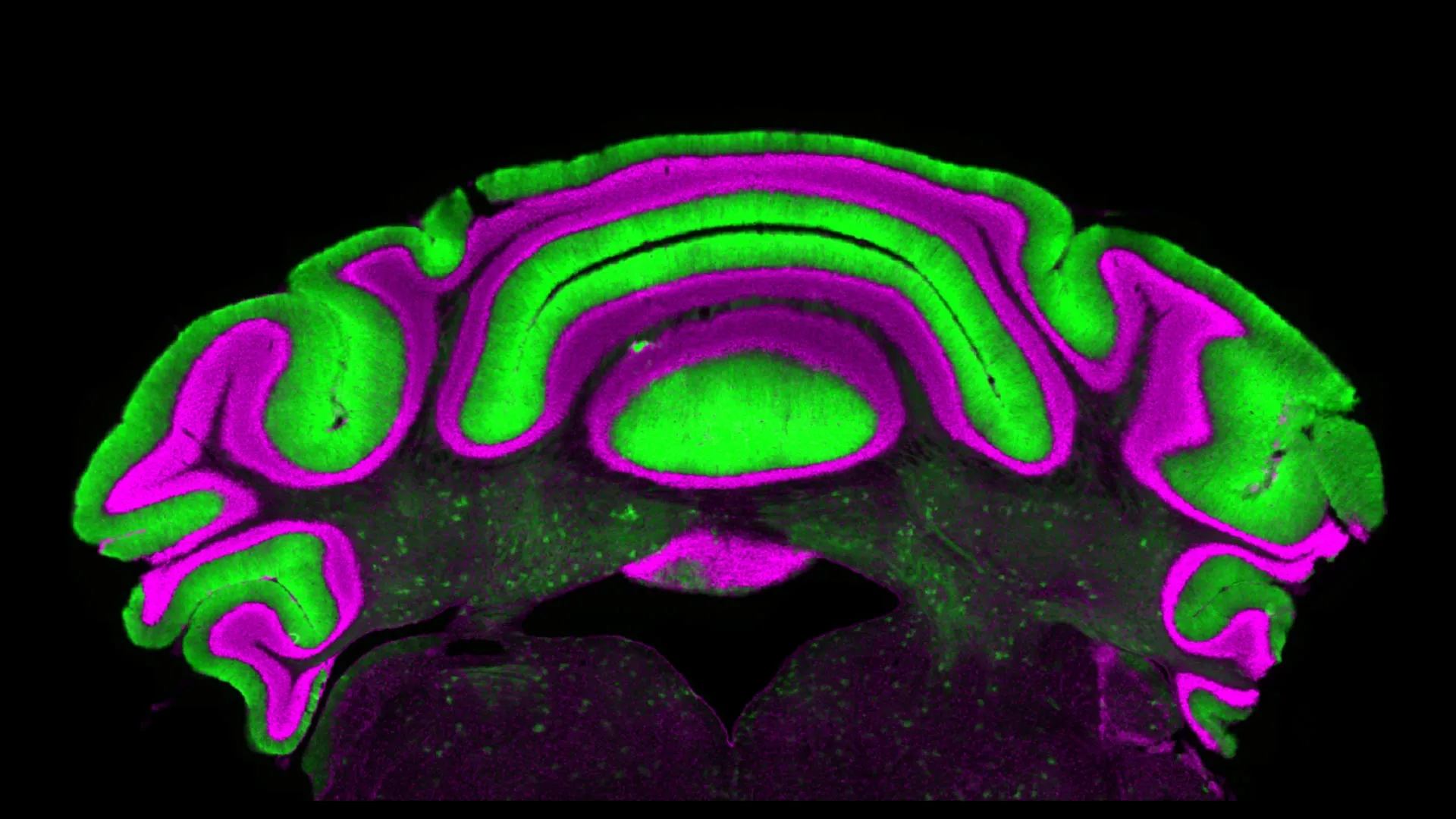
The cerebellum, often referred to as the "little brain," is a densely packed region at the base of the skull responsible for the fine-tuning of motor control, balance, and coordination. While it accounts for only about 10 percent of the brain’s total volume, it contains more than half of its neurons. When the intricate circuitry of this region is disrupted, the results are often debilitating. Patients may suffer from dystonia, characterized by painful, involuntary muscle contractions; ataxia, which leads to a lack of muscle coordination and balance; or tremors, which manifest as uncontrollable rhythmic shaking. For years, the roadmap for understanding these conditions has relied on a specific anatomical hierarchy within the cerebellar cortex.
The Traditional Model of Cerebellar Function
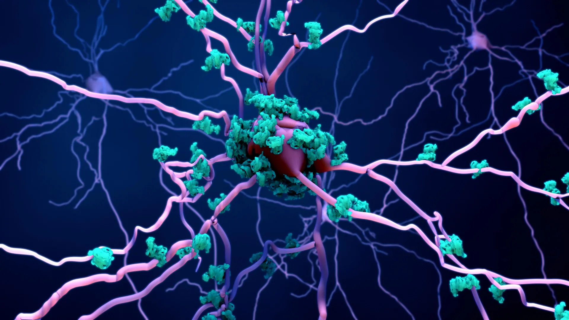
To understand the significance of the new findings, one must first look at the established biological model that has guided neuroscience for nearly half a century. The cerebellar circuit is characterized by a high degree of convergence. At the heart of this circuit are the Purkinje cells, large neurons with extensive dendritic trees that reside in the outer layer of the cerebellum. These cells are unique because they are exclusively inhibitory; they release a neurotransmitter called GABA (gamma-aminobutyric acid), which acts as a "brake" on the neurons they target.
The primary targets of these Purkinje cells are the deep cerebellar nuclei (DCN) cells, located deep within the white matter of the cerebellum. The DCN are the sole output hub of the cerebellum, meaning every instruction the cerebellum sends to the spinal cord or the motor cortex must pass through them. Because Purkinje cells provide the vast majority of the input to the DCN, researchers have long operated under a "linear inhibition" assumption: if Purkinje cells are highly active, the DCN should be suppressed; if Purkinje cells are quiet, the DCN should be highly active.
This assumption turned the Purkinje cell into a convenient biomarker. Because they are located on the surface of the brain, they are significantly easier to record using electrodes or imaging techniques than the deep-seated DCN. Consequently, thousands of studies have used Purkinje cell activity as a proxy for understanding what the cerebellum is "telling" the rest of the body.
Challenging the Proxy: The Van der Heijden Study
The new study, published in the Journal of Physiology, was led by Meike van der Heijden, an assistant professor at the Fralin Biomedical Research Institute and the Virginia Tech School of Neuroscience. Alongside first author Alyssa Lyon, a doctoral candidate in the Translational Biology, Medicine, and Health Graduate Program, the team sought to verify whether this proxy relationship actually held up under the stress of disease states.
The team utilized a comprehensive approach, analyzing a vast database of electrophysiology recordings derived from pre-clinical models of cerebellar disease. Electrophysiology involves the direct measurement of the electrical activity of neurons, providing a millisecond-by-millisecond account of how cells communicate. By comparing the firing patterns of Purkinje cells and DCN cells simultaneously across various conditions, the researchers looked for the expected inverse correlation.
The results were unexpected. The data revealed that there is no significant linear correlation between the activity levels of the two cell populations in diseased states. High levels of Purkinje cell firing did not consistently result in DCN suppression, nor did low levels necessarily lead to DCN excitation.
"We see that there’s not a clear linear relationship between activity in the Purkinje cells and in the deep nuclei cells," stated Van der Heijden. "So there’s very limited predictive power in monitoring one to understand what’s going on in the other."
Understanding the Disconnect in Movement Disorders
The implications of this disconnect are profound for the study of movement disorders. Dystonia, ataxia, and tremor are not just "off" or "on" malfunctions; they are often the result of "noisy" or "erratic" signaling.
- Dystonia: This condition affects approximately 250,000 people in the United States alone. It causes muscles to contract involuntarily, leading to repetitive or twisting movements. In many cases, the cerebellum is suspected of sending "overflow" signals that activate muscles that should remain relaxed.
- Ataxia: Affecting thousands of individuals through genetic predispositions or acquired brain injuries, ataxia disrupts the timing and scaling of movements. It turns smooth actions into jerky, uncoordinated ones.
- Tremor: Essential tremor is one of the most common neurological diseases, affecting millions of older adults. It is characterized by an oscillatory signal that originates within the cerebellar-thalamic circuit.
Historically, if a researcher observed abnormal firing in the Purkinje cells of an ataxic mouse, they would conclude that those Purkinje cells were causing the ataxia by mismanaging the DCN. However, Lyon and Van der Heijden’s research suggests that the DCN might be behaving abnormally for reasons entirely independent of the Purkinje cells, or that the DCN is "ignoring" or "reinterpreting" the Purkinje signals in ways the current models do not account for.
Why the Assumption Persisted
The scientific community’s reliance on Purkinje cells was not merely a matter of oversight, but one of practical necessity and anatomical accessibility. The deep cerebellar nuclei are encased in layers of tissue and located near the brainstem, making them difficult to reach without invasive procedures that can damage the surrounding architecture.
"One reason Purkinje cells have received so much attention is that they are easier to study," the researchers noted. Because they sit in the outer layer, they are the "low-hanging fruit" of cerebellar electrophysiology. For decades, this accessibility bias created a self-reinforcing cycle: because more data existed for Purkinje cells, more theories were built around them, which in turn led to more studies focusing on them.
The Virginia Tech study serves as a "cautionary tale," as Van der Heijden described it, warning that the path of least resistance in data collection does not always lead to the most accurate clinical conclusions.
Implications for Treatment and Drug Development
The most critical impact of this research lies in the realm of therapeutics. Currently, several treatments for movement disorders aim to modulate cerebellar activity. These include pharmacological interventions (drugs) and Deep Brain Stimulation (DBS), where electrodes are implanted into the brain to deliver electrical pulses.
If a pharmaceutical company develops a drug intended to treat dystonia by quieting Purkinje cells, but the DCN cells do not respond to that change in a predictable way, the drug will likely fail in clinical trials. This research suggests that many failed interventions may have been targeting the wrong "lever" in the cerebellar machine.
"Purkinje and cerebellar deep nuclei cell activity is disrupted in a disease state, and a better understanding of the relationship between these neuron types will ultimately help optimize treatments," said Alyssa Lyon.
Furthermore, this study highlights the need for more sophisticated diagnostic tools. If Purkinje cell activity is not a reliable biomarker, then researchers must prioritize the development of technologies that can safely and accurately monitor the deep nuclei in human patients. This could involve high-resolution functional MRI (fMRI) techniques or more refined electrode arrays for use during neurosurgery.
A New Framework for Cerebellar Research
The findings from the Fralin Biomedical Research Institute suggest a shift toward a "DCN-centric" view of cerebellar disease. Instead of viewing the DCN as a passive recipient of Purkinje cell instructions, researchers are now encouraged to view the DCN as an active integrator of multiple signals.
In addition to Purkinje cells, the DCN receive "collateral" inputs from other parts of the brain, including the mossy fibers and climbing fibers. It is possible that in disease states, these collateral inputs become dominant, or that the internal properties of the DCN cells themselves change, rendering them less sensitive to Purkinje cell inhibition.
This research aligns with a growing movement in neuroscience to look at "circuit-wide" dynamics rather than single-cell-type dynamics. The brain is a complex network, and as Van der Heijden’s team has shown, the connections between its nodes are not always as straightforward as an anatomy textbook might suggest.
Future Directions and Scientific Consensus
The publication of this study in the Journal of Physiology is expected to trigger a wave of follow-up research. Scientists will likely look to replicate these findings in different models of disease and explore the specific mechanisms that cause the breakdown in the Purkinje-DCN relationship.
The broader scientific community has long debated the "rate code" versus the "temporal code" of the cerebellum—whether it is the speed of the firing or the timing of the pulses that matters most. This study adds a new layer to that debate by suggesting that neither code may be interpreted by the DCN in the way we previously thought during the progression of neurological disorders.
As the Fralin Biomedical Research Institute continues its work, the focus will likely shift toward identifying the specific "noise" in the DCN that leads to motor symptoms. By bypassing the Purkinje cell proxy and looking directly at the output, researchers hope to find more effective targets for the next generation of neurological therapies.
In the final analysis, the work of Van der Heijden and Lyon serves as a reminder of the fundamental scientific principle: hypotheses must be tested, and even the most "obvious" anatomical connections must be verified through rigorous experimentation. For patients suffering from the debilitating effects of dystonia, ataxia, and tremor, this shift in focus offers a new glimmer of hope that treatments will one day be based on the brain’s actual behavior, rather than our assumptions of it.